Acquired non-inflammatory myopathy

Last updated
Acquired non-inflammatory myopathy
Other namesANIM
Genga 36.jpg
Condition affects skeletal muscle
Specialty Neurology

Acquired non-inflammatory myopathy (ANIM) is a neuromuscular disorder primarily affecting skeletal muscle, most commonly in the limbs of humans, resulting in a weakness or dysfunction in the muscle. [1] A myopathy refers to a problem or abnormality with the myofibrils, which compose muscle tissue. In general, non-inflammatory myopathies are a grouping of muscular diseases not induced by an autoimmune-mediated inflammatory pathway. These muscular diseases usually arise from a pathology within the muscle tissue itself rather than the nerves innervating that tissue. ANIM has a wide spectrum of causes which include drugs and toxins, nutritional imbalances, acquired metabolic dysfunctions such as an acquired defect in protein structure, and infections. [1]

Contents

Acquired non-inflammatory myopathy is a different diagnosis than inflammatory myopathy. Inflammatory myopathies are a direct result of some type of autoimmune mediated pathway whereas ANIM is not the result of a dysfunction of the immune system. [2] In addition, the cause of inflammatory myopathy is relatively unknown, whereas many causal agents for ANIM have been discovered which typically affect the structural integrity and function of the muscle fibers. [2]

Most myopathies are typically first diagnosed and classified as an idiopathic inflammatory myopathy. [2] [3] [4] [5] However, a diagnosis of ANIM occurs when the cause of the myopathy is found to not arise from an autoimmune mechanism. [1]

Symptoms and signs

Patients with acquired non-inflammatory myopathy typically experience weakness, cramping, stiffness, and tetany, most commonly in skeletal muscle surrounding the limbs and upper shoulder girdle. [1]

The most commonly reported symptoms are:

Cause

Acquired non-inflammatory myopathy can be caused by a variety of factors including metabolic abnormalities, drugs, nutritional deficiency, trauma, and upstream abnormalities resulting in decreased function. Two of the most common causes of ANIM are hyperthyroidism and excessive steroid use, while many drugs used to treat rheumatism are known to be inducing agents. Most cases of ANIM can be linked to drugs or dietary abnormalities.[ citation needed ]

Drug-induced myopathy

It is not uncommon for drugs to damage muscle fibers. Particular families of drugs are known to induce myopathies on the molecular level, thus altering organelle function such as the mitochondria. Use of multiple drugs from these families in conjunction with one another can increase the risk of developing a myopathy. [6] Many of the drugs associated with inducing myopathies in patients are found in rheumatology practice.[ citation needed ]

Statins

  • Prescribed statins (HMG-CoA reductase inhibitors) for dyslipidemia are associated with muscle toxicity. Symptoms of this muscle toxicity include combinations of cramping, weakness, aching or tenderness; and are often experienced in the quadriceps, pectoral, biceps, low back, or abdominal region. Symptoms tend to worsen with muscle exercise, and often continue after a patient is removed from statin therapy. [1] Common types of myopathy due to statins include myalgia, myositis, and rhabdomyolysis. Statins induce myopathy by inhibiting protein synthesis within the muscle. [6] Statin therapy tends to not show any histopathological differences, and thus a biopsy does not reveal too much about the damage. Often, the damage is found within the mitochondria. [1]
Colchicine is commonly prescribed for gout treatment. Colchicine.svg
Colchicine is commonly prescribed for gout treatment.

Corticosteroids

  • Corticosteroids often cause muscle weakness to some degree in patients. Symptoms are usually weakness of the proximal muscles, neck flexor, and in extreme cases, respiratory muscle weakness can also occur. [1] Corticosteroids have not only been found to cause some degree of muscle atrophy, but also a local or diffuse cell death. These side effects are more common in women than in men, for reasons that are unknown. [6] EMGs illicit a low amplitude motor potentials, and lack spontaneous electrical activity. [1]

Colchicine

  • Patients who start taking colchicine, and who have compromised renal function, develop a myopathy that shows symptoms in proximal muscle weakness, distal sensory loss and areflexia. A muscle biopsy shows a vacuolar myopathy without significant cell death or inflammation. [1]

Chloroquine/Hydroxychloroquine

  • Chloroquine/hydroxychloroquine prescriptions can cause a development of a progressively slow muscle weakness that begins in the low extremities, and moves to the upper limbs. Muscle enzymes are increased, commonly lactate dehydrogenase (LDH). [1]

Diet and Trauma Induced Myopathy

Many dietary factors and aberrations can induce ANIM. Chemical imbalances brought on by abnormal diets may either affect the muscle directly or induce abnormal functionality in upstream pathways.

Muscular Weakness brought on by ANIM can induce sleep apnea Obstruction ventilation apnee sommeil.svg
Muscular Weakness brought on by ANIM can induce sleep apnea

Trauma to any muscle is also a common cause for acute ANIM. This is due to muscular contusions and partial or complete loss of function for affected muscle groups. [2]

Diagnosis

EMG is often used as a diagnostic tool for ANIM EMG - SIMI.jpg
EMG is often used as a diagnostic tool for ANIM

A patient's history is one of the key factors in diagnosing acquired non-inflammatory myopathy. The history is used not only to analyze the time frame with which the patient began to express symptoms, but to also see if the disease is within the patient's family history, to check medication or drug use history, and to see if the patient has had any trauma due to illness or infection. Basic exams will test for where the muscle weakness is and how weak it is. This is performed by testing for proximal and distal muscle strength, as well as testing for any signs of neurogenic symptoms such as impaired sensation, deep tendon reflexes, and atrophy. [1]

If needed, more advanced equipment can be used to help determine whether a patient has ANIM. This includes:

When examining the serum levels of muscle enzymes, the relative levels of creatine kinase, aldolase, aspartate aminotransferase, alanine aminotransferase, and lactate dehydrogenase are closely examined. Abnormal levels of these proteins are indicative of both inflammatory myopathy and ANIM. [1]

EMGs are particularly useful in locating the affected muscle groups, as well as determining the distribution of the myopathy throughout the cell. EMGs measure several indicators of myopathies such as: [1]

Magnetic Resonance Imaging will elicit edema in inflammatory patients, but it will most likely show nothing in patients with ANIM and if it does, it will show some atrophy. [1]

If an individual's ANIM is a result of a metabolite defect, then additional tests are required. These tests are directed at enzyme function at rest and during exercise, and enzyme intermediates. Molecular genetic testing is often used to determine if there was any predisposition to the expressed symptoms. [1]

Screening

During vigorous ischemic exercise, skeletal muscle functions anaerobically, generating lactate and ammonia a coproduct of muscle myoadenylate deaminase (AMPD) activity. The forearm ischemic exercise test takes advantage of this physiology and has been standardized to screen for disorders of glycogen metabolism and AMPD deficiency. Patients with a glycogen storage disease manifest a normal increase in ammonia but no change from baseline of lactate, whereas in those with AMPD deficiency, lactate levels increase but ammonia levels do not. If ischemic exercise testing gives an abnormal result, enzyme analysis must be performed on muscle to confirm the putative deficiency state because false-positive results can occur.[ citation needed ]

Treatment

Physical Therapy can be used to alleviate atrophy and soreness from ANIM US Navy 111102-N-IZ292-050 Operations Specialist 1st Class Jennifer Funderburk, right, lifts weights during physical therapy in the Physical Therap.jpg
Physical Therapy can be used to alleviate atrophy and soreness from ANIM

Treatment for acquired noninflammatory myopathy is directed towards resolution of the underlying condition, pain management, and muscle rehabilitation. Drug induced ANIMs can be reversed or improved by tapering off of the drugs and finding alternative care. [6] Hyperthyroidism induced ANIM can be treated through anti-thyroid drugs, surgery, and not eating foods high in Iodine such as kelp. Treatment of the hyperthyroidism results in complete recovery of the myopathy. [7] ANIM caused by vitamin D deficiency can easily be resolved by taking vitamin supplements and increasing one's exposure to direct sunlight. [8]

Pain can be managed through massaging affected areas and the use of nonsteroidal anti-inflammatory drugs (NSAIDs). Exercise, physical therapy, and occupational therapy can be used to rehabilitate affected muscle areas and resist the atrophy process. [2] The use of walkers, canes, and braces may assist with the mobility of the affected individual.[ citation needed ]

Research direction

A diagnostic test for statin-associated auto-immune necrotizing myopathy will be available soon in order to differentiate between different types of myopathies during diagnosis. The presence of abnormal spontaneous electrical activity in the resting muscles indicates an irritable myopathy and is postulated to reflect the presence of an active necrotising myopathic process or unstable muscle membrane potential. However, this finding has poor sensitivity and specificity for predicting the presence of an inflammatory myopathy on biopsy. Further research into this spontaneous electrical activity will allow for a more accurate differential diagnosis between the different myopathies. [1]

Currently a muscle biopsy remains a critical test, unless the diagnosis can be secured by genetic testing. Genetic testing is a less invasive test and if it can be improved upon that would be ideal. Molecular genetic testing is now available for many of the more common metabolic myopathies and muscular dystrophies. These tests are costly and are thus best used to confirm rather than screen for a diagnosis of a specific myopathy. It is the hope of researchers that as these testing methods improve in function, both costs and access will become more manageable [6]

The increased study of muscle pathophysiology is of importance to researchers as it helps to better differentiate inflammatory versus non-inflammatory and to aim treatment as part of the differential diagnosis. Certainly classification schemes that better define the wide range of myopathies will help clinicians to gain a better understanding of how to think about these patients. Continued research efforts to help appreciate the pathophysiology will improve clinicians ability to administer the most appropriate therapy based on the particular variety of myopathy. [2]

The mechanism for myopathy in individuals with low vitamin D is not completely understood. A decreased availability of 250HD leads to mishandling of cellular calcium transport to the sarcoplasmic reticulum and mitochondria, and is associated with reduced actomyosin content of myofibrils. [8]

See also

Related Research Articles

Inclusion body myositis (IBM) is the most common inflammatory muscle disease in older adults. The disease is characterized by slowly progressive weakness and wasting of both proximal muscles and distal muscles, most apparent in the finger flexors and knee extensors. IBM is often confused with an entirely different class of diseases, called hereditary inclusion body myopathies (hIBM). The "M" in hIBM is an abbreviation for "myopathy" while the "M" in IBM is for "myositis". In IBM, two processes appear to occur in the muscles in parallel, one autoimmune and the other degenerative. Inflammation is evident from the invasion of muscle fibers by immune cells. Degeneration is characterized by the appearance of holes, deposits of abnormal proteins, and filamentous inclusions in the muscle fibers. sIBM is a rare disease, with a prevalence ranging from 1 to 71 individuals per million.

<span class="mw-page-title-main">Myalgia</span> Muscle pain

Myalgia is the medical term for muscle pain. Myalgia is a symptom of many diseases. The most common cause of acute myalgia is the overuse of a muscle or group of muscles; another likely cause is viral infection, especially when there has been no trauma.

<span class="mw-page-title-main">Adenosine monophosphate deaminase deficiency type 1</span> Medical condition

Adenosine monophosphate deaminase deficiency type 1 or AMPD1, is a human metabolic disorder in which the body consistently lacks the enzyme AMP deaminase, in sufficient quantities. This may result in exercise intolerance, muscle pain and muscle cramping. The disease was formerly known as myoadenylate deaminase deficiency (MADD).

<span class="mw-page-title-main">Benign fasciculation syndrome</span> Involuntary muscle twitching in the voluntary muscles

Benign fasciculation syndrome (BFS) is characterized by fasciculation (twitching) of voluntary muscles in the body. The twitching can occur in any voluntary muscle group but is most common in the eyelids, arms, hands, fingers, legs, and feet. The tongue can also be affected. The twitching may be occasional to continuous. BFS must be distinguished from other conditions that include muscle twitches.

<span class="mw-page-title-main">Glycogen storage disease type V</span> Human disease caused by deficiency of a muscle enzyme

Glycogen storage disease type V, also known as McArdle's disease, is a metabolic disorder, one of the metabolic myopathies, more specifically a muscle glycogen storage disease, caused by a deficiency of myophosphorylase. Its incidence is reported as one in 100,000, roughly the same as glycogen storage disease type I.

<span class="mw-page-title-main">Glycogen storage disease</span> Medical condition

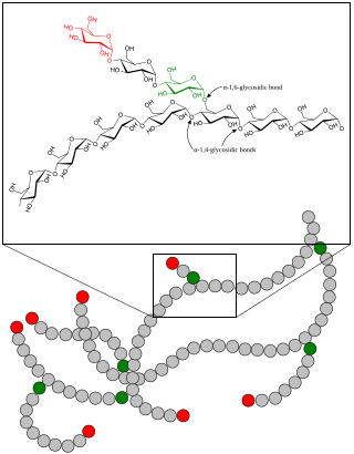
A glycogen storage disease is a metabolic disorder caused by a deficiency of an enzyme or transport protein affecting glycogen synthesis, glycogen breakdown, or glucose breakdown, typically in muscles and/or liver cells.

<span class="mw-page-title-main">Rhabdomyolysis</span> Human disease (condition) in which damaged skeletal muscle breaks down rapidly

Rhabdomyolysis is a condition in which damaged skeletal muscle breaks down rapidly, often due to high intensity exercise over a short period of time. Symptoms may include muscle pains, weakness, vomiting, and confusion. There may be tea-colored urine or an irregular heartbeat. Some of the muscle breakdown products, such as the protein myoglobin, are harmful to the kidneys and can cause acute kidney injury.

<span class="mw-page-title-main">Phosphofructokinase deficiency</span> Medical condition

Phosphofructokinase deficiency is a rare muscular metabolic disorder, with an autosomal recessive inheritance pattern.

In medicine, myopathy is a disease of the muscle in which the muscle fibers do not function properly. Myopathy means muscle disease. This meaning implies that the primary defect is within the muscle, as opposed to the nerves or elsewhere.

<span class="mw-page-title-main">Mitochondrial myopathy</span> Medical condition

Mitochondrial myopathies are types of myopathies associated with mitochondrial disease. Adenosine triphosphate (ATP), the chemical used to provide energy for the cell, cannot be produced sufficiently by oxidative phosphorylation when the mitochondrion is either damaged or missing necessary enzymes or transport proteins. With ATP production deficient in mitochondria, there is an over-reliance on anaerobic glycolysis which leads to lactic acidosis either at rest or exercise-induced.

<span class="mw-page-title-main">Myositis</span> Medical condition

Myositis is a rarely-encountered medical condition characterized by inflammation affecting the muscles. The manifestations of this condition may include skin issues, muscle weakness, and the potential involvement of other organs. Additionally, systemic symptoms like weight loss, fatigue, and low-grade fever can manifest in individuals with myositis.

<span class="mw-page-title-main">MELAS syndrome</span> Medical condition

Mitochondrial encephalopathy, lactic acidosis, and stroke-like episodes (MELAS) is one of the family of mitochondrial diseases, which also include MIDD, MERRF syndrome, and Leber's hereditary optic neuropathy. It was first characterized under this name in 1984. A feature of these diseases is that they are caused by defects in the mitochondrial genome which is inherited purely from the female parent. The most common MELAS mutation is mitochondrial mutation, mtDNA, referred to as m.3243A>G.

<span class="mw-page-title-main">Centronuclear myopathy</span> Medical condition

Centronuclear myopathies (CNM) are a group of congenital myopathies where cell nuclei are abnormally located in the center of muscle cells instead of their normal location at the periphery.

<span class="mw-page-title-main">Congenital muscular dystrophy</span> Medical condition

Congenital muscular dystrophies are autosomal recessively-inherited muscle diseases. They are a group of heterogeneous disorders characterized by muscle weakness which is present at birth and the different changes on muscle biopsy that ranges from myopathic to overtly dystrophic due to the age at which the biopsy takes place.

Thyrotoxic myopathy (TM) is a neuromuscular disorder that develops due to the overproduction of the thyroid hormone thyroxine. Also known as hyperthyroid myopathy, TM is one of many myopathies that lead to muscle weakness and muscle tissue breakdown. Evidence indicates the onset may be caused by hyperthyroidism. Physical symptoms of TM may include muscle weakness, the breakdown of muscle tissue, fatigue, and heat intolerance. Physical acts such as lifting objects and climbing stairs may become increasingly difficult. If untreated, TM can be an extremely debilitating disorder that can, in extreme rare cases, lead to death. If diagnosed and treated properly the effects can be controlled and in most cases reversed leaving no lasting effects.

<span class="mw-page-title-main">Inflammatory myopathy</span> Medical condition

Inflammatory myopathy, also known as idiopathic inflammatory myopathy (IIM), is disease featuring muscle weakness, inflammation of muscles (myositis), and in some types, muscle pain. The cause of much inflammatory myopathy is unknown (idiopathic), and such cases are classified according to their symptoms and signs, electromyography, MRI, and laboratory findings. It can also be associated with underlying cancer. The main classes of idiopathic inflammatory myopathy are polymyositis (PM), dermatomyositis (DM), inclusion-body myositis (IBM), immune-mediated necrotising myopathy (IMNM), and focal autoimmune myositis.

<span class="mw-page-title-main">Metabolic myopathy</span> Type of myopathies

Metabolic myopathies are myopathies that result from defects in biochemical metabolism that primarily affect muscle. They are generally genetic defects that interfere with muscle's ability to create energy, causing a low ATP reservoir within the muscle cell.

<span class="mw-page-title-main">Brody myopathy</span> Medical condition

Brody myopathy, also called Brody disease, is a rare disorder that affects skeletal muscle function. BD was first characterized in 1969 by Dr. Irwin A. Brody at Duke University Medical Center. Individuals with BD have difficulty relaxing their muscles after exercise. This difficulty in relaxation leads to symptoms including cramps, stiffness, and discomfort in the muscles of the limbs and face. Symptoms are heightened by exercise and commonly progress in severity throughout adulthood.

Statin-associated autoimmune myopathy (SAAM), also known as anti-HMGCR myopathy, is a very rare form of muscle damage caused by the immune system in people who take statin medications. However, there are cases of SAAM in patients who have not taken statin medication, and this can be explained by the exposure to natural sources of statin such as red yeast rice, which is statin rich. This theory is supported by the higher prevalence of statin-naive SAAM patients in Asian cohorts, who have statin-rich diets.

Autophagic vacuolar myopathy (AVM) consists of multiple rare genetic disorders with common histological and pathological features on muscle biopsy. The features highlighted are vacuolar membranes of the autophagic vacuoles having sarcolemmal characteristics and an excess of autophagic vacuoles. There are currently five types of AVM identified. The signs and symptoms become more severe over the course of the disease. It begins with an inability to pick up small objects and progresses to difficulty in walking. The age of onset varies from early childhood to late adulthood, affecting people of all ages.

References

  1. 1 2 3 4 5 6 7 8 9 10 11 12 13 14 15 16 17 18 19 20 21 22 23 24 25 26 27 Baer AN, Wortmann RL (May 2013). "Noninflammatory myopathies". Rheumatic Disease Clinics of North America. 39 (2): 457–479. doi:10.1016/j.rdc.2013.02.006. PMID   23597974.
  2. 1 2 3 4 5 6 Castro C, Gourley M (April 2012). "Diagnosis and treatment of inflammatory myopathy: issues and management". Therapeutic Advances in Musculoskeletal Disease. 4 (2): 111–120. doi:10.1177/1759720x11425092. PMC   3383519 . PMID   22870499.
  3. Bhai SF, Dimachkie MM, de Visser M (June 2022). "Is it really myositis? Mimics and pitfalls". Best Practice & Research. Clinical Rheumatology. 36 (2): 101764. doi:10.1016/j.berh.2022.101764. PMID   35752578. S2CID   250012951.
  4. Szczęsny P, Świerkocka K, Olesińska M (2018). "Differential diagnosis of idiopathic inflammatory myopathies in adults - the first step when approaching a patient with muscle weakness". Reumatologia. 56 (5): 307–315. doi:10.5114/reum.2018.79502. PMC   6263305 . PMID   30505013.
  5. Tarnopolsky MA, Hatcher E, Shupak R (May 2016). "Genetic Myopathies Initially Diagnosed and Treated as Inflammatory Myopathy". The Canadian Journal of Neurological Sciences. Le Journal Canadien des Sciences Neurologiques. 43 (3): 381–384. doi: 10.1017/cjn.2015.386 . PMID   26911292. S2CID   25515951.
  6. 1 2 3 4 5 Owczarek J, Jasińska M, Orszulak-Michalak D (2005). "Drug-induced myopathies. An overview of the possible mechanisms". Pharmacological Reports. 57 (1): 23–34. PMID   15849374.
  7. 1 2 3 Wilkinson I, Lennox G (2005). Essential Neurology (4th ed.). Wiley-Blackwell. p. 171. ISBN   978-1-4051-1867-5.
  8. 1 2 3 4 McCarty DE, Chesson AL, Jain SK, Marino AA (August 2014). "The link between vitamin D metabolism and sleep medicine". Sleep Medicine Reviews. 18 (4): 311–319. doi:10.1016/j.smrv.2013.07.001. PMID   24075129.