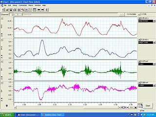
Spasticity is a feature of altered skeletal muscle performance with a combination of paralysis, increased tendon reflex activity, and hypertonia. It is also colloquially referred to as an unusual "tightness", stiffness, or "pull" of muscles.

Muscle spindles are stretch receptors within the body of a skeletal muscle that primarily detect changes in the length of the muscle. They convey length information to the central nervous system via afferent nerve fibers. This information can be processed by the brain as proprioception. The responses of muscle spindles to changes in length also play an important role in regulating the contraction of muscles, for example, by activating motor neurons via the stretch reflex to resist muscle stretch.

Sir Charles Scott Sherrington was an eminent English neurophysiologist. His experimental research established many aspects of contemporary neuroscience, including the concept of the spinal reflex as a system involving connected neurons, and the ways in which signal transmission between neurons can be potentiated or depotentiated. Sherrington himself coined the word "synapse" to define the connection between two neurons. His book The Integrative Action of the Nervous System (1906) is a synthesis of this work, in recognition of which he was awarded the Nobel Prize in Physiology or Medicine in 1932.

Stretching is a form of physical exercise in which a specific muscle or tendon is deliberately flexed or stretched in order to improve the muscle's felt elasticity and achieve comfortable muscle tone. The result is a feeling of increased muscle control, flexibility, and range of motion. Stretching is also used therapeutically to alleviate cramps and to improve function in daily activities by increasing range of motion.

The patellar reflex, also called the knee reflex or knee-jerk, is a stretch reflex which tests the L2, L3, and L4 segments of the spinal cord.
René Descartes (1596–1650) was one of the first to conceive a model of reciprocal innervation as the principle that provides for the control of agonist and antagonist muscles. Reciprocal innervation describes skeletal muscles as existing in antagonistic pairs, with contraction of one muscle producing forces opposite to those generated by contraction of the other. For example, in the human arm, the triceps acts to extend the lower arm outward while the biceps acts to flex the lower arm inward. To reach optimum efficiency, contraction of opposing muscles must be inhibited while muscles with the desired action are excited. This reciprocal innervation occurs so that the contraction of a muscle results in the simultaneous relaxation of its corresponding antagonist.
In physiology, medicine, and anatomy, muscle tone is the continuous and passive partial contraction of the muscles, or the muscle's resistance to passive stretch during resting state. It helps to maintain posture and declines during REM sleep.
Reciprocal inhibition describes the relaxation of muscles on one side of a joint to accommodate contraction on the other side. In some allied health disciplines, this is known as reflexive antagonism. The central nervous system sends a message to the agonist muscle to contract. The tension in the antagonist muscle is activated by impulses from motor neurons, causing it to relax.
Closed kinetic chain exercises or closed chain exercises (CKC) are physical exercises performed where the hand or foot is fixed in space and cannot move. The extremity remains in constant contact with the immobile surface, usually the ground or the base of a machine.
Muscle Energy Techniques (METs) describes a broad class of manual therapy techniques directed at improving musculoskeletal function or joint function, and improving pain. METs are commonly used by manual therapists, physical therapists, occupational therapist, chiropractors, athletic trainers, osteopathic physicians, and massage therapists. Muscle energy requires the patient to actively use his or her muscles on request to aid in treatment. Muscle energy techniques are used to treat somatic dysfunction, especially decreased range of motion, muscular hypertonicity, and pain.

Muscle coactivation occurs when agonist and antagonist muscles surrounding a joint contract simultaneously to provide joint stability. It is also known as muscle cocontraction, since two muscle groups are contracting at the same time. It is able to be measured using electromyography (EMG) from the contractions that occur. The general mechanism of it is still widely unknown. It is believed to be important in joint stabilization, as well as general motor control.

Muscle contractures can occur for many reasons, such as paralysis, muscular atrophy, and forms of muscular dystrophy. Fundamentally, the muscle and its tendons shorten, resulting in reduced flexibility. For example, in the case of partial paralysis the loss of strength and muscle control tend to be greater in some muscles than in others, leading to an imbalance between the various muscle groups around specific joints. Case in point: when the muscles which dorsiflex are less functional than the muscles which plantarflex a contracture occurs, giving the foot a progressively downward angle and loss of flexibility. Various interventions can slow, stop, or even reverse muscle contractures, ranging from physical therapy to surgery. A common cause for having the ankle lose its flexibility in this manner is from having sheets tucked in at the foot of the bed when sleeping. The weight of the sheets keep the feet plantarflexed all night. Correcting this by not tucking the sheets in at the foot of the bed, or by sleeping with the feet hanging off the bed when in the prone position, is part of correcting this imbalance.
The Golgi tendon reflex (also called inverse stretch reflex, autogenic inhibition, tendon reflex) is an inhibitory effect on the muscle resulting from the muscle tension stimulating Golgi tendon organs (GTO) of the muscle, and hence it is self-induced. The reflex arc is a negative feedback mechanism preventing too much tension on the muscle and tendon. When the tension is extreme, the inhibition can be so great it overcomes the excitatory effects on the muscle's alpha motoneurons causing the muscle to suddenly relax. This reflex is also called the inverse myotatic reflex, because it is the inverse of the stretch reflex.

Proprioception, also referred to as kinaesthesia, is the sense of self-movement, force, and body position. It is sometimes described as the "sixth sense".
Normal aging movement control in humans is about the changes in the muscles, motor neurons, nerves, sensory functions, gait, fatigue, visual and manual responses, in men and women as they get older but who do not have neurological, muscular or neuromuscular disorder. With aging, neuromuscular movements are impaired, though with training or practice, some aspects may be prevented.
Ballistic movement can be defined as muscle contractions that exhibit maximum velocities and accelerations over a very short period of time. They exhibit high firing rates, high force production, and very brief contraction times.
Williams flexion exercises (WFE) – also called Williams lumbar flexion exercises – are a set or system of related physical exercises intended to enhance lumbar flexion, avoid lumbar extension, and strengthen the abdominal and gluteal musculature in an effort to manage low back pain non-surgically. The system was first devised in 1937 by Dallas orthopedic surgeon Dr. Paul C. Williams.

A spinal interneuron, found in the spinal cord, relays signals between (afferent) sensory neurons, and (efferent) motor neurons. Different classes of spinal interneurons are involved in the process of sensory-motor integration. Most interneurons are found in the grey column, a region of grey matter in the spinal cord.

Anatomical terminology is used to uniquely describe aspects of skeletal muscle, cardiac muscle, and smooth muscle such as their actions, structure, size, and location.
Proprioception refers to the sensory information relayed from muscles, tendons, and skin that allows for the perception of the body in space. This feedback allows for more fine control of movement. In the brain, proprioceptive integration occurs in the somatosensory cortex, and motor commands are generated in the motor cortex. In the spinal cord, sensory and motor signals are integrated and modulated by motor neuron pools called central pattern generators (CPGs). At the base level, sensory input is relayed by muscle spindles in the muscle and Golgi tendon organs (GTOs) in tendons, alongside cutaneous sensors in the skin.


