Agouti-related peptide

Last updated
AGRP
1hyk.png
Available structures
PDB Ortholog search: PDBe RCSB
Identifiers
Aliases AGRP , agouti related neuropeptide, AGRT, ART, ASIP2, AgRP
External IDs OMIM: 602311 MGI: 892013 HomoloGene: 7184 GeneCards: AGRP
Orthologs
SpeciesHumanMouse
Entrez
Ensembl
UniProt
RefSeq (mRNA)

NM_001138

NM_001271806
NM_007427

RefSeq (protein)

NP_001129

NP_001258735
NP_031453

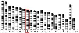
Location (UCSC) Chr 16: 67.48 – 67.48 Mb Chr 8: 106.29 – 106.36 Mb
PubMed search [3] [4]
Wikidata
View/Edit Human View/Edit Mouse

Agouti-related protein (AgRP), also called agouti-related peptide, is a neuropeptide produced in the brain by the AgRP/NPY neuron. It is synthesized in neuropeptide Y (NPY)-containing cell bodies located in the ventromedial part of the arcuate nucleus in the hypothalamus. [5] AgRP is co-expressed with NPY and acts to increase appetite and decrease metabolism and energy expenditure. It is one of the most potent and long-lasting of appetite stimulators. In humans, the agouti-related peptide is encoded by the AGRP gene. [6] [7]

Contents

Structure

AgRP is a paracrine signaling molecule made of 112 amino acids (the gene product of 132 amino acids is processed by removal of the N-terminal 20-residue signal peptide domain). It was independently identified by two teams in 1997 based on its sequence similarity with agouti signalling peptide (ASIP), a protein synthesized in the skin controlling coat colour. [6] [7] AgRP is approximately 25% identical to ASIP. The murine homologue of AgRP consists of 111 amino acids (precursor is 131 amino acids) and shares 81% amino acid identity with the human protein. Biochemical studies indicate AgRP to be very stable to thermal denaturation and acid degradation. Its secondary structure consists mainly of random coils and β-sheets [8] that fold into an inhibitor cystine knot motif. [9] AGRP maps to human chromosome 16q22 and Agrp to mouse chromosome 8D1-D2.

Function

Agouti-related protein is expressed primarily in the adrenal gland, subthalamic nucleus, and hypothalamus, with lower levels of expression in the testis, kidneys, and lungs. The appetite-stimulating effects of AgRP are inhibited by the hormone leptin and activated by the hormone ghrelin. Adipocytes secrete leptin in response to food intake. This hormone acts in the arcuate nucleus and inhibits the AgRP/NPY neuron from releasing orexigenic peptides. [10] Ghrelin has receptors on NPY/AgRP neurons that stimulate the secretion of NPY and AgRP to increase appetite. AgRP is stored in intracellular secretory granules and is secreted via a regulated pathway. [11] The transcriptional and secretory action of AgRP is regulated by inflammatory signals. [12] Levels of AgRP are increased during periods of fasting. It has been found that AgRP stimulates the hypothalamic-pituitary-adrenocortical axis to release ACTH, cortisol and prolactin. It also enhances the ACTH response to IL-1-beta, suggesting it may play a role in the modulation of neuroendocrine response to inflammation. [13] Conversely, AgRP-secreting neurons inhibit the release of TRH from the paraventricular nucleus (PVN), which may contribute to conservation of energy in starvation. [14] This pathway is part of a feedback loop, since TRH-secreting neurons from PVN stimulate AgRP neurons. [15]

Mechanism

AGRP has been demonstrated to be a competitive antagonist of melanocortin receptors, specifically MC3-R and MC4-R. The melanocortin receptors, MC3-R and MC4-R, are directly linked to metabolism and body weight control. These receptors are activated by the peptide hormone α-MSH (melanocyte-stimulating hormone) and antagonized by the agouti-related protein. [16] Whereas α-MSH acts broadly on most members of the MCR family (with the exception of MC2-R), AGRP is highly specific for only MC3-R and MC4-R. This inverse agonism not only antagonizes the action of melanocortin agonists such as α-MSH but also further decreases the cAMP produced by the affected cells. The exact mechanism by which AgRP inhibits melanocortin-receptor signalling is not completely clear. It has been suggested that Agouti-related protein binds MSH receptors and acts as a competitive antagonist of ligand binding. [17] Studies of Agouti protein in B16 melanoma cells supported this logic. The expression of AgRP in the adrenal gland is regulated by glucocorticoids. The protein blocks α-MSH-induced secretion of corticosterone. [18]

History

Orthologs of AgRP, ASIP, MCIR, and MC4R have been found in mammalian, teleost fish, and avian genomes. This suggests that the agouti-melanocortin system evolved by gene duplication from individual ligand and receptor genes in the last 500 million years. [16]

Role in obesity

AgRP induces obesity by chronic antagonism of the MC4-R. [19] Overexpression of AgRP in transgenic mice (or intracerebroventricular injection) causes hyperphagia and obesity, [20] whilst AgRP plasma levels have been found to be elevated in obese human males. [21] Understanding the role AgRP plays in weight gain may assist in developing pharmaceutical models for treating obesity. AgRP mRNA levels have been found to be down regulated following an acute stressful event. Studies suggest that systems involved in the regulation of stress response and of energy balance are highly integrated. Loss or gain of AgRP function may result in inadequate adaptive behavioural responses to environmental events, such as stress, and have potential to contribute to the development of eating disorders. It has been shown that polymorphisms in the AgRP gene have been linked with anorexia nervosa [22] as well as obesity. Some studies suggest that inadequate signalling of AgRP during stress may result in binge eating. Starvation-induced hypothalamic autophagy generates free fatty acids, which in turn regulate neuronal AgRP levels. [23]

Agouti protein
1mr0.png
Identifiers
SymbolAgouti
Pfam PF05039
Pfam clan CL0083
InterPro IPR007733
PROSITE PDOC60024
SCOP2 1hyk / SCOPe / SUPFAM
OPM superfamily 112
OPM protein 1mr0
Available protein structures:
Pfam   structures / ECOD  
PDB RCSB PDB; PDBe; PDBj
PDBsum structure summary

Role in hunger circuitry

According to Mark L. Andermann and Bradford B. Lowell: "...AgRP neurons and the wiring diagram within which they operate can be viewed as the physical embodiment of the intervening variable, hunger." [24] Stimulation of neurons expressing AgRP can induce robust feeding behavior in mice that will trigger: increased food consumption, [25] increased willingness to work for food, [26] and increased investigation of food odors. [27]

Despite this, AgRP neurons are rapidly inhibited upon food presentation and the onset of eating. [28] One mechanism which may account for this discrepancy is the fact that AgRP neurons signal with Neuropeptide Y in order to allow for sustained feeding behavior that outlasts the activation of the neurons. [29]

AgRP neurons are also sensitive to satiety and hunger hormonal signals. One is an appetite stimulant, ghrelin which makes AgRP neurons more excitable through interactions with specialized ghrelin receptors. [30] Another is a satiety signal, leptin, which modulates AgRP activity through inwardly rectifying potassium channels, which alter the excitability of the neurons. [31] Leptin can also decrease the ability of AgRP neurons to carry out other physiological functions, such as triggering Long Term Potentiation of adjacent neurons. [32]

Although AgRP neurons can drive many different phases of feeding behavior, separate AgRP neurons project to different areas of the brain, demonstrating a parallel organizational structure. [33] This is evidenced by different projections of AgRP neurons to various areas of the brain driving different food related behaviors; for example, certain projections will promote increased food consumption, but not increased food odor investigation. [27]

Human proteins containing this domain

AGRP; ASIP

See also

Related Research Articles

<span class="mw-page-title-main">Proopiomelanocortin</span> Mammalian protein found in Homo sapiens

Pro-opiomelanocortin (POMC) is a precursor polypeptide with 241 amino acid residues. POMC is synthesized in corticotrophs of the anterior pituitary from the 267-amino-acid-long polypeptide precursor pre-pro-opiomelanocortin (pre-POMC), by the removal of a 26-amino-acid-long signal peptide sequence during translation. POMC is part of the central melanocortin system.

<span class="mw-page-title-main">Leptin</span> Hormone that inhibits hunger

Leptin is a protein hormone predominantly made by adipose cells and its primary role is likely to regulate long-term energy balance.

<span class="mw-page-title-main">Orexin</span> Neuropeptide that regulates arousal, wakefulness, and appetite.

Orexin, also known as hypocretin, is a neuropeptide that regulates arousal, wakefulness, and appetite. The most common form of narcolepsy, type 1, in which the individual experiences brief losses of muscle tone, is caused by a lack of orexin in the brain due to destruction of the cells that produce it. It exists in the forms of orexin-A and orexin-B.
<span class="mw-page-title-main">Arcuate nucleus</span>

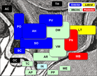
The arcuate nucleus of the hypothalamus is an aggregation of neurons in the mediobasal hypothalamus, adjacent to the third ventricle and the median eminence. The arcuate nucleus includes several important and diverse populations of neurons that help mediate different neuroendocrine and physiological functions, including neuroendocrine neurons, centrally projecting neurons, and astrocytes. The populations of neurons found in the arcuate nucleus are based on the hormones they secrete or interact with and are responsible for hypothalamic function, such as regulating hormones released from the pituitary gland or secreting their own hormones. Neurons in this region are also responsible for integrating information and providing inputs to other nuclei in the hypothalamus or inputs to areas outside this region of the brain. These neurons, generated from the ventral part of the periventricular epithelium during embryonic development, locate dorsally in the hypothalamus, becoming part of the ventromedial hypothalamic region. The function of the arcuate nucleus relies on its diversity of neurons, but its central role is involved in homeostasis. The arcuate nucleus provides many physiological roles involved in feeding, metabolism, fertility, and cardiovascular regulation.

<span class="mw-page-title-main">Ghrelin</span> Peptide hormone involved in appetite regulation

Ghrelin is a hormone produced by enteroendocrine cells of the gastrointestinal tract, especially the stomach, and is often called a "hunger hormone" because it increases the drive to eat. Blood levels of ghrelin are highest before meals when hungry, returning to lower levels after mealtimes. Ghrelin may help prepare for food intake by increasing gastric motility and stimulating the secretion of gastric acid.

<span class="mw-page-title-main">Neuropeptide Y</span> Mammalian protein found in Homo sapiens

Neuropeptide Y (NPY) is a 36 amino-acid neuropeptide that is involved in various physiological and homeostatic processes in both the central and peripheral nervous systems. It is secreted alongside other neurotransmitters such as GABA and glutamate. 

The melanocortins are a family of neuropeptide hormones which are the ligands of the melanocortin receptors The melanocortin system consists of melanocortin receptors, ligands, and accessory proteins. The genes of the melanocortin system are found in chordates. Melanocortins were originally named so because their earliest known function was in melanogenesis. It is now known that the melanocortin system regulates diverse functions throughout the body, including inflammatory response, fibrosis, melanogenesis, steroidogenesis, energy homeostasis, sexual function, and exocrine gland function.

<span class="mw-page-title-main">Agouti-signaling protein</span> Protein-coding gene in the species Homo sapiens

Agouti-signaling protein is a protein that in humans is encoded by the ASIP gene. It is responsible for the distribution of melanin pigment in mammals. Agouti interacts with the melanocortin 1 receptor to determine whether the melanocyte produces phaeomelanin, or eumelanin. This interaction is responsible for making distinct light and dark bands in the hairs of animals such as the agouti, which the gene is named after. In other species such as horses, agouti signalling is responsible for determining which parts of the body will be red or black. Mice with wildtype agouti will be grey, with each hair being partly yellow and partly black. Loss of function mutations in mice and other species cause black fur coloration, while mutations causing expression throughout the whole body in mice cause yellow fur and obesity.

<span class="mw-page-title-main">Peptide YY</span> Peptide released from cells in the ileum and colon in response to feeding

Peptide YY (PYY) also known as peptide tyrosine tyrosine is a peptide that in humans is encoded by the PYY gene. Peptide YY is a short peptide released from cells in the ileum and colon in response to feeding. In the blood, gut, and other elements of periphery, PYY acts to reduce appetite; similarly, when injected directly into the central nervous system, PYY is also anorexigenic, i.e., it reduces appetite.

Nesfatin-1 is a neuropeptide produced in the hypothalamus of mammals. It participates in the regulation of hunger and fat storage. Increased nesfatin-1 in the hypothalamus contributes to diminished hunger, a 'sense of fullness', and a potential loss of body fat and weight.

<span class="mw-page-title-main">Growth hormone secretagogue receptor</span> Protein-coding gene in the species Homo sapiens

Growth hormone secretagogue receptor(GHS-R), also known as ghrelin receptor, is a G protein-coupled receptor that binds growth hormone secretagogues (GHSs), such as ghrelin, the "hunger hormone". The role of GHS-R is thought to be in regulating energy homeostasis and body weight. In the brain, they are most highly expressed in the hypothalamus, specifically the ventromedial nucleus and arcuate nucleus. GSH-Rs are also expressed in other areas of the brain, including the ventral tegmental area, hippocampus, and substantia nigra. Outside the central nervous system, too, GSH-Rs are also found in the liver, in skeletal muscle, and even in the heart.

<span class="mw-page-title-main">KiSS1-derived peptide receptor</span> Mammalian protein found in Homo sapiens

The KiSS1-derived peptide receptor is a G protein-coupled receptor which binds the peptide hormone kisspeptin (metastin). Kisspeptin is encoded by the metastasis suppressor gene KISS1, which is expressed in a variety of endocrine and gonadal tissues. Activation of the kisspeptin receptor is linked to the phospholipase C and inositol trisphosphate second messenger cascades inside the cell.

<span class="mw-page-title-main">Melanocortin 4 receptor</span> Mammalian protein found in Homo sapiens

Melanocortin 4 receptor (MC4R) is a melanocortin receptor that in humans is encoded by the MC4R gene. It encodes the MC4R protein, a G protein-coupled receptor (GPCR) that binds α-melanocyte stimulating hormone (α-MSH). In mouse models, MC4 receptors have been found to be involved in feeding behaviour, the regulation of metabolism, sexual behaviour, and male erectile function.

<span class="mw-page-title-main">Melanocortin 3 receptor</span> Mammalian protein found in Homo sapiens

Melanocortin 3 receptor (MC3R) is a protein that in humans is encoded by the MC3R gene.

<span class="mw-page-title-main">Pancreatic polypeptide receptor 1</span> Protein-coding gene in the species Homo sapiens

Pancreatic polypeptide receptor 1, also known as Neuropeptide Y receptor type 4, is a protein that in humans is encoded by the PPYR1 gene.

The central melanocortin system is defined anatomically as a collection of central nervous system circuits which include:

Hunger is a sensation that motivates the consumption of food. The sensation of hunger typically manifests after only a few hours without eating and is generally considered to be unpleasant. Satiety occurs between 5 and 20 minutes after eating. There are several theories about how the feeling of hunger arises. The desire to eat food, or appetite, is another sensation experienced with regards to eating.

<span class="mw-page-title-main">Teleost leptins</span>

Teleost leptins are a family of peptide hormones found in fish (teleostei) that are orthologs of the mammalian hormone leptin. The teleost and mammalian leptins appear to have similar functions, namely, regulation of energy intake and expenditure.

<span class="mw-page-title-main">MRAP2</span> Protein

Melanocortin 2 receptor accessory protein 2 is a protein that in humans is encoded by the MRAP2 gene. MRAP2 is a transmembrane accessory protein to a family of five receptors called the melanocortin receptors (MC1-5). Human genome sequencing analysis led to the discovery of MRAP2, Human MRAP2 gene is located in chromosome 6q14.3, a different chromosomal location from that of human MRAP which is a paralogy to MRAP2. MRAP2 is thought to be involved in regulating the expression of the melanocortin (MC1-5) and some non-melanocortin receptors such as ghrelin receptor (GHSR-1a), orexin (OX1R) receptor and prokineticin receptor (PKR-1).

<span class="mw-page-title-main">Pathophysiology of obesity</span>

Pathophysiology of obesity is the study of disordered physiological processes that cause, result from, or are otherwise associated with obesity. A number of possible pathophysiological mechanisms have been identified which may contribute in the development and maintenance of obesity.

References

  1. 1 2 3 GRCh38: Ensembl release 89: ENSG00000159723 - Ensembl, May 2017
  2. 1 2 3 GRCm38: Ensembl release 89: ENSMUSG00000005705 - Ensembl, May 2017
  3. "Human PubMed Reference:". National Center for Biotechnology Information, U.S. National Library of Medicine.
  4. "Mouse PubMed Reference:". National Center for Biotechnology Information, U.S. National Library of Medicine.
  5. Bäckberg M, Madjid N, Ogren SO, Meister B (June 2004). "Down-regulated expression of agouti-related protein (AGRP) mRNA in the hypothalamic arcuate nucleus of hyperphagic and obese tub/tub mice". Brain Research. Molecular Brain Research. 125 (1–2): 129–39. doi:10.1016/j.molbrainres.2004.03.012. PMID   15193430.
  6. 1 2 Shutter JR, Graham M, Kinsey AC, Scully S, Lüthy R, Stark KL (March 1997). "Hypothalamic expression of ART, a novel gene related to agouti, is up-regulated in obese and diabetic mutant mice". Genes & Development. 11 (5): 593–602. doi: 10.1101/gad.11.5.593 . PMID   9119224.
  7. 1 2 Ollmann MM, Wilson BD, Yang YK, Kerns JA, Chen Y, Gantz I, Barsh GS (October 1997). "Antagonism of central melanocortin receptors in vitro and in vivo by agouti-related protein". Science. 278 (5335): 135–8. doi:10.1126/science.278.5335.135. PMID   9311920.
  8. Rosenfeld RD, Zeni L, Welcher AA, Narhi LO, Hale C, Marasco J, et al. (November 1998). "Biochemical, biophysical, and pharmacological characterization of bacterially expressed human agouti-related protein". Biochemistry. 37 (46): 16041–52. doi:10.1021/bi981027m. PMID   9819197.
  9. Jackson PJ, McNulty JC, Yang YK, Thompson DA, Chai B, Gantz I, et al. (June 2002). "Design, pharmacology, and NMR structure of a minimized cystine knot with agouti-related protein activity". Biochemistry. 41 (24): 7565–72. doi:10.1021/bi012000x. PMID   12056887.
  10. Enriori PJ, Evans AE, Sinnayah P, Jobst EE, Tonelli-Lemos L, Billes SK, et al. (March 2007). "Diet-induced obesity causes severe but reversible leptin resistance in arcuate melanocortin neurons". Cell Metabolism. 5 (3): 181–94. doi: 10.1016/j.cmet.2007.02.004 . PMID   17339026.
  11. Creemers JW, Pritchard LE, Gyte A, Le Rouzic P, Meulemans S, Wardlaw SL, et al. (April 2006). "Agouti-related protein is posttranslationally cleaved by proprotein convertase 1 to generate agouti-related protein (AGRP)83-132: interaction between AGRP83-132 and melanocortin receptors cannot be influenced by syndecan-3". Endocrinology. 147 (4): 1621–31. doi:10.1210/en.2005-1373. PMID   16384863.
  12. Scarlett JM, Zhu X, Enriori PJ, Bowe DD, Batra AK, Levasseur PR, et al. (October 2008). "Regulation of agouti-related protein messenger ribonucleic acid transcription and peptide secretion by acute and chronic inflammation". Endocrinology. 149 (10): 4837–45. doi:10.1210/en.2007-1680. PMC   2582916 . PMID   18583425.
  13. Xiao E, Xia-Zhang L, Vulliémoz NR, Ferin M, Wardlaw SL (May 2003). "Agouti-related protein stimulates the hypothalamic-pituitary-adrenal (HPA) axis and enhances the HPA response to interleukin-1 in the primate". Endocrinology. 144 (5): 1736–41. doi: 10.1210/en.2002-220013 . PMID   12697678.
  14. Nillni EA (April 2010). "Regulation of the hypothalamic thyrotropin releasing hormone (TRH) neuron by neuronal and peripheral inputs". Frontiers in Neuroendocrinology. 31 (2): 134–56. doi:10.1016/j.yfrne.2010.01.001. PMC   2849853 . PMID   20074584.
  15. Krashes MJ, Shah BP, Madara JC, Olson DP, Strochlic DE, Garfield AS, et al. (March 2014). "An excitatory paraventricular nucleus to AgRP neuron circuit that drives hunger". Nature. 507 (7491): 238–42. doi:10.1038/nature12956. PMC   3955843 . PMID   24487620.
  16. 1 2 Jackson PJ, Douglas NR, Chai B, Binkley J, Sidow A, Barsh GS, Millhauser GL (December 2006). "Structural and molecular evolutionary analysis of Agouti and Agouti-related proteins". Chemistry & Biology. 13 (12): 1297–305. doi:10.1016/j.chembiol.2006.10.006. PMC   2907901 . PMID   17185225.
  17. Ollmann MM, Lamoreux ML, Wilson BD, Barsh GS (February 1998). "Interaction of Agouti protein with the melanocortin 1 receptor in vitro and in vivo". Genes & Development. 12 (3): 316–30. doi:10.1101/gad.12.3.316. PMC   316484 . PMID   9450927.
  18. Dhillo WS, Small CJ, Gardiner JV, Bewick GA, Whitworth EJ, Jethwa PH, et al. (January 2003). "Agouti-related protein has an inhibitory paracrine role in the rat adrenal gland". Biochemical and Biophysical Research Communications. 301 (1): 102–7. doi:10.1016/S0006-291X(02)02991-1. PMID   12535647.
  19. Huszar D, Lynch CA, Fairchild-Huntress V, Dunmore JH, Fang Q, Berkemeier LR, et al. (January 1997). "Targeted disruption of the melanocortin-4 receptor results in obesity in mice". Cell. 88 (1): 131–41. doi: 10.1016/S0092-8674(00)81865-6 . PMID   9019399. S2CID   14528879.
  20. Graham M, Shutter JR, Sarmiento U, Sarosi I, Stark KL (November 1997). "Overexpression of Agrt leads to obesity in transgenic mice". Nature Genetics. 17 (3): 273–4. doi:10.1038/ng1197-273. PMID   9354787. S2CID   36127101.
  21. Katsuki A, Sumida Y, Gabazza EC, Murashima S, Tanaka T, Furuta M, et al. (May 2001). "Plasma levels of agouti-related protein are increased in obese men". The Journal of Clinical Endocrinology and Metabolism. 86 (5): 1921–4. doi: 10.1210/jcem.86.5.7458 . PMID   11344185.
  22. Vink T, Hinney A, van Elburg AA, van Goozen SH, Sandkuijl LA, Sinke RJ, et al. (May 2001). "Association between an agouti-related protein gene polymorphism and anorexia nervosa". Molecular Psychiatry. 6 (3): 325–8. doi: 10.1038/sj.mp.4000854 . PMID   11326303. S2CID   6755288.
  23. Kaushik S, Rodriguez-Navarro JA, Arias E, Kiffin R, Sahu S, Schwartz GJ, et al. (August 2011). "Autophagy in hypothalamic AgRP neurons regulates food intake and energy balance". Cell Metabolism. 14 (2): 173–83. doi:10.1016/j.cmet.2011.06.008. PMC   3148494 . PMID   21803288.
  24. Andermann ML, Lowell BB (August 2017). "Toward a Wiring Diagram Understanding of Appetite Control". Neuron. 95 (4): 757–778. doi: 10.1016/j.neuron.2017.06.014 . PMC   5657399 . PMID   28817798.
  25. Aponte Y, Atasoy D, Sternson SM (March 2011). "AGRP neurons are sufficient to orchestrate feeding behavior rapidly and without training". Nature Neuroscience. 14 (3): 351–5. doi:10.1038/nn.2739. PMC   3049940 . PMID   21209617.
  26. Krashes MJ, Koda S, Ye C, Rogan SC, Adams AC, Cusher DS, et al. (April 2011). "Rapid, reversible activation of AgRP neurons drives feeding behavior in mice". The Journal of Clinical Investigation. 121 (4): 1424–8. doi:10.1172/JCI46229. PMC   3069789 . PMID   21364278.
  27. 1 2 Horio N, Liberles SD (April 2021). "Hunger enhances food-odour attraction through a neuropeptide Y spotlight". Nature. 592 (7853): 262–266. doi:10.1038/s41586-021-03299-4. PMC   8035273 . PMID   33658716.
  28. Chen Y, Lin YC, Kuo TW, Knight ZA (February 2015). "Sensory detection of food rapidly modulates arcuate feeding circuits". Cell. 160 (5): 829–841. doi:10.1016/j.cell.2015.01.033. PMC   4373539 . PMID   25703096.
  29. Chen Y, Essner RA, Kosar S, Miller OH, Lin YC, Mesgarzadeh S, Knight ZA (April 2019). Palmiter RD, Dulac C, Krashes MJ (eds.). "Sustained NPY signaling enables AgRP neurons to drive feeding". eLife. 8: e46348. doi: 10.7554/eLife.46348 . PMC   6513552 . PMID   31033437.
  30. Wang Q, Liu C, Uchida A, Chuang JC, Walker A, Liu T, et al. (February 2014). "Arcuate AgRP neurons mediate orexigenic and glucoregulatory actions of ghrelin". Molecular Metabolism. 3 (1): 64–72. doi:10.1016/j.molmet.2013.10.001. PMC   3929914 . PMID   24567905.
  31. Baver SB, Hope K, Guyot S, Bjørbaek C, Kaczorowski C, O'Connell KM (April 2014). "Leptin modulates the intrinsic excitability of AgRP/NPY neurons in the arcuate nucleus of the hypothalamus". The Journal of Neuroscience. 34 (16): 5486–96. doi:10.1523/JNEUROSCI.4861-12.2014. PMC   4298648 . PMID   24741039.
  32. Wang C, Zhou W, He Y, Yang T, Xu P, Yang Y, et al. (January 2021). "AgRP neurons trigger long-term potentiation and facilitate food seeking". Translational Psychiatry. 11 (1): 11. doi:10.1038/s41398-020-01161-1. PMC   7791100 . PMID   33414382.
  33. Betley JN, Cao ZF, Ritola KD, Sternson SM (December 2013). "Parallel, redundant circuit organization for homeostatic control of feeding behavior". Cell. 155 (6): 1337–50. doi:10.1016/j.cell.2013.11.002. PMC   3970718 . PMID   24315102.

Further reading