
Medical ultrasound includes diagnostic techniques using ultrasound, as well as therapeutic applications of ultrasound. In diagnosis, it is used to create an image of internal body structures such as tendons, muscles, joints, blood vessels, and internal organs, to measure some characteristics or to generate an informative audible sound. The usage of ultrasound to produce visual images for medicine is called medical ultrasonography or simply sonography, or echography. The practice of examining pregnant women using ultrasound is called obstetric ultrasonography, and was an early development of clinical ultrasonography. The machine used is called an ultrasound machine, a sonograph or an echograph. The visual image formed using this technique is called an ultrasonogram, a sonogram or an echogram.

Obstetric ultrasonography, or prenatal ultrasound, is the use of medical ultrasonography in pregnancy, in which sound waves are used to create real-time visual images of the developing embryo or fetus in the uterus (womb). The procedure is a standard part of prenatal care in many countries, as it can provide a variety of information about the health of the mother, the timing and progress of the pregnancy, and the health and development of the embryo or fetus.

Sonic and ultrasonic weapons (USW) are weapons of various types that use sound to injure or incapacitate an opponent. Some sonic weapons make a focused beam of sound or of ultrasound; others produce an area field of sound. As of 2023 military and police forces make some limited use of sonic weapons.
A sonographer is an allied healthcare professional who specializes in the use of ultrasonic imaging devices to produce diagnostic images, scans, videos or three-dimensional volumes of anatomy and diagnostic data. The requirements for clinical practice vary greatly by country. Sonography requires specialized education and skills to acquire, analyze and optimize information in the image. Due to the high levels of decisional latitude and diagnostic input, sonographers have a high degree of responsibility in the diagnostic process. Many countries require medical sonographers to have professional certification. Sonographers have core knowledge in ultrasound physics, cross-sectional anatomy, physiology, and pathology.
The American College of Radiology (ACR), founded in 1923, is a professional medical society representing nearly 40,000 diagnostic radiologists, radiation oncologists, interventional radiologists, nuclear medicine physicians and medical physicists.
Asim Kurjak is the President of International Academy of Perinatal Medicine and director of Ian Donald Inter-University School of Medical Ultrasound. He is a regular fellow of World Academy of Art and Science, European Academy of Sciences and Art, International Academy for Human Reproduction, Italian Academy of Science and Art of Reggio Puglia, Academy of Medical Sciences of Catalonia; honorary member of American Institute of Ultrasound in Medicine and Biology; regular member of Russian Academy of Science and Art.
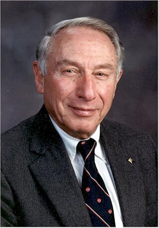
Roger Cobban Sanders is an American doctor specializing in Ultrasound and Radiology. Originally from the United Kingdom, he obtained a degree in physiology at Oxford University, from the Oxford Clinical Medical School. In 1970 Sanders traveled to the United States to begin a one-year teaching position at Johns Hopkins University in Baltimore, Maryland. During this time the University was presented a bistable ultrasound system, at the time the only one in Maryland, which Sanders was asked to oversee and use. Eventually he became professor of Radiology, Urology, Obstetric, and Gynecology, as well as director of Ultrasound at the University. He helped develop the department within the University, and it has grown into one of the foremost radiology institutions in the United States, having been named the "Cream of the Crop" by Medical Imaging Magazine in April 2007.
Floyd Dunn was an American electrical engineer who made contributions to all aspects of the interaction of ultrasound and biological media. Dunn was a member of Scientific Committee 66 of the National Council on Radiation Protection and Measurements as well as many FDA, NIH, AIUM, and ASA committees. He collaborated with scientists in the UK, Japan, China and Post-Soviet states.
The American Registry for Diagnostic Medical Sonography (ARDMS), incorporated in June 1975, is an independent nonprofit organization that administers examinations and awards credentials in the areas of diagnostic medical sonography, diagnostic cardiac sonography, vascular technology, physicians’ vascular interpretation, musculoskeletal sonography and midwifery ultrasound. ARDMS has over 90,000 certified individuals in the U.S., Canada and throughout the world. ARDMS provides certifications, resources, and career information to healthcare practitioners and students practicing medical sonography.
Fetal echocardiography, or Fetal echocardiogram, is the name of the test used to diagnose cardiac conditions in the fetal stage. Cardiac defects are amongst the most common birth defects. Their diagnosis is important in the fetal stage as it might help provide an opportunity to plan and manage the baby as and when the baby is born. Not all pregnancies need to undergo fetal echo.
Marnix van Holsbeeck is the director of musculoskeletal radiology in the Department of Radiology and director of radiology in the Department of Orthopaedic Surgery at the Henry Ford Health System. He is an authority in the field of musculoskeletal radiology.
The IEEE Biomedical Engineering Award is a Technical Field Award of the IEEE given annually for outstanding contributions to the field of biomedical engineering. It was established in 2010.

An ultrasonic toothbrush is an electric toothbrush designed for daily home use that operates by generating ultrasound in order to aid in removing plaque and rendering plaque bacteria harmless. It typically operates on a frequency of 1.6 MHz, which translates to 96,000,000 pulses or 192,000,000 movements per minute. Ultrasound is defined as a series of acoustic pressure waves generated at a frequency beyond human hearing.
The American Association of Clinical Endocrinology (AACE), formerly known as the American Association of Clinical Endocrinologists, is a professional community of physicians specializing in endocrinology, diabetes, and metabolism. AACE's mission is elevating clinical endocrinology to improve global health. The association is headquartered in Jacksonville, Florida, US.
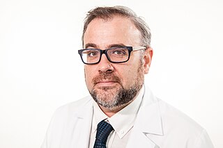
Fernando Alfageme Roldán is a Spanish dermatologist. He introduced the diagnostic technique of cutaneous ultrasound in Spain, is an associate professor at the Autonomous University of Madrid, and is responsible for the Dermatological Ultrasound Unit at the Puerta de Hierro University Hospital in Majadahonda. He has authored several essays, manuals and academic articles about dermatology.
Kathryn Radabaugh Nightingale is an American biomedical engineer in the field of medical ultrasound. She is the Theo Pilkington Professor of Biomedical Engineering at Duke University in Durham, North Carolina. Nightingale is also a Member of the Duke Cancer Institute and Bass Fellow in the Duke University Pratt School of Engineering.

Shelby Kutty is an Indian-born American cardiologist, a professor of pediatrics and internal medicine at the Johns Hopkins University School of Medicine. He holds the Helen B. Taussig endowed professorship at Johns Hopkins and is Director of the Helen B. Taussig Heart Center at the Johns Hopkins Hospital. Prior to this, he held the title of assistant dean for research and development and vice chair of pediatrics at the University of Nebraska Medical Center College of Medicine.
A specific branch of contrast-enhanced ultrasound, acoustic angiography is a minimally invasive and non-ionizing medical imaging technique used to visualize vasculature. Acoustic angiography was first developed by the Dayton Laboratory at North Carolina State University and provides a safe, portable, and inexpensive alternative to the most common methods of angiography such as Magnetic Resonance Angiography and Computed Tomography Angiography. Although ultrasound does not traditionally exhibit the high resolution of MRI or CT, high-frequency ultrasound (HFU) achieves relatively high resolution by sacrificing some penetration depth. HFU typically uses waves between 20 and 100 MHz and achieves resolution of 16-80μm at depths of 3-12mm. Although HFU has exhibited adequate resolution to monitor things like tumor growth in the skin layers, on its own it lacks the depth and contrast necessary for imaging blood vessels. Acoustic angiography overcomes the weaknesses of HFU by combining contrast-enhanced ultrasound with the use of a dual-element ultrasound transducer to achieve high resolution visualization of blood vessels at relatively deep penetration levels.

Jasjit S. Suri is an American engineer who works in the fields of biomedical engineering, computer science and clinical engineering. His work is focused on the implementation of artificial intelligence in biomedicine, and healthcare.
Beryl Rice Benacerraf was an American radiologist and professor of obstetrics, gynecology and reproductive biology and radiology at Harvard Medical School. She was a pioneer in the use of prenatal ultrasound to diagnose fetal abnormalities, including Down syndrome. In 2021, she was recognized as a "Giant in Obstetrics and Gynecology" by the American Journal of Obstetrics & Gynecology.