Related Research Articles
The dinoflagellates are a monophyletic group of single-celled eukaryotes constituting the phylum Dinoflagellata and are usually considered protists. Dinoflagellates are mostly marine plankton, but they are also common in freshwater habitats. Their populations vary with sea surface temperature, salinity, and depth. Many dinoflagellates are photosynthetic, but a large fraction of these are in fact mixotrophic, combining photosynthesis with ingestion of prey.

Microsporidia are a group of spore-forming unicellular parasites. These spores contain an extrusion apparatus that has a coiled polar tube ending in an anchoring disc at the apical part of the spore. They were once considered protozoans or protists, but are now known to be fungi, or a sister group to fungi. These fungal microbes are obligate eukaryotic parasites that use a unique mechanism to infect host cells. They have recently been discovered in a 2017 Cornell study to infect Coleoptera on a large scale. So far, about 1500 of the probably more than one million species are named. Microsporidia are restricted to animal hosts, and all major groups of animals host microsporidia. Most infect insects, but they are also responsible for common diseases of crustaceans and fish. The named species of microsporidia usually infect one host species or a group of closely related taxa. Approximately 10 percent of the species are parasites of vertebrates —several species, most of which are opportunistic, can infect humans, in whom they can cause microsporidiosis.
Perkinsus is a genus of alveolates in the phylum Perkinsozoa. The genus was erected in 1978 to better treat its type species, Perkinsus marinus, known formerly as Dermocystidium marinum. These are parasitic protozoans that infect molluscs, at least some of which cause disease and mass mortality. P. marinus is the most notorious, causing the disease perkinsosis, or dermo, in wild and farmed oysters.
Predatory dinoflagellates are predatory heterotrophic or mixotrophic alveolates that derive some or most of their nutrients from digesting other organisms. About one half of dinoflagellates lack photosynthetic pigments and specialize in consuming other eukaryotic cells, and even photosynthetic forms are often predatory.
Hematodinium perezi is a pathogenic dinoflagellate parasite that infects crustaceans, including the Blue Crab and Norway Lobster and has been observed to have a significant impact on crustacean fisheries. Infected crustaceans frequently show signs of weakness and lethargy, and often die due to stress-related handling from fishing as well as metabolic exhaustion due to reduced feeding. This parasite is known to be quite transmissible between various crustacean hosts.
Rhytidocystis is a genus of apicomplexans. It is the only genus within the monotypic family Rhytidocystidae. The species of this genus are parasitic protozoa found in marine annelids.
Mesodinium chamaeleon is a ciliate of the genus Mesodinium. It is known for being able to consume and maintain algae endosymbiotically for days before digesting the algae. It has the ability to eat red and green algae, and afterwards using the chlorophyll granules from the algae to generate energy, turning itself from being a heterotroph into an autotroph. The species was discovered in January 2012 outside the coast of Nivå, Denmark by professor Øjvind Moestrup.

Parvilucifera is a genus of marine alveolates that behave as endoparasites of dinoflagellates. It was described in 1999 by biologists Fredrik Norén and Øjvind Moestrup, who identified the genus among collections of Dinophysis dinoflagellates off the coast of Sweden. Initially mistaken for products of sexual reproduction, the round bodies found within these collections were eventually recognized as sporangia, spherical structures that generate zoospores of a parasitic protist. This organism was later identified as P. infectans, the type species. The examination of this organism and its close genetic relationship to Perkinsus led to the creation of the Perkinsozoa phylum within the Alveolata group.

Polykrikos kofoidii is a species of phagotrophic marine pseudocolonial dinoflagellates that can capture and engulf other protist prey, including the toxic dinoflagellate, Alexandrium tamarense. P. kofoidii is of scientific interest due to its status as a predator of other dinoflagellates, a behavior that is significant in the control of algal blooms. It has a complex life cycle of both vegetative (asexual) and sexual reproduction complicated by its pseudocolonial structure.
Luciella masanensis is a species of heterotrophic marine dinoflagellates.
The Platyproteum are a genus of parasitic alveolates in the phylum Apicomplexa. Species in this genus infect marine invertebrates.
The Polykrikaceae are a family of athecate dinoflagellates of the order Gymnodiniales. Members of the family are known as polykrikoids. The family contains two genera: Polykrikos and Pheopolykrikos.

Dinoflagellates are eukaryotic plankton, existing in marine and freshwater environments. Previously, dinoflagellates had been grouped into two categories, phagotrophs and phototrophs. Mixotrophs, however include a combination of phagotrophy and phototrophy. Mixotrophic dinoflagellates are a sub-type of planktonic dinoflagellates and are part of the phylum Dinoflagellata. They are flagellated eukaryotes that combine photoautotrophy when light is available, and heterotrophy via phagocytosis. Dinoflagellates are one of the most diverse and numerous species of phytoplankton, second to diatoms.

Mesodinium is a genus of ciliates that are widely distributed and are abundant in marine and brackish waters.
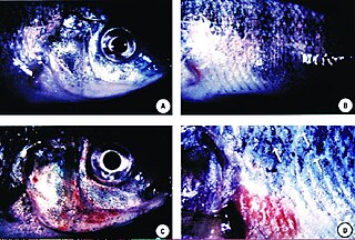
Miamiensis avidus is a species of unicellular marine eukaryote that is a parasite of many different types of fish. It is one of several organisms known to cause the fish disease scuticociliatosis and is considered an economically significant pathogen of farmed fish. M. avidus is believed to be the cause of a 2017 die-off of fish and sharks in the San Francisco Bay.
Coccidinium is a genus of parasitic syndinian dinoflagellates that infect the nucleus and cytoplasm of other marine dinoflagellates. Coccidinium, along with two other dinoflagellate genera, Amoebophyra and Duboscquella, contain species that are the primary endoparasites of marine dinoflagellates. While numerous studies have been conducted on the genus Amoebophyra, specifically Amoebophyra ceratii, little is known about Coccidinium. These microscopic organisms have gone relatively unstudied after the initial observations of Édouard Chatton and Berthe Biecheler in 1934 and 1936.

Gregarina garnhami is a eukaryotic unicellular organism belonging to the Apicomplexa described in 1956 by Canning as a parasite found in several locusts, such as the desert locust, African migratory locust, and Egyptian locust. Especially, the desert locust is the host for this species, as up to 100% of animals can become infected. An estimated thousands of different species of gregarines can be in insects and 99% of these gregarines still need to be described. Each insect is said to host multiple species. A remarkable feature of G. garnhami is its autofluorescence.

Haplozoon (/hæploʊ’zoʊən/) are unicellular endo-parasites, primarily infecting maldanid polychaetes. They belong to Dinoflagellata but differ from typical dinoflagellates. Most dinoflagellates are free-living and possess two flagella. Instead, Haplozoon belong to a 5% minority of parasitic dinoflagellates that are not free-living. Additionally, the Haplozoon trophont stage is particularly unique due to an apparent lack of flagella. The presence of flagella or remnant structures is the subject of ongoing research.
Euduboscquella (juˌduːboʊˈskwɛlə) is a genus of early branching dinoflagellates found in coastal waters around the globe. The members of this genus are all intracellular parasites that primarily infect Tintinnids. Euduboscquella are commonly found in marine environments, either infecting a host or in a resting stage in search of a new host, but there are a few freshwater and terrestrial species. Euduboscquella possess a multi-grooved shield separating their cytoplasm from the host’s cytoplasm, which is used by researchers to taxonomically identify them. The genus Euduboscquella contains nine species.

The cortical alveolum is a cellular organelle consisting of a vesicle located under the cytoplasmic membrane, to which they give support. The term "corticate" comes from an evolutionary hypothesis about the common origin of kingdoms Plantae and Chromista, because both kingdoms have cortical alveoli in at least one phylum. At least three protist lineages exhibit these structures: Telonemia, Alveolata and Glaucophyta.
References
- ↑ Salomon, P. S.; Janson, S.; Granéli, E. (2003). "Multiple species of the dinophagous dinoflagellate genus Amoebophrya infect the same host species". Environmental Microbiology. 5 (11): 1046–1052. doi:10.1046/j.1462-2920.2003.00511.x. PMID 14641584.
- ↑ Miller, John J.; Delwiche, Charles F.; Coats, D. Wayne (September 2012). "Ultrastructure of Amoebophrya sp. and its Changes during the Course of Infection". Protist. 163 (5): 720–745. doi:10.1016/j.protis.2011.11.007. PMID 22209083.
- ↑ Sunjun, Kim (2006-12-23). "Patterns in host range for two strains of Amoebophrya (Dinophyta) infecting thecate dinoflagellates: Amoebophrya spp. ex Alexandrium affine and ex Gonyaulax polygramma". Biosis. 42 (6): 1170–1173. doi:10.1111/j.1529-8817.2006.00277.x. S2CID 84697048.
- ↑ Sunju, Kim; Park, Myung Gil; Kim, Keun-Yong; Kim, Chang-Hoon; Yih, Wonho; Park, Jong Soo; Coats, D. Wayne (Jan 2008). "Genetic Diversity of Parasitic Dinoflagellates in the Genus Amoebophrya and Its Relationship to Parasite Biology and Biogeography". Journal of Eukaryotic Microbiology. 55 (1): 1–8. doi: 10.1111/j.1550-7408.2007.00295.x . PMID 18251796.
- ↑ Yih, W; Coats, DW (September 2000). "Infection of Gymnodinium sanguineum by the Dinoflagellate Amoebophrya sp.: Effect of Nutrient Environment on Parasite Generation Time, Reproduction, and Infectivity". Journal of Eukaryotic Microbiology. 47 (5): 504–510. doi:10.1111/j.1550-7408.2000.tb00082.x. PMID 11001148. S2CID 27962913.
- ↑ Sunju, Kim; Park, Myung Gil; Kim, Keun-Yong; Kim, Chang-Hoon; Yih, Wonho; Park, Jong Soo; Coats, D. Wayne (Jan 2008). "Genetic Diversity of Parasitic Dinoflagellates in the Genus Amoebophrya and Its Relationship to Parasite Biology and Biogeography". Journal of Eukaryotic Microbiology. 55 (1): 1–8. doi: 10.1111/j.1550-7408.2007.00295.x . PMID 18251796.
- ↑ Velo-Suárez, Lourdes; Brosnahan, Michael L.; Anderson, Donald M.; McGillicuddy, Jr., Dennis J. (Dec 2013). "A Quantitative Assessment of the Role of the Parasite Amoebophrya in the Termination of Alexandrium fundyense Blooms within a Small Coastal Embayment". PLOS ONE. 8 (12): e81150. doi: 10.1371/journal.pone.0081150 . PMC 3852033 . PMID 24324668.
- ↑ Sunju, Kim; Park, Myung Gil; Kim, Keun-Yong; Kim, Chang-Hoon; Yih, Wonho; Park, Jong Soo; Coats, D. Wayne (Jan 2008). "Genetic Diversity of Parasitic Dinoflagellates in the Genus Amoebophrya and Its Relationship to Parasite Biology and Biogeography". Journal of Eukaryotic Microbiology. 55 (1): 1–8. doi: 10.1111/j.1550-7408.2007.00295.x . PMID 18251796.
- ↑ Yih, Wonho; Coats, D. Wayne (2000). "Infection of Gymnodinium sanguineum by the Dinoflagellate Amoebophrya sp.: Effect of Nutrient Environment on Parasite Generation Time, Reproduction, and Infectivity". Journal of Eukaryotic Microbiology. 45 (5): 504–510. doi:10.1111/j.1550-7408.2000.tb00082.x. PMID 11001148. S2CID 27962913.
- ↑ Yih, W; Coats, DW (September 2000). "Infection of Gymnodinium sanguineum by the Dinoflagellate Amoebophrya sp.: Effect of Nutrient Environment on Parasite Generation Time, Reproduction, and Infectivity". Journal of Eukaryotic Microbiology. 47 (5): 504–510. doi:10.1111/j.1550-7408.2000.tb00082.x. PMID 11001148. S2CID 27962913.
- ↑ Yih, Wonho; Coats, D. Wayne (2000). "Infection of Gymnodinium sanguineum by the Dinoflagellate Amoebophrya sp.: Effect of Nutrient Environment on Parasite Generation Time, Reproduction, and Infectivity". Journal of Eukaryotic Microbiology. 45 (5): 504–510. doi:10.1111/j.1550-7408.2000.tb00082.x. PMID 11001148. S2CID 27962913.