Anterior chamber angle
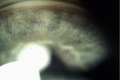
Last updated

The anterior chamber angle is a part of the eye located between the cornea and iris which contains the trabecular meshwork. The size of this angle is an important determinant of the rate aqueous humour flows out of the eye, and thus, the intraocular pressure. The anterior chamber angle is the structure which determines the anterior chamber depth. An extremely narrow anterior chamber angle is a feature of angle closure glaucoma. [1] [2]

Related Research Articles

Rib cage is a "box" created of ribs, that protects the vital organs and great vessels

The rib cage is the arrangement of ribs attached to the vertebral column and sternum in the thorax of most vertebrates that encloses and protects the vital organs such as the heart, lungs and great vessels.

Glaucoma Eye disease in which high intraocular pressure damages the optic nerve

Glaucoma is a group of eye diseases which result in damage to the optic nerve and cause vision loss. The most common type is open-angle glaucoma, in which the drainage angle for fluid within the eye remains open, with less common types including closed-angle glaucoma and normal-tension glaucoma. Open-angle glaucoma develops slowly over time and there is no pain. Peripheral vision may begin to decrease, followed by central vision, resulting in blindness if not treated. Closed-angle glaucoma can present gradually or suddenly. The sudden presentation may involve severe eye pain, blurred vision, mid-dilated pupil, redness of the eye, and nausea. Vision loss from glaucoma, once it has occurred, is permanent. Eyes affected by glaucoma are referred to as being glaucomatous.

Vitreous body Gel in eyeballs

The vitreous body is the clear gel that fills the space between the lens and the retina of the eyeball of humans and other vertebrates. It is often referred to as the vitreous humor or simply "the vitreous".

Aqueous humour Fluid in the anterior segment of the eye

The aqueous humour is a transparent water-like fluid similar to plasma, but containing low protein concentrations. It is secreted from the ciliary body, a structure supporting the lens. It fills both the anterior and the posterior chambers of the eye, and is not to be confused with the vitreous humour, which is located in the space between the lens and the retina, also known as the posterior cavity or vitreous chamber.

Ciliary body Part of the eye

The ciliary body is a part of the eye that includes the ciliary muscle, which controls the shape of the lens, and the ciliary epithelium, which produces the aqueous humor. The aqueous humor is produced in the non-pigmented portion of the ciliary body. The ciliary body is part of the uvea, the layer of tissue that delivers oxygen and nutrients to the eye tissues. The ciliary body joins the ora serrata of the choroid to the root of the iris.

Ciliary muscle Eye muscle which is used for focussing

The ciliary muscle is an intrinsic muscle of the eye formed as a ring of smooth muscle in the eye's middle layer. It controls accommodation for viewing objects at varying distances and regulates the flow of aqueous humor into Schlemm's canal. It also changes the shape of the lens within the eye but not the size of the pupil which is carried out by the sphincter pupillae muscle and dilator pupillae.

Trabecular meshwork Area of tissue in the eye

The trabecular meshwork is an area of tissue in the eye located around the base of the cornea, near the ciliary body, and is responsible for draining the aqueous humor from the eye via the anterior chamber.

Hip Anatomical region between the torso and the legs, holding the buttocks and genital region

In vertebrate anatomy, hip refers to either an anatomical region or a joint.

Anterior chamber of eyeball

The anterior chamber (AC) is the aqueous humor-filled space inside the eye between the iris and the cornea's innermost surface, the endothelium. Hyphema, anterior uveitis and glaucoma are three main pathologies in this area. In hyphema, blood fills the anterior chamber as a result of a hemorrhage, most commonly after a blunt eye injury. Anterior uveitis is an inflammatory process affecting the iris and ciliary body, with resulting inflammatory signs in the anterior chamber. In glaucoma, blockage of the trabecular meshwork prevents the normal outflow of aqueous humour, resulting in increased intraocular pressure, progressive damage to the optic nerve head, and eventually blindness.

Gonioscopy

Gonioscopy is using a goniolens together with a slit lamp or operating microscope to view the iridocorneal angle, or the anatomical angle formed between the eye's cornea and iris. Its use is important in diagnosing and monitoring various eye conditions associated with glaucoma.

Anatomical terms of motion Terms describing animal motion

Motion, the process of movement, is described using specific anatomical terms. Motion includes movement of organs, joints, limbs, and specific sections of the body. The terminology used describes this motion according to its direction relative to the anatomical position of the joints. Anatomists use a unified set of terms to describe most of the movements, although other, more specialized terms are necessary for describing the uniqueness of the movements such as those of the hands, feet, and eyes.

Superior ophthalmic vein

The superior ophthalmic vein begins at the inner angle of the orbit in a vein named the nasofrontal which communicates anteriorly with the angular vein; it does not pursue the same course as the ophthalmic artery and receives tributaries corresponding to the branches of that vessel.

Calcarine sulcus

The calcarine sulcus is an anatomical landmark located at the caudal end of the medial surface of the brain of humans and other primates. Its name comes from the Latin "calcar" meaning "spur". It is very deep and known as a complete sulcus.

Zygomatic process

The zygomatic processes are three processes (protrusions) from other bones of the skull which each articulate with the zygomatic bone. The three processes are:

Angular artery

The angular artery is the terminal part of the facial artery; it ascends to the medial angle of the eye's orbit, imbedded in the fibers of the angular head of the quadratus labii superioris, and accompanied by the angular vein.

Posterior chamber of eyeball

The posterior chamber is a narrow space behind the peripheral part of the iris, and in front of the suspensory ligament of the lens and the ciliary processes. The posterior chamber consists of small space directly posterior to the iris but anterior to the lens. The posterior chamber is part of the anterior segment and should not be confused with the vitreous chamber.

Vitreous chamber

The vitreous chamber is the space in the eye occupied by vitreous humor.

Scleral spur An annular structure composed of collagen in the human eye

The scleral spur is an annular structure composed of collagen in the human eye, a protrusion of the sclera into the anterior chamber.

Anterior spinal veins

Anterior spinal veins are veins that receive blood from the anterior spinal cord.

The pars plicata is the folded and most anterior portion of the ciliary body of an eye. The ciliary body is a part of the uvea, one of the three layers that comprise the eye. The pars plicata is located anterior to the pars plana portion of the ciliary body, and posterior to the iris. The lens zonules that are used to control accommodation are attached to the pars plana.

References

  1. Snell, Richard S.; Lemp, Michael A. (April 9, 2013). Clinical Anatomy of the Eye. John Wiley & Sons. ISBN   9781118691007 via Google Books.
  2. Bron, A.; Tripathi, R.; Tripathi, B. (September 4, 1998). Wolff's Anatomy of the Eye and Orbit, 8Ed. Taylor & Francis. ISBN   9780412410109 via Google Books.