Anti-neutrophil cytoplasmic antibody

Last updated
Perinuclear staining typical of p-ANCA P anca.jpg
Perinuclear staining typical of p-ANCA
The granular, cytoplasmic staining pattern of c-ANCA C anca.jpg
The granular, cytoplasmic staining pattern of c-ANCA
Immunofluorescence staining pattern of ANCA. Top left - PR3 antibodies on ethanol-fixed neutrophils (c-ANCA pattern). Bottom left - PR3 antibodies on formalin-fixed neutrophils(c-ANCA pattern). Top right - MPO antibodies on ethanol-fixed neutrophils (p-ANCA pattern). Bottom right - MPO antibodies on formalin-fixed neutrophils (c-ANCA pattern).(FITC conjugate) ANCA ETHANOL AND FORMALIN.JPEG
Immunofluorescence staining pattern of ANCA. Top left – PR3 antibodies on ethanol-fixed neutrophils (c-ANCA pattern). Bottom left – PR3 antibodies on formalin-fixed neutrophils(c-ANCA pattern). Top right – MPO antibodies on ethanol-fixed neutrophils (p-ANCA pattern). Bottom right – MPO antibodies on formalin-fixed neutrophils (c-ANCA pattern).(FITC conjugate)

Anti-neutrophil cytoplasmic antibodies (ANCAs) are a group of autoantibodies, mainly of the IgG type, against antigens in the cytoplasm of neutrophils (the most common type of white blood cell) and monocytes. They are detected as a blood test in a number of autoimmune disorders, but are particularly associated with systemic vasculitis, so called ANCA-associated vasculitides (AAV).[ citation needed ]

Contents

ANCA IF patterns

Immunofluorescence (IF) on ethanol-fixed neutrophils is used to detect ANCA, although formalin-fixed neutrophils may be used to help differentiate ANCA patterns. ANCA can be divided into four patterns when visualised by IF; cytoplasmic ANCA (c-ANCA), C-ANCA (atypical), perinuclear ANCA (p-ANCA) and atypical ANCA (a-ANCA), also known as x-ANCA. c-ANCA shows cytoplasmic granular fluorescence with central interlobular accentuation. C-ANCA (atypical) shows cytoplasmic staining that is usually uniform and has no interlobular accentuation. p-ANCA has three subtypes, classical p-ANCA, p-ANCA without nuclear extension and granulocyte specific-antinuclear antibody (GS-ANA). Classical p-ANCA shows perinuclear staining with nuclear extension, p-ANCA without nuclear extension has perinuclear staining without nuclear extension and GS-ANA shows nuclear staining on granulocytes only. a-ANCA often shows combinations of both cytoplasmic and perinuclear staining. [1]

ANCA antigens

The c-ANCA antigen is specifically proteinase 3 (PR3). p-ANCA antigens include myeloperoxidase (MPO) and bacterial permeability increasing factor Bactericidal/permeability-increasing protein (BPI). Other antigens exist for c-ANCA (atypical), however many are as yet unknown. Classical p-ANCA occurs with antibodies directed to MPO. p-ANCA without nuclear extension occurs with antibodies to BPI, cathepsin G, elastase, lactoferrin and lysozyme. GS-ANA are antibodies directed to granulocyte specific nuclear antigens. Atypical ANCA are thought to be antigens similar to that of the p-ANCAs, however may occur due to differences in neutrophil processing. [1]

Other less common antigens include HMG1 (p-ANCA pattern), HMG2 (p-ANCA pattern), alpha enolase (p and c-ANCA pattern), catalase (p and c-ANCA pattern), beta glucuronidase (p-ANCA pattern), azurocidin (p and c-ANCA pattern), actin (p and a-ANCA) and h-lamp-2 (c-ANCA). [1]

ELISA

Enzyme-linked immunosorbent assay (ELISA) is used in diagnostic laboratories to detect ANCAs. Although IF can be used to screen for many ANCAs, ELISA is used to detect antibodies to individual antigens. The most common antigens used on an ELISA microtitre plate are MPO and PR3, which are usually tested for after a positive IF test. [2]

Development

It is poorly understood how ANCA are developed, although several hypotheses have been suggested. There is probably a genetic contribution, particularly in genes controlling the level of immune response – although genetic susceptibility is likely to be linked to an environmental factor, some possible factors including vaccination or exposure to silicates. Two possible mechanisms of ANCA development are postulated, although neither of these theories answers the question of how the different ANCA specificities are developed, and there is much research still being undertaken on the development of ANCA. [3]

Theory of molecular mimicry

Microbial superantigens are molecules expressed by bacteria and other microorganisms that have the power to stimulate a strong immune response by activation of T-cells. These molecules generally have regions that resemble self-antigens that promote a residual autoimmune response – this is the theory of molecular mimicry. Staphylococcal and streptococcal superantigens have been characterized in autoimmune diseases – the classical example in post group A streptococcal rheumatic heart disease, where there is similarity between M proteins of Streptococcus pyogenes to cardiac myosin and laminin. It has also been shown that up to 70% of patients with granulomatosis with polyangiitis are chronic nasal carriers of Staphylococcus aureus , with carriers having an eight times increased risk of relapse. [3] This would therefore be considered a type II hypersensitivity reaction.[ citation needed ]

Theory of defective apoptosis

Neutrophil apoptosis, or programmed cell death, is vital in controlling the duration of the early inflammatory response, thus restricting damage to tissues by the neutrophils. ANCA may be developed either via ineffective apoptosis or ineffective removal of apoptotic cell fragments, leading to the exposure of the immune system to molecules normally sequestered inside the cells. This theory solves the paradox of how it could be possible for antibodies to be raised against the intracellular antigenic targets of ANCA. [3]

Role in disease

Disease associations

ANCAs are associated with small vessel vasculitides including granulomatosis with polyangiitis, microscopic polyangiitis, primary pauci-immune necrotizing crescentic glomerulonephritis (a type of renal-limited microscopic polyangiitis), eosinophilic granulomatosis with polyangiitis and drug induced vasculitides. ANCA-associated vasculitides (AAV) have new classification criteria, updated in 2022. [4] [5] PR3 directed c-ANCA is present in 80-90% of granulomatosis with polyangiitis, 20-40% of microscopic polyangiitis, 20-40% of pauci-immune crescentic glomerulonephritis and 35% of eosinophilic granulomatosis with polyangiitis. c-ANCA (atypical) is present in 80% of cystic fibrosis (with BPI as the target antigen) and also in inflammatory bowel disease, primary sclerosing cholangitis and rheumatoid arthritis (with antibodies to multiple antigenic targets). p-ANCA with MPO specificity is found in 50% of microscopic polyangiitis, 50% of primary pauci-immune necrotizing crescentic glomerulonephritis and 35% of eosinophilic granulomatosis with polyangiitis. p-ANCA with specificity to other antigens are associated with inflammatory bowel disease, rheumatoid arthritis, drug-induced vasculitis, autoimmune liver disease, drug induced syndromes and parasitic infections. Atypical ANCA is associated with drug-induced systemic vasculitis, inflammatory bowel disease and rheumatoid arthritis. [2] [6] The ANCA‐positive rate is much higher in patients with type 1 diabetes mellitus than in healthy individuals. [7]

Levamisole, which is a common adulterant of cocaine, can cause an ANCA positive vasculitis. [8]

The presence or absence of ANCA cannot indicate presence or absence of disease and results are correlated with clinical features. The association of ANCA and disease activity remains controversial; however, the reappearance of ANCA after treatment can indicate a relapse. [9] [10]

Pathogenesis

Although the pathogenic role of ANCA is still controversial, in vitro and animal models support the idea that the antibodies have a direct pathological role in the formation of small vessel vasculitides. MPO and PR3 specific ANCA can activate neutrophils and monocytes through their Fc and Fab'2 receptors, which can be enhanced by cytokines which cause neutrophils to display MPO and PR3 on their surface. Aberrant glycosylation of the MPO and PR3 specific ANCA enhances their ability to interact with activating Fc receptors on neutrophils. [11] The activated neutrophils can then adhere to endothelial cells where degranulation occurs. This releases free oxygen radicals and lytic enzymes, resulting in damage to the endothelium via the induction of necrosis and apoptosis. Furthermore, neutrophils release chemoattractive signalling molecules that recruit more neutrophils to the endothelium, acting as a positive feedback loop. Animal models have shown that MPO antibodies can induce necrotizing crescentic glomerulonephritis and systemic small vessel vasculitis. In these animal models the formation of glomerulonephritis and vasculitis can occur in the absence of T-cells, however neutrophils must be present. [12] [13] [14] [15] Although ANCA titres have been noted to have limited correlation with disease activity, except for kidney disease, and with risk of relapse, this is explained by differences in the epitopes and affinity of ANCAs. [16] ANCAs induce excess activation of neutrophils, resulting in the production of neutrophil extracellular traps (NETs), which cause damage to small blood vessels. [16] In addition, in patients with active disease, treated with Rituximab, an anti-CD20 antibody which remove circulating B-cells, clinical remission correlates more to the decreasing number of circulating B-cells than decrease in ANCA titre, which in some patient does not change during treatment. The same study found that clinical relapse in some patients were in association with the return of circulating B-cells. [17] Based on the above observations and that ANCA reactive B-cells can be found in circulation in patients with AAV, an alternative hypothesis have been proposed assigning a direct pathogenic role of these cells, whereby activated neutrophils and ANCA-reactive B-cells engage in intercellular cross-talk, which leads not only to neutrophil degranulation and inflammation but also to the proliferation and differentiation of ANCA-reactive B-cells. [18] However, this hypothesis remains to be tested.

Treatment

Avacopan was approved for medical use in the United States to treat anti-neutrophil cytoplasmic autoantibody-associated vasculitis in October 2021. [19]

History

ANCAs were originally described in Davies et al. in 1982 in segmental necrotising glomerulonephritis. [16] [20] The Second International ANCA Workshop, held in The Netherlands in May 1989, fixed the nomenclature on perinuclear vs. cytoplasmic patterns, and the antigens MPO and PR3 were discovered in 1988 and 1989, respectively. [21] International ANCA Workshops have been carried out every two years.[ citation needed ]

Related Research Articles

<span class="mw-page-title-main">Antinuclear antibody</span> Autoantibody that binds to contents of the cell nucleus

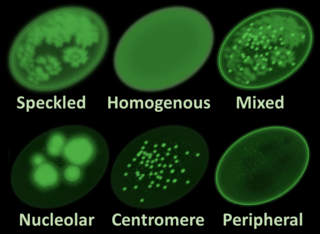
Antinuclear antibodies are autoantibodies that bind to contents of the cell nucleus. In normal individuals, the immune system produces antibodies to foreign proteins (antigens) but not to human proteins (autoantigens). In some cases, antibodies to human antigens are produced.

<span class="mw-page-title-main">Vasculitis</span> Medical disorders that destroy blood vessels by inflammation

Vasculitis is a group of disorders that destroy blood vessels by inflammation. Both arteries and veins are affected. Lymphangitis is sometimes considered a type of vasculitis. Vasculitis is primarily caused by leukocyte migration and resultant damage. Although both occur in vasculitis, inflammation of veins (phlebitis) or arteries (arteritis) on their own are separate entities.

<span class="mw-page-title-main">Granulomatosis with polyangiitis</span> Medical condition

Granulomatosis with polyangiitis (GPA), also known as Wegener's granulomatosis (WG), after the German physician Friedrich Wegener, is a rare long-term systemic disorder that involves the formation of granulomas and inflammation of blood vessels (vasculitis). It is an autoimmune disease and a form of vasculitis that affects small- and medium-size vessels in many organs but most commonly affects the upper respiratory tract, lungs and kidneys. The signs and symptoms of GPA are highly varied and reflect which organs are supplied by the affected blood vessels. Typical signs and symptoms include nosebleeds, stuffy nose and crustiness of nasal secretions, and inflammation of the uveal layer of the eye. Damage to the heart, lungs and kidneys can be fatal.

<span class="mw-page-title-main">Eosinophilic granulomatosis with polyangiitis</span> Medical condition

Eosinophilic granulomatosis with polyangiitis (EGPA), formerly known as allergic granulomatosis, is an extremely rare autoimmune condition that causes inflammation of small and medium-sized blood vessels (vasculitis) in persons with a history of airway allergic hypersensitivity (atopy).

<span class="mw-page-title-main">Polyarteritis nodosa</span> Medical condition

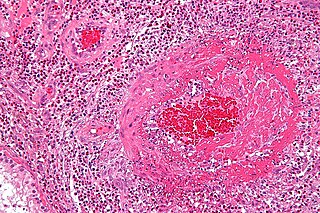
Polyarteritis nodosa (PAN) is a systemic necrotizing inflammation of blood vessels (vasculitis) affecting medium-sized muscular arteries, typically involving the arteries of the kidneys and other internal organs but generally sparing the lungs' circulation. Small aneurysms are strung like the beads of a rosary, therefore making this "rosary sign" an important diagnostic feature of the vasculitis. PAN is sometimes associated with infection by the hepatitis B or hepatitis C virus. The condition may be present in infants.

<span class="mw-page-title-main">Glomerulonephritis</span> Term for several kidney diseases

Glomerulonephritis (GN) is a term used to refer to several kidney diseases. Many of the diseases are characterised by inflammation either of the glomeruli or of the small blood vessels in the kidneys, hence the name, but not all diseases necessarily have an inflammatory component.

<span class="mw-page-title-main">Myeloperoxidase</span> Mammalian protein found in Homo sapiens

Myeloperoxidase (MPO) is a peroxidase enzyme that in humans is encoded by the MPO gene on chromosome 17. MPO is most abundantly expressed in neutrophils, and produces hypohalous acids to carry out their antimicrobial activity, including hypochlorous acid, the sodium salt of which is the chemical in bleach. It is a lysosomal protein stored in azurophilic granules of the neutrophil and released into the extracellular space during degranulation. Neutrophil myeloperoxidase has a heme pigment, which causes its green color in secretions rich in neutrophils, such as mucus and sputum. The green color contributed to its outdated name verdoperoxidase.

<span class="mw-page-title-main">Nephritic syndrome</span> Medical condition

Nephritic syndrome is a syndrome comprising signs of nephritis, which is kidney disease involving inflammation. It often occurs in the glomerulus, where it is called glomerulonephritis. Glomerulonephritis is characterized by inflammation and thinning of the glomerular basement membrane and the occurrence of small pores in the podocytes of the glomerulus. These pores become large enough to permit both proteins and red blood cells to pass into the urine. By contrast, nephrotic syndrome is characterized by proteinuria and a constellation of other symptoms that specifically do not include hematuria. Nephritic syndrome, like nephrotic syndrome, may involve low level of albumin in the blood due to the protein albumin moving from the blood to the urine.

Microscopic polyangiitis is an autoimmune disease characterized by a systemic, pauci-immune, necrotizing, small-vessel vasculitis without clinical or pathological evidence of granulomatous inflammation.

<span class="mw-page-title-main">Proteinase 3</span> Mammalian protein found in Homo sapiens

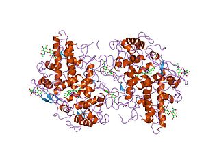
Proteinase 3, also known as PRTN3, is an enzyme that in humans is encoded by the PRTN3 gene.

<span class="mw-page-title-main">Friedrich Wegener</span> German pathologist (1907–1990

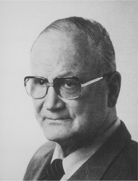
Friedrich Wegener was a German pathologist who is notable for his description of a rare disease originally referred to Wegener disease and now referred to as granulomatosis with polyangiitis. Although this disease was known before Wegener's description, from the 1950s onwards it was generally referred to as Wegener's granulomatosis.

c-ANCA Type of autoantibody

c-ANCAs, or PR3-ANCA, or antineutrophil cytoplasmic antibodies, are a type of autoantibody, an antibody produced by the body that acts against one of its own proteins. These antibodies show a diffusely granular, cytoplasmic staining pattern under microscopy. This pattern results from binding of ANCAs to antigen targets throughout the neutrophil cytoplasm, the most common protein target being proteinase 3 (PR3). For example, PR3 is the most common antigen target of ANCA in patients with granulomatosis with polyangiitis. In active granulomatosis with polyangiitis, c-ANCA is found over 90% of the time. Other antigens may also occasionally result in a c-ANCA pattern.

p-ANCA Type of autoantibody

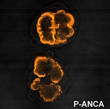
p-ANCA, or MPO-ANCA, or perinuclear anti-neutrophil cytoplasmic antibodies, are antibodies that stain the material around the nucleus of a neutrophil. They are a special class of anti-neutrophil cytoplasmic antibodies.

<span class="mw-page-title-main">Rapidly progressive glomerulonephritis</span> Medical condition

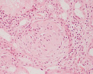
Rapidly progressive glomerulonephritis (RPGN) is a syndrome of the kidney that is characterized by a rapid loss of kidney function, with glomerular crescent formation seen in at least 50% or 75% of glomeruli seen on kidney biopsies. If left untreated, it rapidly progresses into acute kidney failure and death within months. In 50% of cases, RPGN is associated with an underlying disease such as Goodpasture syndrome, systemic lupus erythematosus or granulomatosis with polyangiitis; the remaining cases are idiopathic. Regardless of the underlying cause, RPGN involves severe injury to the kidneys' glomeruli, with many of the glomeruli containing characteristic glomerular crescents.

Pulmonary-renal syndrome (PRS) is a rare medical syndrome in which respiratory failure involving bleeding in the lungs and kidney failure (glomerulonephritis) occur. PRS is associated with a high rate of morbidity and death. The term was first used by Goodpasture in 1919 to describe the association of respiratory and kidney failure.

Pauci-immune vasculitis is a form of vasculitis that is associated with minimal evidence of hypersensitivity upon immunofluorescent staining for IgG. Often, this is discovered in the setting of the kidney.

<span class="mw-page-title-main">Systemic vasculitis</span> Medical condition

Necrotizing vasculitis, also called systemic necrotizing vasculitis, is a general term for the inflammation of veins and arteries that develops into necrosis and narrows the vessels.

Ronald Jonathan Falk, MD, FACP, FASN is the Nan and Hugh Cullman Eminent Professor and Chair of the Department of Medicine at the University of North Carolina-Chapel Hill (UNC). He is a clinical nephrologist and internationally recognized expert in anti-neutrophil cytoplasmic autoantibody (ANCA)-induced vasculitis and autoimmune kidney disease. His career as a translational physician-scientist spans more than three decades. His clinical practice and translational research focus on characterizing the cell, tissue and physiologic changes in the development of specific autoimmune kidney diseases and developing new approaches for studying autoimmunity, inflammation and basic neutrophil/monocyte biology. He was Chief of the UNC Division of Nephrology and Hypertension from 1993-2015. He co-founded the UNC Kidney Center in 2005 and continues as Co-Director. Falk is a Past-President of the American Society of Nephrology (ASN). Since 2015, he has served as Chair of the Department of Medicine at UNC.

Vasculitic neuropathy is a peripheral neuropathic disease. In a vasculitic neuropathy there is damage to the vessels that supply blood to the nerves. It can be as part of a systemic problem or can exist as a single-organ issue only affecting the peripheral nervous system (PNS). It is diagnosed with the use of electrophysiological testing, blood tests, nerve biopsy and clinical examination. It is a serious medical condition that can cause prolonged morbidity and disability and generally requires treatment. Treatment depends on the type but it is mostly with corticosteroids or immunomodulating therapies.

<span class="mw-page-title-main">Avacopan</span> Chemical compound

Avacopan, sold under the brand name Tavneos, is a medication used to treat anti-neutrophil cytoplasmic autoantibody-associated vasculitis. Avacopan is a complement 5a receptor antagonist and a cytochrome P450 3A4 inhibitor.

References

  1. 1 2 3 Mead A.R. Bradwell, R.P. Stokes, G.P. (1999). Advanced atlas of autoantibody patterns. Birmingham: The Binding Site. ISBN   978-0-7044-8510-5.{{cite book}}: CS1 maint: multiple names: authors list (link)
  2. 1 2 Savige J, Davies, D, Falk, RJ, Jennette, JC, Wiik, A (Mar 2000). "Antineutrophil cytoplasmic antibodies and associated diseases: a review of the clinical and laboratory features". Kidney International. 57 (3): 846–62. doi: 10.1046/j.1523-1755.2000.057003846.x . PMID   10720938. S2CID   14521707.
  3. 1 2 3 Reumaux D, Duthilleul P, Roos D (2004). "Pathogenesis of diseases associated with antineutrophil cytoplasm autoantibodies". Hum Immunol. 65 (1): 1–12. doi:10.1016/j.humimm.2003.09.013. PMID   14700590.
  4. Robson JC (2022). "2022 American College of Rheumatology/European Alliance of Associations for Rheumatology classification criteria for granulomatosis with polyangiitis". Annals of the Rheumatic Diseases. 81 (3): 315–320. doi:10.1136/annrheumdis-2021-221795. hdl: 1983/ba2d3ba8-c9a1-4606-a765-459e1eb54e36 . PMID   35110333 . Retrieved 27 September 2023.
  5. Suppiah R, Robson JC, Grayson PC, Ponte C, Craven A, Khalid S, et al. (2022). "2022 American College of Rheumatology/European Alliance of Associations for Rheumatology classification criteria for microscopic polyangiitis". Annals of the Rheumatic Diseases. 81 (3): 321–326. doi:10.1136/annrheumdis-2021-221796. hdl: 1983/129ff4b5-ffde-474d-abc0-339895d912d9 . PMID   35110332.
  6. Bossuyt X (February 2006). "Serologic markers in inflammatory bowel disease". Clinical Chemistry. 52 (2): 171–81. doi: 10.1373/clinchem.2005.058560 . PMID   16339302.
  7. Omura T (September 2019). "Oldest-old type 1 diabetes patient receiving insulin pump treatment with positive myeloperoxidase-antineutrophil cytoplasmic antibody complication: A case report". Geriatrics & Gerontology International. 19 (9): 957–58. doi:10.1111/ggi.13683. PMID   31490005. S2CID   201845957.
  8. Tran H, Tan D, Marnejon TP (February 2013). "Cutaneous vasculopathy associated with levamisole-adulterated cocaine". Clinical Medicine & Research. 11 (1): 26–30. doi:10.3121/cmr.2012.1085. PMC   3573092 . PMID   22723468.
  9. Sinclair D, Stevens, JM (Sep 2007). "Role of antineutrophil cytoplasmic antibodies and glomerular basement membrane antibodies in the diagnosis and monitoring of systemic vasculitides". Annals of Clinical Biochemistry. 44 (Pt 5): 432–42. doi:10.1258/000456307781646049. PMID   17761028. S2CID   28849057.
  10. Stegeman CA (Nov 2005). "Predictive value of antineutrophil cytoplasmic antibodies in small-vessel vasculitis: is the glass half full or half empty?". The Journal of Rheumatology. 32 (11): 2075–7. PMID   16265681.
  11. Maverakis E, Kim K, Shimoda M, Gershwin M, Patel F, Wilken R, et al. (2015). "Glycans in the immune system and The Altered Glycan Theory of Autoimmunity". J Autoimmun. 57 (6): 1–13. doi:10.1016/j.jaut.2014.12.002. PMC   4340844 . PMID   25578468.
  12. Falk RJ, Jennette, JC (May 2010). "ANCA disease: where is this field heading?". Journal of the American Society of Nephrology. 21 (5): 745–52. doi: 10.1681/ASN.2009121238 . PMID   20395376.
  13. Seo P, Stone, JH (Jul 1, 2004). "The antineutrophil cytoplasmic antibody-associated vasculitides". The American Journal of Medicine. 117 (1): 39–50. doi:10.1016/j.amjmed.2004.02.030. PMID   15210387.
  14. Jennette JC, Xiao, H, Falk, RJ (May 2006). "Pathogenesis of vascular inflammation by anti-neutrophil cytoplasmic antibodies". Journal of the American Society of Nephrology. 17 (5): 1235–42. doi: 10.1681/ASN.2005101048 . PMID   16624929.
  15. Falk RJ, Terrell RS, Charles LA, Jennette JC (1990). "Anti-neutrophil cytoplasmic autoantibodies induce neutrophils to degranulate and produce oxygen radicals in vitro". Proc Natl Acad Sci U S A. 87 (11): 4115–4119. Bibcode:1990PNAS...87.4115F. doi: 10.1073/pnas.87.11.4115 . PMC   54058 . PMID   2161532.
  16. 1 2 3 Nakazawa D, Masuda S, Tomaru U, Ishizu A (February 2019). "Pathogenesis and therapeutic interventions for ANCA-associated vasculitis" (PDF). Nature Reviews Rheumatology. 15 (2): 91–101. doi:10.1038/s41584-018-0145-y. hdl: 2115/74654 . PMID   30542206. S2CID   54474335.
  17. Jayne DR JR, Ferraro AJ, Chaudhry AN, Brogan P, Salama AD, Smith KG, et al. (2009-07-01). "A multicenter survey of rituximab therapy for refractory antineutrophil cytoplasmic antibody-associated vasculitis". Arthritis Rheum. 60 (7): 2156–68. doi:10.1002/art.24637. PMID   19565480.
  18. Hurtado P, Nitschke J, Hurtado-Perez E, Peh C (April 2013). "ANCA reactive B cells and neutrophils cross-talk in the pathogenesis of AAV: A model proposal". La Presse Médicale. 42 (4 Part 2): 720. doi:10.1016/j.lpm.2013.02.256.
  19. "ChemoCentryx Announces FDA Approval of Tavneos (avacopan) in ANCA-Associated Vasculitis". ChemoCentryx, Inc. (Press release). 8 October 2021. Archived from the original on 8 October 2021. Retrieved 11 October 2021.
  20. Davies DJ, Moran, JE, Niall, JF, Ryan, GB (Aug 28 – Sep 4, 1982). "Segmental necrotising glomerulonephritis with antineutrophil antibody: possible arbovirus aetiology?". British Medical Journal (Clinical Research Ed.). 285 (6342): 606. doi:10.1136/bmj.285.6342.606. PMC   1499415 . PMID   6297657.
  21. Jennette JC, Hoidal JR, Falk RJ (Jun 1, 1990). "Specificity of anti-neutrophil cytoplasmic autoantibodies for proteinase 3". Blood. 75 (11): 2263–4. doi: 10.1182/blood.V75.11.2263.2263 . PMID   2189509.