Aortic valve repair

Last updated
Aortic valve repair
Blausen 0039 AorticRegurgitation.png
Other namesAortic valve reconstruction
Specialty Cardiology
ICD-9-CM 35.9

Aortic valve repair or aortic valve reconstruction is the reconstruction of both form and function of a dysfunctional aortic valve. Most frequently it is used for the treatment of aortic regurgitation. [1] It can also become necessary for the treatment of aortic aneurysm, less frequently for congenital aortic stenosis.

Contents

Background

An aortic valve repair will realistically be possible in the absence of calcification or shrinking (retraction) of the aortic valve. Thus, congenital aortic stenosis may be treated by aortic valve repair. [2] In acquired aortic stenosis valve replacement will be the only realistic option. In most instances, aortic valve repair will be performed for aortic regurgitation (insufficiency). [3] Aortic valve repair may also be performed in the treatment of aortic aneurysm or aortic dissection if either aneurysm or dissection involves the aorta close to the valve. [4]

Indications for aortic valve repair:

Repair versus replacement

The goal of the operation is the improvement of life expectancy and treatment of heart failure as the consequence of dysfunction of the aortic valve. The goal may also be to avert complications of the aorta (rupture or dissection) in the treatment of aneurysm. Repair is a more recent alternative to replacement; in many instances replacement will be the only realistic option because of severe destruction of the valve. [6]

While replacement of the aortic valve is a safe and reproducible procedure it may still be associated with the long-term occurrence of so-called valve-related complications. The probability of these complications depends on the age of the patient and the type of operation. [7] Typical complications are blood clot formation on the valve or dislodgment of thrombus (embolism); bleeding complications are commonly a consequence of "blood-thinning" medication needed to prevent clots (anticoagulation). Biologic/tissue replacement valves have a tendency to degenerate, and there is also an increased risk of infections of valve prosthesis (prosthetic valve endocarditis).

Compared to the results of valve replacement there will be a minimal tendency towards clot formation after aortic valve repair, and anticoagulation is commonly not necessary, thus minimizing the possibility of bleeding complications. The likelihood of infection of the repaired aortic valve is much lower compared to what is seen after aortic valve replacement. [8] A repair procedure may not last forever, but in many instances the durability of an aortic valve repair will markedly exceed that of a biological prosthesis. [6]

Surgical technique

The details of the aortic valve repair procedure depend on the possibility of congenital malformation of the valve, the type and degree of secondary deformation, and the existence of an aortic aneurysm. The goal of the procedure is the restoration of a normal form of the aortic valve, which will then lead to near-normal function and good durability of the repair. A transesophageal echocardiogram during the operation and prior to the repair will be important to define the exact deformation of the aortic valve and thus the mechanism of regurgitation.[ citation needed ]

In order to best accommodate the complex geometry of the aortic valve, these procedures are generally performed through open-heart surgery. Minimally invasive procedures limit the ability to precisely judge the form of the aortic valve and will lead to a higher uncertainty regarding function and durability of aortic valve repair. As for aortic valve replacement, the heart-lung machine is usually connected to the patient via aorta and right atrium. The heart is arrested through cardioplegia, and the form of the aortic valve is carefully analyzed. Currently documented predicted values for certain aspects of the form of the aortic valve. [9] are available. Using these parameters and a good transesophageal echocardiogram the precise mechanism of regurgitation can be determined in most cases. [10]

Aortic valve stenosis

Congenital aortic valve stenosis can be treated by aortic valve repair if there is no relevant calcification. [2] In this scenario the aortic valve will almost always be unicuspid and the valve configuration must be altered as part of the procedure in order to improve opening of the valve. Because of the unicuspid form of the valve the repair concept will be similar to that of the regurgitant unicuspid valve.

The traditional treatment of congenital aortic stenosis is balloon valvuloplasty or surgical commissurotomy. Both approaches will frequently not eliminate the narrowing of the valve; in addition, they will lead to a variable degree of aortic valve regurgitation which places an additional burden on the heart. In both interventions some of the valve tissue is opened; the peculiar form aspects of unicuspid aortic valves are not taken into consideration. [11] The repair approach differs from commissurotomy mainly in that not only valve tissue is divided to improve opening, but also at least an additional commissure (suspension point of the valve) is created for the aortic valve. Thus, a bicuspid valve is created which results in near-normal function of the aortic valve.

The most reproducible concept is the creation of a bicuspid aortic valve with two normal commissures and two cusps. Tissue of the aortic valve is removed or detached from the aorta in places where it is clearly abnormal. The location of a second commissure of normal height is determined; using a patch or the original cusp tissue the cusps are then sutured to the aortic wall in order to create cusps of sufficient tissue and adequate form reaching the new commissure.

Aortic regurgitation

Tricuspid aortic valve

In tricuspid aortic valves the anatomy is principally normal; if there is an aneurysm of the ascending aorta the principles of aortic aneurysm will have to be applied. Without aneurysm, the cause of regurgitation is frequently stretching of one or two of the valve components (cusps). Such stretching can be combined with the presence of congenital tissue fenestrations. Additionally, enlargement of the aortic annulus can contribute to valve dysfunction. Shrinkage of the cusps is less frequent in industrial countries; this is currently not well treatable by repair.[ citation needed ]

In surgical treatment, the extent of cusp stretching is exactly determined and then corrected by sutures. Enlargement of the annulus requires its size reduction and stabilization by an annuloplasty. In the case of annular dilatation, the annulus has to be reduced; currently, the largest experience exists with a strong suture that is placed around the annulus and tied to the desired size. Stretching is corrected by plicating sutures to the point that all cusps have a normal configuration. At the end of the operation, the cusp margins should be at an identical height.[ citation needed ]

Bicuspid aortic valve

Bicuspid aortic valve which had to be operated on for severe regurgitation. Two of the cusps (upper side right and left) are grown together (fused) since birth. The lack of closure is seen in the central part of the valve, it is caused by stretching of the fused cusp. Aortenklappe praoperativ.tif
Bicuspid aortic valve which had to be operated on for severe regurgitation. Two of the cusps (upper side right and left) are grown together (fused) since birth. The lack of closure is seen in the central part of the valve, it is caused by stretching of the fused cusp.

In bicuspid aortic valve anatomy, there is congenital fusion of two cusps. This fused cusp is exposed to higher than normal stress and will stretch over time as a consequence. This results in aortic valve regurgitation. Annular enlargement is very frequent in this context, and it increases the tendency to leak. As a result of long-standing dysfunction also the normal cusp may undergo deformation and stretch. In half of the affected individuals there is also an aneurysm of the ascending aorta which has to be treated appropriately.[ citation needed ]

Repaired bicuspid aortic valve. The stretching of the fused cusp has been corrected by sutures, the correct coaptation of the cusps is easily visible. Aortenklappe postoperativ.tif
Repaired bicuspid aortic valve. The stretching of the fused cusp has been corrected by sutures, the correct coaptation of the cusps is easily visible.

Since the bicuspid anatomy commonly has an almost normal valve function (unless deformed) it is left bicuspid; the repair procedure simply corrects the secondary deformations that led to regurgitation. Similar to tricuspid aortic valves, the cusps must be measured to rule out shrinkage. The annulus is commonly enlarged, it must be reduced and stabilized by an annuloplasty. [12] Tissue redundancy through stretching is corrected by sutures.

Unicuspid aortic valve

The unicuspid aortic valve may not only result in relevant stenosis (narrowing), it may also primarily lead to regurgitation. In a proportion of the affected individuals, an aneurysm of the ascending aorta may be present which may need treatment as well. The repair procedure will change the configuration of the valve by creating at least one additional commissure. Commonly the unicuspid valve is changed into a bicuspid configuration; the resulting valve function will be close to normal. Cusp tissue is resected where it is grossly abnormal. Using patch tissue, the cusps are enlarged so they reach the second (new) commissure. If the annulus is enlarged it must be reduced and stabilized. [12]

Quadricuspid aortic valve

Aortic regurgitation in a quadricuspid valve is commonly caused by the additional (4th) commissure, which holds back cusp tissue and keeps it from closing adequately. Currently, the most reliable concept for repair of a quadricuspid valve seems to be its conversion into a tricuspid valve. [13] In some cases a bicuspid configuration may be appropriate. In order to achieve this cusp, tissue is detached from the aorta and the valve is then brought into adequate form.

Aneurysm of the ascending aorta

The enlargement of the ascending aorta may lead to aortic valve regurgitation because the outward tension on the cusps prevents their adequate closure. Regurgitation may also (in part) be due to congenital malformation of the aortic valve or concomitant stretching of a tricuspid aortic valve. Life expectancy may be limited by severe aortic regurgitation. The aneurysm of the ascending aorta may also become so large that it can develop rupture or dissection as life-threatening complications.[ citation needed ]

The operation must address the aneurysm by replacing the enlarged part of the aorta. Since the aortic valve is very sensitive in its form and function to any changes of the aortic dimensions, the operation will in most cases also have to address the valve, i.e. apply the principles of aortic valve repair. This principle applies to tricuspid valves as well as bicuspid or unicuspid aortic valves. [14] [15]

The goal of the operation is to eliminate the aneurysm and to preserve or repair the aortic valve. The operation may include replacement of the aortic root. Replacement of the root is usually not necessary if its diameter is less than 40 to 45 mm. In those instances replacement of the ascending aorta is sufficient. If root diameter exceeds 45 mm it will have to be replaced in many instances. There are mainly 2 operative techniques currently used, [16] [17] and both lead to similar results. [18] [19] With both techniques the aortic valve must be carefully assessed after replacement of the root; repair of any aortic valve abnormalities is necessary in order to achieve good and durable valve function. [19] [20]

Operative details

There are two options: tubular ascending aortic replacement or replacement of the aortic root.

Tubular ascending aortic replacement

The aorta is divided above the aortic valve and root. The avascular graft is then sutured to the aortic root. The form of the aortic valve may have been changed by this maneuver, it thus has to be carefully checked. Often stretching of a cusp becomes apparent at that point, and this will have to correct by sutures (see 3.3.1, 3.3.2).

Replacement of the aortic root

After the heart has been arrested, the enlarged aorta is removed close to the insertion line of the aortic valve cusps. The origins of the coronary arteries must be detached from the aorta. For the procedure, according to Magdi Yacoub [16] a graft is tailored to create 3 tongues that replace the aneurysmatic aortic wall in the root. The graft is then sutured to the cusp insertion lines. Some surgeons combine this procedure with an annuloplasty. [12] [21] For the procedure according to Tirone David, [17] the aortic valve is mobilized even further from the surrounding tissues. The avascular graft is then positioned around the valve, and the valve is fixed inside the graft with sutures.

With both techniques, the form of the aortic valve must be carefully assessed after completed root replacement. [22] In most instances some cusp stretching will be found which would result in prolapse and relevant regurgitation afterward if uncorrected. Thus an aortic valve repair procedure will frequently be necessary according to the principles of tricuspid or bicuspid aortic valve repair.

Postoperative treatment

Contrary to valve replacement with mechanical prostheses inhibition of the blood clotting system (anticoagulation) is not necessary after aortic valve repair. Blood-thinning may only be necessary if atrial fibrillation occurs or persists in order to prevent blood clot formation in the left atrium.[ citation needed ]

Following aortic valve replacement, prophylactic administration of antibiotics is recommended for interventions involving mouth and throat (e.g. dental surgery). [4] It is unclear whether this is also necessary after aortic valve repair.

History

First attempts at aortic valve repair were undertaken even before heart valve prostheses were developed. In 1912 the French surgeon Theodore Tuffier widened a stenotic (narrowed) aortic valve. The colleagues of Dwight Harken reported in 1958 on their experience with aortic valve repair for aortic regurgitation by narrowing the annulus of the aortic valve. [23] In those times, both surgeons and cardiologists had minimal information on the exact nature and severity of dysfunction of the aortic valve. This changed with the development of echocardiography by Inge Edler and Carl Hellmuth Hertz in the early 1950s. Nonetheless, the development of heart valve prostheses made replacement the standard approach because of its reproducibility. The first ball-cage valve was implanted in 1961 by the American surgeons Albert Starr and Lowell Edwards, [24] and in the next decades many mechanical and biological prostheses were developed and used. The positive results with the repair of the mitral valve stimulated surgeons in the 1980s and 1990s to develop surgical techniques that could be applied for the different causes of aortic regurgitation. Stepwise improvements were introduced in the subsequent years; today many regurgitant aortic valves can be treated by repair.

Related Research Articles

<span class="mw-page-title-main">Aortic stenosis</span> Medical condition

Aortic stenosis is the narrowing of the exit of the left ventricle of the heart, such that problems result. It may occur at the aortic valve as well as above and below this level. It typically gets worse over time. Symptoms often come on gradually with a decreased ability to exercise often occurring first. If heart failure, loss of consciousness, or heart related chest pain occur due to AS the outcomes are worse. Loss of consciousness typically occurs with standing or exercising. Signs of heart failure include shortness of breath especially when lying down, at night, or with exercise, and swelling of the legs. Thickening of the valve without narrowing is known as aortic sclerosis.

<span class="mw-page-title-main">Heart valve</span> A flap of tissue that prevent backflow of blood around the heart

A heart valve is a one-way valve that allows blood to flow in one direction through the chambers of the heart. Four valves are usually present in a mammalian heart and together they determine the pathway of blood flow through the heart. A heart valve opens or closes according to differential blood pressure on each side.

<span class="mw-page-title-main">Aortic valve</span> Valve in the human heart between the left ventricle and the aorta

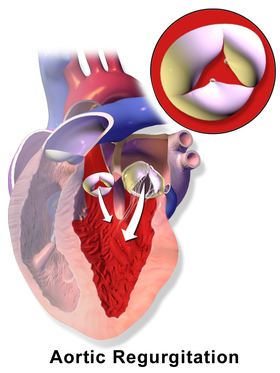
The aortic valve is a valve in the heart of humans and most other animals, located between the left ventricle and the aorta. It is one of the four valves of the heart and one of the two semilunar valves, the other being the pulmonary valve. The aortic valve normally has three cusps or leaflets, although in 1–2% of the population it is found to congenitally have two leaflets. The aortic valve is the last structure in the heart the blood travels through before stopping the flow through the systemic circulation.

<span class="mw-page-title-main">Mitral valve</span> Valve in the heart connecting the left atrium and left ventricle

The mitral valve, also known as the bicuspid valve or left atrioventricular valve, is one of the four heart valves. It has two cusps or flaps and lies between the left atrium and the left ventricle of the heart. The heart valves are all one-way valves allowing blood flow in just one direction. The mitral valve and the tricuspid valve are known as the atrioventricular valves because they lie between the atria and the ventricles.

<span class="mw-page-title-main">Aortic dissection</span> Injury to the innermost layer of the aorta

Aortic dissection (AD) occurs when an injury to the innermost layer of the aorta allows blood to flow between the layers of the aortic wall, forcing the layers apart. In most cases, this is associated with a sudden onset of severe chest or back pain, often described as "tearing" in character. Also, vomiting, sweating, and lightheadedness may occur. Other symptoms may result from decreased blood supply to other organs, such as stroke, lower extremity ischemia, or mesenteric ischemia. Aortic dissection can quickly lead to death from insufficient blood flow to the heart or complete rupture of the aorta.

<span class="mw-page-title-main">Bicuspid aortic valve</span> Medical condition

Bicuspid aortic valve is a form of heart disease in which two of the leaflets of the aortic valve fuse during development in the womb resulting in a two-leaflet (bicuspid) valve instead of the normal three-leaflet (tricuspid) valve. BAV is the most common cause of heart disease present at birth and affects approximately 1.3% of adults. Normally, the mitral valve is the only bicuspid valve and this is situated between the heart's left atrium and left ventricle. Heart valves play a crucial role in ensuring the unidirectional flow of blood from the atrium to the ventricles, or from the ventricle to the aorta or pulmonary trunk. BAV is normally inherited.

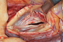
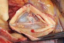
Aortic valve replacement is a procedure whereby the failing aortic valve of a patient's heart is replaced with an artificial heart valve. The aortic valve may need to be replaced because:

A transthoracic echocardiogram (TTE) is the most common type of echocardiogram, which is a still or moving image of the internal parts of the heart using ultrasound. In this case, the probe is placed on the chest or abdomen of the subject to get various views of the heart. It is used as a non-invasive assessment of the overall health of the heart, including a patient's heart valves and degree of heart muscle contraction. The images are displayed on a monitor for real-time viewing and then recorded.

<span class="mw-page-title-main">Valvular heart disease</span> Disease in the valves of the heart

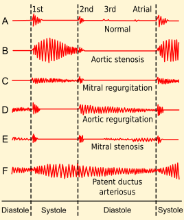
Valvular heart disease is any cardiovascular disease process involving one or more of the four valves of the heart. These conditions occur largely as a consequence of aging, but may also be the result of congenital (inborn) abnormalities or specific disease or physiologic processes including rheumatic heart disease and pregnancy.

Cor triatriatum is a congenital heart defect where the left atrium or right atrium is subdivided by a thin membrane, resulting in three atrial chambers.

<span class="mw-page-title-main">Arterial switch operation</span>

Arterial switch operation (ASO) or arterial switch, is an open heart surgical procedure used to correct dextro-transposition of the great arteries (d-TGA); its development was pioneered by Canadian cardiac surgeon William Mustard and it was named for Brazilian cardiac surgeon Adib Jatene, who was the first to use it successfully. It was the first method of d-TGA repair to be attempted, but the last to be put into regular use because of technological limitations at the time of its conception.

<span class="mw-page-title-main">Ascending aorta</span> Part of the heart

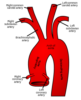
The ascending aorta (AAo) is a portion of the aorta commencing at the upper part of the base of the left ventricle, on a level with the lower border of the third costal cartilage behind the left half of the sternum.

The Dor procedure is a medical technique used as part of heart surgery and originally introduced by the French cardiac surgeon Vincent Dor (b.1932). It is also known as endoventricular circular patch plasty (EVCPP).

Valve-sparing aortic root replacement is a cardiac surgery procedure which is used to treat Aortic aneurysms and to prevent Aortic dissection. It involves replacement of the aortic root without replacement of the aortic valve. Two similar procedures were developed, one by Sir Magdi Yacoub, and another by Tirone David.

The Bentall procedure is a type of cardiac surgery involving composite graft replacement of the aortic valve, aortic root, and ascending aorta, with re-implantation of the coronary arteries into the graft. This operation is used to treat combined disease of the aortic valve and ascending aorta, including lesions associated with Marfan syndrome. The Bentall procedure was first described in 1968 by Hugh Bentall and Antony De Bono. It is considered a standard for individuals who require aortic root replacement, and the vast majority of individuals who undergo the surgery receive mechanical valves.

The following outline is provided as an overview of and topical guide to cardiology, the branch of medicine dealing with disorders of the human heart. The field includes medical diagnosis and treatment of congenital heart defects, coronary artery disease, heart failure, valvular heart disease and electrophysiology. Physicians who specialize in cardiology are called cardiologists.

<span class="mw-page-title-main">Apicoaortic Conduit</span> Cardiothoracic surgical process

Apicoaortic Conduit (AAC), also known as Aortic Valve Bypass (AVB), is a cardiothoracic surgical procedure that alleviates symptoms caused by blood flow obstruction from the left ventricle of the heart. Left ventricular outflow tract obstruction (LVOTO) is caused by narrowing of the aortic valve and other valve disorders. AAC, or AVB, relieves the obstruction to blood flow by adding a bioprosthetic valve to the circulatory system to decrease the load on the aortic valve. When an apicoaortic conduit is implanted, blood continues to flow from the heart through the aortic valve. In addition, blood flow bypasses the native valve and exits the heart through the implanted valved conduit. The procedure is effective at relieving excessive pressure gradient across the natural valve. High pressure gradient across the aortic valve can be congenital or acquired. A reduction in pressure gradient results in relief of symptoms.

<span class="mw-page-title-main">Open aortic surgery</span> Surgical technique

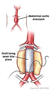
Open aortic surgery (OAS), also known as open aortic repair (OAR), describes a technique whereby an abdominal, thoracic or retroperitoneal surgical incision is used to visualize and control the aorta for purposes of treatment, usually by the replacement of the affected segment with a prosthetic graft. OAS is used to treat aneurysms of the abdominal and thoracic aorta, aortic dissection, acute aortic syndrome, and aortic ruptures. Aortobifemoral bypass is also used to treat atherosclerotic disease of the abdominal aorta below the level of the renal arteries. In 2003, OAS was surpassed by endovascular aneurysm repair (EVAR) as the most common technique for repairing abdominal aortic aneurysms in the United States.

<span class="mw-page-title-main">Hans-Joachim Schäfers</span> German surgeon, as well as cardiac, thoracic, and vascular surgeon

Hans-Joachim Schäfers is a German surgeon, as well as cardiac, thoracic, and vascular surgeon and university professor. He is director of the department of Thoracic and Cardiovascular Surgery at the Saarland University Medical Center in Homburg/Saar, Germany. He is known for his activities in aortic valve repair, aortic surgery, and pulmonary endarterectomy.

References

  1. "Aortic Regurgitation". The Lecturio Medical Concept Library. Retrieved 29 June 2021.
  2. 1 2 Schäfers HJ, et al. Bicuspidization of the unicuspid aortic valve: a new reconstructive approach. Ann Thorac Surg. 2008 Jun;85(6):2012-8.
  3. Aicher D, Schäfers HJ. Aortic valve repair - current status, indications, and outcomes. Semin Thorac Cardiovasc Surg. 2012;24(3):195-201
  4. 1 2 Vahanian A, et al. Guidelines on the management of valvular heart disease (version 2012). Eur Heart J. 2012 Oct;33(19):2451-96
  5. Ahmed, T; Puckett, Y (2021). "Aortic Valve Repair". National Center for Biotechnology Information, U.S. National Library of Medicine. PMID   32644365 . Retrieved 29 June 2021.
  6. 1 2 Regeer, M; Versteegh, M; Klautz, R; Stijnen, T; Schalij, M; Bax, J; Marsan, N; Delgado, V (17 October 2014). "Aortic valve repair versus replacement for aortic regurgitation: effects on left ventricular remodeling". Cardiothoracic Surgery Journals. 30 (1): 13–9. doi:10.1111/jocs.12457. PMID   25327584. S2CID   20937340 . Retrieved 29 June 2021.
  7. Hammermeister K, et al. Outcomes 15 years after valve replacement with a mechanical versus a bioprosthetic valve: final report of the Veterans Affairs randomized trial. J Am Coll Cardiol. 2000;36:1152-8.
  8. Aicher D, et al. Aortic valve repair leads to a low incidence of valve-related complications. Eur J Cardiothorac Surg. 2010;37:127-32
  9. Hans-Joachim Schäfers: Current treatment of aortic regurgitation. UNI-MED Science, Bremen, London, Boston 2013, ISBN   978-3-8374-1406-6
  10. Langer F, et al. Aortic valve repair using a differentiated surgical strategy. Circulation. 2004;110:II67-73
  11. Anderson RH. Understanding the structure of the unicuspid and unicommissural aortic valve. J Heart Valve Dis. 2003 Nov;12(6):670-3
  12. 1 2 3 Aicher D, Schneider U, Schmied W, Kunihara T, Tochii M, Schäfers HJ. Early results with annular support in reconstruction of the bicuspid aortic valve. J Thorac Cardiovasc Surg. 2013 Mar;145(3 Suppl):p30-4
  13. Schmidt KI, et al. Tricuspidization of the quadricuspid aortic valve. Ann Thorac Surg. 2008 Mar;85(3):1087-9
  14. Schäfers HJ, et al. (2000). "Remodeling of the aortic root and reconstruction of the bicuspid aortic valve". Ann Thorac Surg. 70 (2): 542–546. doi:10.1016/s0003-4975(00)01457-0. PMID   10969677.
  15. Franciulli M, et al. (Sep 2014). "Root remodeling and aortic valve repair for unicuspid aortic valve". Ann Thorac Surg. 98 (3): 823–829. doi:10.1016/j.athoracsur.2014.05.024. PMID   25085562.
  16. 1 2 Sarsam MA, Yacoub M (1993). "Remodeling of the aortic valve annulus". J Thorac Cardiovasc Surg. 105 (3): 435–438. doi: 10.1016/S0022-5223(19)34225-4 . PMID   8445922.
  17. 1 2 David TE, Feindel CM (Apr 1992). "An aortic valve-sparing operation for patients with aortic incompetence and aneurysm of the ascending aorta". J Thorac Cardiovasc Surg. 103 (4): 617–621. doi: 10.1016/S0022-5223(19)34942-6 . PMID   1532219.
  18. Schäfers HJ, et al. (Feb 2015). "Reexamining remodeling". J Thorac Cardiovasc Surg. 149 (2 Suppl): S30-36. doi: 10.1016/j.jtcvs.2014.09.048 . PMID   25439784.
  19. 1 2 David TE (Autumn 2014). "Current readings: Aortic valve-sparing operations". Semin Thorac Cardiovasc Surg. 26 (3): 231–238. doi:10.1053/j.semtcvs.2014.10.002. PMID   25527017.
  20. Kunihara T, et al. (Jun 2012). "Preoperative aortic root geometry and postoperative cusp configuration primarily determine long-term outcome after valve-preserving aortic root repair". J Thorac Cardiovasc Surg. 143 (6): 1389–1395. doi: 10.1016/j.jtcvs.2011.07.036 . PMID   21855091.
  21. Lansac E, et al. (2010). "An aortic ring: From physiologic reconstruction of the root to a standardized approach for aortic valve repair". J Thorac Cardiovasc Surg. 140 (6): S28-35. doi: 10.1016/j.jtcvs.2010.08.004 . PMID   21092793.
  22. Schäfers HJ, Bierbach B, Aicher D (2006). "A new approach to the assessment of aortic cusp geometry". J Thorac Cardiovasc Surg. 132 (2): 436–438. doi: 10.1016/j.jtcvs.2006.04.032 . PMID   16872982.
  23. Taylor WJ, et al. The surgical correction of aortic insufficiency by circumcision. J Thorac Cardiovasc Surg. 1958;35:192-231.
  24. L. Wi Stephenson: History of Cardiac Surgery. In: L. H. Cohn, L. H. Edmunds Jr. (Hrsg.): Cardiac Surgery in the Adult. McGraw-Hill, New York (USA) 2003, S. 3–29.

Further reading