Astigmatism (optical systems)

Last updated
Astigmatism: The image of point G is at point BM for rays in plane M, and at point BS for rays in plane S. Meridional+SagittalPlane.png
Astigmatism: The image of point G is at point BM for rays in plane M, and at point BS for rays in plane S.

An optical system with astigmatism is one where rays that propagate in two perpendicular planes have different foci. If an optical system with astigmatism is used to form an image of a cross, the vertical and horizontal lines will be in sharp focus at two different distances. The term comes from the Greek α- (a-) meaning "without" and στίγμα (stigma), "a mark, spot, puncture". [1]

Contents

Forms of astigmatism

There are two distinct forms of astigmatism. The first is a third-order aberration, which occurs for objects (or parts of objects) away from the optical axis. This form of aberration occurs even when the optical system is perfectly symmetrical. This is often referred to as a "monochromatic aberration", because it occurs even for light of a single wavelength. This terminology may be misleading, however, as the amount of aberration can vary strongly with wavelength in an optical system.

The second form of astigmatism occurs when the optical system is not symmetric about the optical axis. This may be by design (as in the case of a cylindrical lens), or due to manufacturing error in the surfaces of the components or misalignment of the components. In this case, astigmatism is observed even for rays from on-axis object points. This form of astigmatism is extremely important in vision science and eye care, since the human eye often exhibits this aberration due to imperfections in the shape of the cornea or the lens.

Third-order astigmatism

Page explaining and illustrating astigmatism Astigmatism.png
Page explaining and illustrating astigmatism

In the analysis of this form of astigmatism, it is most common to consider rays from a given point on the object, which propagate in two particular planes. The first plane is the tangential plane. This is the plane which includes both the object point under consideration and the axis of symmetry. Rays that propagate in this plane are called tangential rays. Planes that include the optical axis are meridional planes. It is common to simplify problems in radially-symmetric optical systems by choosing object points in the vertical ("y") plane only. This plane is then sometimes referred to as the meridional plane.

The second plane used in the analysis is the sagittal plane. This is defined as the plane, orthogonal to the tangential plane, which contains the object point being considered and intersects the optical axis at the entrance pupil of the optical system. This plane contains the chief ray, but does not contain the optic axis. It is therefore a skew plane, in other words not a meridional plane. Rays propagating in this plane are called sagittal rays.
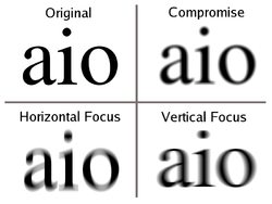
In third-order astigmatism, the sagittal and transverse rays form foci at different distances along the optic axis. These foci are called the sagittal focus and the transverse focus, respectively. In the presence of astigmatism, an off-axis point on the object is not sharply imaged by the optical system. Instead, sharp lines are formed at the sagittal and transverse foci. The image at the transverse focus is a short line, oriented in the direction of the sagittal plane; images of circles centered on the optic axis, or lines tangential to such circles, will be sharp in this plane. The image at the sagittal focus is a short line, oriented in the tangential direction; images of spokes radiating from the center are sharp at this focus. In between these two foci, a round but "blurry" image is formed. This is called the medial focus or circle of least confusion . This plane often represents the best compromise image location in a system with astigmatism.

The amount of aberration due to astigmatism is proportional to the square of the angle between the rays from the object and the optical axis of the system. With care, an optical system can be designed to reduce or eliminate astigmatism. Such systems are called anastigmats.

Astigmatism in systems that are not rotationally symmetric

Visual astigmatism Astigmatism.svg
Visual astigmatism
Blur from astigmatic lens at different distances. Astigmatism text blur.png
Blur from astigmatic lens at different distances.

If an optical system is not axisymmetric, either due to an error in the shape of the optical surfaces or due to misalignment of the components, astigmatism can occur even for on-axis object points. This effect is often used deliberately in complex optical systems, especially certain types of telescope. Some telescopes deliberately use non-spherical optics to overcome this phenomenon.[ why? ] [3] [ failed verification ]

In the analysis of these systems, it is common to consider tangential rays (as defined above), and rays in a meridional plane (a plane containing the optic axis) perpendicular to the tangential plane. This plane is called either the sagittal meridional plane or, confusingly, just the sagittal plane.

Ophthalmic astigmatism

In optometry and ophthalmology, the vertical and horizontal planes are identified as tangential and sagittal meridians, respectively. Ophthalmic astigmatism is a refraction error of the eye in which there is a difference in degree of refraction in different meridians. [4] It is typically characterized by an aspherical, non-figure of revolution cornea in which the corneal profile slope and refractive power in one meridian is less than that of the perpendicular axis.

Astigmatism causes difficulties in seeing fine detail. Astigmatism can be often corrected by glasses with a lens that has different radii of curvature in different planes (a cylindrical lens), contact lenses, or refractive surgery. [5]

Astigmatism is quite common. Studies have shown that about one in three people suffers from it. [6] [7] [8] The prevalence of astigmatism increases with age. [9] Although a person may not notice mild astigmatism, higher amounts of astigmatism may cause blurry vision, squinting, asthenopia, fatigue, or headaches. [10] [11] [12]

There are a number of tests that are used by ophthalmologists and optometrists during eye examinations to determine the presence of astigmatism and to quantify the amount and axis of the astigmatism. [13] A Snellen chart or other eye chart may initially reveal reduced visual acuity. A keratometer may be used to measure the curvature of the steepest and flattest meridians in the cornea's front surface. [14] Corneal topography may also be used to obtain a more accurate representation of the cornea's shape. [15] An autorefractor or retinoscopy may provide an objective estimate of the eye's refractive error and the use of Jackson cross cylinders in a phoropter may be used to subjectively refine those measurements. [16] [17] [18] An alternative technique with the phoropter requires the use of a "clock dial" or "sunburst" chart to determine the astigmatic axis and power. [19] [20]

Astigmatism may be corrected with eyeglasses, contact lenses, or refractive surgery. [21] [22] [23] Various considerations involving ocular health, refractive status, and lifestyle frequently determine whether one option may be better than another. In those with keratoconus, toric contact lenses often enable patients to achieve better visual acuities than eyeglasses. If the astigmatism is caused by a problem such as deformation of the eyeball due to a chalazion, treating the underlying cause will resolve the astigmatism.

Misaligned or malformed lenses and mirrors

Grinding and polishing of precision optical parts, either by hand or machine, typically employs significant downward pressure, which in turn creates significant frictional side pressures during polishing strokes that can combine to locally flex and distort the parts. These distortions generally do not possess figure-of-revolution symmetry and are thus astigmatic, and slowly become permanently polished into the surface if the problems causing the distortion are not corrected. Astigmatic, distorted surfaces potentially introduce serious degradations in optical system performance.

Surface distortion due to grinding or polishing increases with the aspect ratio of the part (diameter to thickness ratio). To a first order, glass strength increases as the cube of the thickness. Thick lenses at 4:1 to 6:1 aspect ratios will flex much less than high aspect ratio parts, such as optical windows, which can have aspect ratios of 15:1 or higher. The combination of surface or wavefront error precision requirements and part aspect ratio drives the degree of back support uniformity required, especially during the higher down pressures and side forces during polishing. Optical working typically involves a degree of randomness that helps greatly in preserving figure-of-revolution surfaces, provided the part is not flexing during the grind/polish process.

Deliberate astigmatism in optical systems

Compact disc players use an astigmatic lens for focusing. When one axis is more in focus than the other, dot-like features on the disc project to oval shapes. The orientation of the oval indicates which axis is more in focus, and thus which direction the lens needs to move. A square arrangement of only four sensors can observe this bias and use it to bring the read lens to best focus, without being fooled by oblong pits or other features on the disc surface.[ citation needed ]

In 3D PALM/STORM, a type of optical super-resolution microscopy, a cylindrical lens can be introduced into the imaging system to create astigmatism, which allows measurement of the Z position of a diffraction-limited light source. [24]

Laser line levels use a cylindrical lens to spread a laser beam from a point into a line.

See also

Related Research Articles

<span class="mw-page-title-main">Optical aberration</span> Deviation from perfect paraxial optical behavior

In optics, aberration is a property of optical systems, such as lenses, that causes light to be spread out over some region of space rather than focused to a point. Aberrations cause the image formed by a lens to be blurred or distorted, with the nature of the distortion depending on the type of aberration. Aberration can be defined as a departure of the performance of an optical system from the predictions of paraxial optics. In an imaging system, it occurs when light from one point of an object does not converge into a single point after transmission through the system. Aberrations occur because the simple paraxial theory is not a completely accurate model of the effect of an optical system on light, rather than due to flaws in the optical elements.

<span class="mw-page-title-main">Lens</span> Optical device which transmits and refracts light

A lens is a transmissive optical device that focuses or disperses a light beam by means of refraction. A simple lens consists of a single piece of transparent material, while a compound lens consists of several simple lenses (elements), usually arranged along a common axis. Lenses are made from materials such as glass or plastic and are ground, polished, or molded to the required shape. A lens can focus light to form an image, unlike a prism, which refracts light without focusing. Devices that similarly focus or disperse waves and radiation other than visible light are also called "lenses", such as microwave lenses, electron lenses, acoustic lenses, or explosive lenses.

<span class="mw-page-title-main">Corrective lens</span> Type of lens

A corrective lens is a lens that is typically worn in front of the eye to improve daily vision. The most common use is to treat refractive errors: myopia, hypermetropia, astigmatism, and presbyopia. Glasses or "spectacles" are worn on the face a short distance in front of the eye. Contact lenses are worn directly on the surface of the eye. Intraocular lenses are surgically implanted most commonly after cataract removal but can be used for purely refractive purposes.

<span class="mw-page-title-main">Reflecting telescope</span> Telescopes which utilize curved mirrors to form an image

A reflecting telescope is a telescope that uses a single or a combination of curved mirrors that reflect light and form an image. The reflecting telescope was invented in the 17th century by Isaac Newton as an alternative to the refracting telescope which, at that time, was a design that suffered from severe chromatic aberration. Although reflecting telescopes produce other types of optical aberrations, it is a design that allows for very large diameter objectives. Almost all of the major telescopes used in astronomy research are reflectors. Many variant forms are in use and some employ extra optical elements to improve image quality or place the image in a mechanically advantageous position. Since reflecting telescopes use mirrors, the design is sometimes referred to as a catoptric telescope.

<span class="mw-page-title-main">LASIK</span> Corrective ophthalmological surgery

LASIK or Lasik, commonly referred to as laser eye surgery or laser vision correction, is a type of refractive surgery for the correction of myopia, hyperopia, and an actual cure for astigmatism, since it is in the cornea. LASIK surgery is performed by an ophthalmologist who uses a laser or microkeratome to reshape the eye's cornea in order to improve visual acuity.

<span class="mw-page-title-main">Eyeglass prescription</span> Order written by an eyewear prescriber

An eyeglass prescription is an order written by an eyewear prescriber, such as an optometrist, that specifies the value of all parameters the prescriber has deemed necessary to construct and/or dispense corrective lenses appropriate for a patient. If an eye examination indicates that corrective lenses are appropriate, the prescriber generally provides the patient with an eyewear prescription at the conclusion of the exam.

<span class="mw-page-title-main">Coma (optics)</span> Aberration inherent to certain optical designs or due to imperfection in the lens

In optics, the coma, or comatic aberration, in an optical system refers to aberration inherent to certain optical designs or due to imperfection in the lens or other components that results in off-axis point sources such as stars appearing distorted, appearing to have a tail (coma) like a comet. Specifically, coma is defined as a variation in magnification over the entrance pupil. In refractive or diffractive optical systems, especially those imaging a wide spectral range, coma can be a function of wavelength, in which case it is a form of chromatic aberration.

<span class="mw-page-title-main">Refractive surgery</span> Surgery to treat common vision disorders

Refractive surgery is optional eye surgery used to improve the refractive state of the eye and decrease or eliminate dependency on glasses or contact lenses. This can include various methods of surgical remodeling of the cornea (keratomileusis), lens implantation or lens replacement. The most common methods today use excimer lasers to reshape the curvature of the cornea. Refractive eye surgeries are used to treat common vision disorders such as myopia, hyperopia, presbyopia and astigmatism.

<span class="mw-page-title-main">Intraocular lens</span> Lens implanted in the eye to treat cataracts or myopia

An Intraocular lens (IOL) is a lens implanted in the eye usually as part of a treatment for cataracts or for correcting other vision problems such as short sightedness and long sightedness, a form of refractive surgery. If the natural lens is left in the eye, the IOL is known as phakic, otherwise it is a pseudophakic lens. Both kinds of IOLs are designed to provide the same light-focusing function as the natural crystalline lens. This can be an alternative to LASIK, but LASIK is not an alternative to an IOL for treatment of cataracts.

<span class="mw-page-title-main">Refractive error</span> Problem with focusing light accurately on the retina due to the shape of the eye

Refractive error, also known as refraction error, is a problem with focusing light accurately on the retina due to the shape of the eye and or cornea. The most common types of refractive error are near-sightedness, far-sightedness, astigmatism, and presbyopia. Near-sightedness results in far away objects being blurry, far-sightedness and presbyopia result in close objects being blurry, and astigmatism causes objects to appear stretched out or blurry. Other symptoms may include double vision, headaches, and eye strain.

<span class="mw-page-title-main">Ray (optics)</span> Idealized model of light

In optics, a ray is an idealized geometrical model of light or other electromagnetic radiation, obtained by choosing a curve that is perpendicular to the wavefronts of the actual light, and that points in the direction of energy flow. Rays are used to model the propagation of light through an optical system, by dividing the real light field up into discrete rays that can be computationally propagated through the system by the techniques of ray tracing. This allows even very complex optical systems to be analyzed mathematically or simulated by computer. Ray tracing uses approximate solutions to Maxwell's equations that are valid as long as the light waves propagate through and around objects whose dimensions are much greater than the light's wavelength. Ray optics or geometrical optics does not describe phenomena such as diffraction, which require wave optics theory. Some wave phenomena such as interference can be modeled in limited circumstances by adding phase to the ray model.

In Gaussian optics, the cardinal points consist of three pairs of points located on the optical axis of a rotationally symmetric, focal, optical system. These are the focal points, the principal points, and the nodal points. For ideal systems, the basic imaging properties such as image size, location, and orientation are completely determined by the locations of the cardinal points; in fact only four points are necessary: the focal points and either the principal or nodal points. The only ideal system that has been achieved in practice is the plane mirror, however the cardinal points are widely used to approximate the behavior of real optical systems. Cardinal points provide a way to analytically simplify a system with many components, allowing the imaging characteristics of the system to be approximately determined with simple calculations.

<span class="mw-page-title-main">Corneal topography</span> Medical imaging technique

Corneal topography, also known as photokeratoscopy or videokeratography, is a non-invasive medical imaging technique for mapping the anterior curvature of the cornea, the outer structure of the eye. Since the cornea is normally responsible for some 70% of the eye's refractive power, its topography is of critical importance in determining the quality of vision and corneal health.

<span class="mw-page-title-main">Astigmatism</span> Type of eye defect

Astigmatism is a type of refractive error due to rotational asymmetry in the eye's refractive power. This results in distorted or blurred vision at any distance. Other symptoms can include eyestrain, headaches, and trouble driving at night. Astigmatism often occurs at birth and can change or develop later in life. If it occurs in early life and is left untreated, it may result in amblyopia.

Aniseikonia is an ocular condition where there is a significant difference in the perceived size of images. It can occur as an overall difference between the two eyes, or as a difference in a particular meridian. If the ocular image size in both eyes are equal, the condition is known as iseikonia.

<span class="mw-page-title-main">Cylindrical lens</span>

A cylindrical lens is a lens which focuses light into a line instead of a point, as a spherical lens would. The curved face or faces of a cylindrical lens are sections of a cylinder, and focus the image passing through it into a line parallel to intersection of the surface of the lens and a plane tangent to it along the cylinder's axis. The lens converges or diverges the image in the direction perpendicular to this line, and leaves it unaltered in the direction parallel to its cylinder's axis.

<span class="mw-page-title-main">Toric lens</span> Type of lens

A toric lens is a lens with different optical power and focal length in two orientations perpendicular to each other. One of the lens surfaces is shaped like a "cap" from a torus, and the other one is usually spherical. Such a lens behaves like a combination of a spherical lens and a cylindrical lens. Toric lenses are used primarily in eyeglasses, contact lenses and intraocular lenses to correct astigmatism.

The Alpins Method is a system to plan and analyze the results of refractive surgical procedures, such as laser in-situ keratomileus (LASIK). The Alpins Method is also used to plan cataract/toric intraocular lens (IOL) surgical procedures.

The eye, like any other optical system, suffers from a number of specific optical aberrations. The optical quality of the eye is limited by optical aberrations, diffraction and scatter. Correction of spherocylindrical refractive errors has been possible for nearly two centuries following Airy's development of methods to measure and correct ocular astigmatism. It has only recently become possible to measure the aberrations of the eye and with the advent of refractive surgery it might be possible to correct certain types of irregular astigmatism.

Stokes lens also known as variable power cross cylinder lens is a lens used to diagnose a type of refractive error known as astigmatism.

References

  1. Harper, Douglas (2001). "Online Etymology Dictionary" . Retrieved 2007-12-29.
  2. Frederic Eugene Wright (1911). The Methods of Petrographic-microscopic Research, Their Relative Accuracy and Range of Application. Carnegie institution of Washington.
  3. Sacek, Vladimir (July 14, 2006). "Telescope astigmatism". Amateur Telescope Optics. Archived from the original on 19 September 2008. Retrieved Oct 16, 2008.
  4. "Facts About Astigmatism | National Eye Institute". nei.nih.gov. Archived from the original on 2016-10-02. Retrieved 2019-05-06.
  5. "Astigmatism Laser Eye Surgery". The Irish Times. Archived from the original on 2012-10-24. Retrieved 2011-08-29.
  6. Kleinstein RN, Jones LA, Hullett S, Kwon S, et al. (2003). "Refractive Error and Ethnicity in Children". Arch. Ophthalmol. 121 (8): 1141–47. doi: 10.1001/archopht.121.8.1141 . PMID   12912692.
  7. Garcia CA, Oréfice F, Nobre GF, Souza Dde B, Rocha ML, Vianna RN (2005). "[Prevalence of refractive errors in students in Northeastern Brazil.]". Arq Bras Oftalmol (in Portuguese). 68 (3): 321–25. doi: 10.1590/S0004-27492005000300009 . PMID   16059562.
  8. Bourne RR, Dineen BP, Ali SM, Noorul Huq DM, Johnson GJ (June 2004). "Prevalence of refractive error in Bangladeshi adults: results of the National Blindness and Low Vision Survey of Bangladesh". Ophthalmology. 111 (6): 1150–60. doi:10.1016/j.ophtha.2003.09.046. PMID   15177965.
  9. Asano K, Nomura H, Iwano M, et al. (2005). "Relationship between astigmatism and aging in middle-aged and elderly Japanese". Jpn. J. Ophthalmol. 49 (2): 127–33. doi:10.1007/s10384-004-0152-1. PMID   15838729. S2CID   20925765.
  10. Eyetopics.com
  11. Medicinenet.com
  12. Hipusa.com Archived May 1, 2006, at the Wayback Machine
  13. Hipusa.com Archived April 26, 2006, at the Wayback Machine
  14. Stlukeseye.com Archived March 23, 2006, at the Wayback Machine
  15. Emedicine.com Archived February 18, 2006, at the Wayback Machine
  16. Graff T (June 1962). "[Control of the determination of astigmatism with the Jackson cross cylinder.]". Klin Monatsblätter Augenheilkd Augenarztl Fortbild (in German). 140: 702–08. PMID   13900989.
  17. Del Priore LV, Guyton DL (November 1986). "The Jackson cross cylinder. A reappraisal". Ophthalmology. 93 (11): 1461–65. doi:10.1016/s0161-6420(86)33545-0. PMID   3808608.
  18. Brookman KE (May 1993). "The Jackson crossed cylinder: historical perspective". J Am Optom Assoc. 64 (5): 329–31. PMID   8320415.
  19. Quantumoptical.com
  20. Nova.edu
  21. "Contact Lenses for Vision Correction". American Academy of Ophthalmology. 2018-09-01. Retrieved 2019-05-06.
  22. "Eyeglasses for Vision Correction". American Academy of Ophthalmology. 2015-12-12. Retrieved 2019-05-06.
  23. "LASIK eye surgery". Mayo Clinic. Retrieved 2019-05-06.
  24. Huang, Bo (Feb 8, 2008). "Three-dimensional Super-resolution Imaging by Stochastic Optical Reconstruction Microscopy". Science. 319 (5864): 810–13. Bibcode:2008Sci...319..810H. doi:10.1126/science.1153529. PMC   2633023 . PMID   18174397.