The endomembrane system is composed of the different membranes that are suspended in the cytoplasm within a eukaryotic cell. These membranes divide the cell into functional and structural compartments, or organelles. In eukaryotes the organelles of the endomembrane system include: the nuclear membrane, the endoplasmic reticulum, the Golgi apparatus, lysosomes, vesicles, endosomes, and plasma (cell) membrane among others. The system is defined more accurately as the set of membranes that form a single functional and developmental unit, either being connected directly, or exchanging material through vesicle transport. Importantly, the endomembrane system does not include the membranes of chloroplasts or mitochondria, but might have evolved from the latter.

Endocytosis is a cellular process in which substances are brought into the cell. The material to be internalized is surrounded by an area of plasma membrane, which then buds off inside the cell to form a vesicle containing the ingested material. Endocytosis includes pinocytosis and phagocytosis. It is a form of active transport.

A lysosome is a membrane-bound organelle found in many animal cells and most plant cells. They are spherical vesicles that contain hydrolytic enzymes that can break down many kinds of biomolecules. A lysosome has a specific composition, of both its membrane proteins, and its lumenal proteins. The lumen's pH (4.5–5.0) is optimal for the enzymes involved in hydrolysis, analogous to the activity of the stomach. Besides degradation of polymers, the lysosome is involved in various cell processes, including secretion, plasma membrane repair, cell signaling, and energy metabolism.

In cell biology, a vesicle is a large structure within a cell, or extracellular, consisting of liquid enclosed by a lipid bilayer. Vesicles form naturally during the processes of secretion (exocytosis), uptake (endocytosis) and transport of materials within the plasma membrane. Alternatively, they may be prepared artificially, in which case they are called liposomes. If there is only one phospholipid bilayer, they are called unilamellar liposome vesicles; otherwise they are called multilamellar. The membrane enclosing the vesicle is also a lamellar phase, similar to that of the plasma membrane and vesicles can fuse with the plasma membrane to release their contents outside the cell. Vesicles can also fuse with other organelles within the cell.

Exocytosis is a form of active transport and bulk transport in which a cell transports molecules out of the cell by expelling them through an energy-dependent process. Exocytosis and its counterpart, endocytosis, are used by all cells because most chemical substances important to them are large polar molecules that cannot pass through the hydrophobic portion of the cell membrane by passive means. It is a process of which the contents of a vacuole are released, since the plant cell has large amounts of content in its vacuole, when exocytosis is in process a large amount of molecules are released thus making it a form of bulk transport.
Secretion is the movement of material from one point to another, e.g. secreted chemical substance from a cell or gland. In contrast, excretion, is the removal of certain substances or waste products from a cell or organism. The classical mechanism of cell secretion is via secretory portals at the cell plasma membrane called porosomes. Porosomes are permanent cup-shaped lipoprotein structure at the cell plasma membrane, where secretory vesicles transiently dock and fuse to release intra-vesicular contents from the cell.

In cell biology, a phagosome is a vesicle formed around a particle engulfed by a phagocyte via phagocytosis. Professional phagocytes include macrophages, neutrophils, and dendritic cells (DCs). A phagosome is formed by the fusion of the cell membrane around a microorganism, a senescent cell or an apoptotic cell. Phagosomes have membrane-bound proteins to recruit and fuse with lysosomes to form mature phagolysosomes. The lysosomes contain hydrolytic enzymes and reactive oxygen species (ROS) which kill and digest the pathogens. Phagosomes can also form in non-professional phagocytes, but they can only engulf a smaller range of particles, and do not contain ROS. The useful materials from the digested particles are moved into the cytosol, and waste is removed by exocytosis. Phagosome formation is crucial for tissue homeostasis and both innate and adaptive host defense against pathogens.

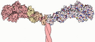
SNARE proteins — "SNAP" REceptor" — are a large protein complex consisting of at least 24 members in yeasts and more than 60 members in mammalian cells. The primary role of SNARE proteins is to mediate vesicle fusion, that is, the fusion of vesicles with their target membrane bound compartments. The best studied SNAREs are those that mediate docking of synaptic vesicles with the presynaptic membrane in neurons. These SNAREs are the targets of the bacterial neurotoxins responsible for botulism and tetanus.
Two-pore channels (TPCs) are eukaryotic intracellular voltage-gated and ligand gated cation selective ion channels. There are two known paralogs in the human genome, TPC1s and TPC2s. In humans, TPC1s are sodium selective and TPC2s conduct sodium ions, calcium ions and possibly hydrogen ions. Plant TPC1s are non-selective channels. Expression of TPCs are found in both plant vacuoles and animal acidic organelles. These organelles consist of endosomes and lysosomes. TPCs are formed from two transmembrane non-equivalent tandem Shaker-like, pore-forming subunits, dimerized to form quasi-tetramers. Quasi-tetramers appear very similar to tetramers, but are not quite the same. Some key roles of TPCs include calcium dependent responses in muscle contraction(s), hormone secretion, fertilization, and differentiation. Disorders linked to TPCs include membrane trafficking, Parkinson’s disease, Ebola, and fatty liver.

In biology, a phagolysosome, or endolysosome, is a cytoplasmic body formed by the fusion of a phagosome with a lysosome in a process that occurs during phagocytosis. Formation of phagolysosomes is essential for the intracellular destruction of microorganisms and pathogens. It takes place when the phagosome's and lysosome's membranes 'collide', at which point the lysosomal contents—including hydrolytic enzymes—are discharged into the phagosome in an explosive manner and digest the particles that the phagosome had ingested. Some products of the digestion are useful materials and are moved into the cytoplasm; others are exported by exocytosis.

Microvesicles are a type of extracellular vesicle, between 50 and 1,000 nanometers (nm) in diameter, found in many types of body fluids as well as the interstitial space between cells. Microvesicles are membrane-bound vesicles containing phospholipids, ranging from 100 nm to 1000 nm shed from almost all cell types. Not to be confused with smaller intracellularly generated extracellular vesicles known as exosomes. Microvesicles play a role in intercellular communication and can transport mRNA, miRNA, and proteins between cells. Microvesicles have been implicated in the process of a remarkable anti-tumor reversal effect in cancer, tumor immune suppression, metastasis, tumor-stroma interactions and angiogenesis along with having a primary role in tissue regeneration. They originate directly from the plasma membrane of the cell and reflect the antigenic content of the cells from which they originate.
They remove misfolded proteins, cytotoxic agents and metabolic waste from the cell.

Synaptosomal-associated protein 23 is a protein that in humans is encoded by the SNAP23 gene. Two alternative transcript variants encoding different protein isoforms have been described for this gene.

Syntaxin-7 is a protein that in humans is encoded by the STX7 gene.

Syntaxin-6 is a protein that in humans is encoded by the STX6 gene.

Porosomes are cup-shaped supramolecular structures in the cell membranes of eukaryotic cells where secretory vesicles transiently dock in the process of vesicle fusion and secretion. The transient fusion of secretory vesicle membrane at the porosome base via SNARE proteins, result in the formation of a fusion pore or continuity for the release of intravesicular contents from the cell. After secretion is complete, the fusion pore temporarily formed at the base of the porosome is sealed. The porosomes are few nanometers in size and contain many different types of protein, especially chloride and calcium channels, actin, and SNARE proteins that mediate the docking and fusion of the vesicles with the cell membrane. Once the vesicles have docked with the SNARE proteins, they swell, which increases their internal pressure. They then transiently fuse at the base of the porosome, and these pressurized contents are ejected from the cell. Examination of cells following secretion using electron microscopy, demonstrate increased presence of partially empty vesicles following secretion. This suggested that during the secretory process, only a portion of the vesicular contents are able to exit the cell. This could only be possible if the vesicle were to temporarily establish continuity with the cell plasma membrane, expel a portion of its contents, then detach, reseal, and withdraw into the cytosol (endocytose). In this way, the secretory vesicle could be reused for subsequent rounds of exo-endocytosis, until completely empty of its contents.

In membrane biology, fusion is the process by which two initially distinct lipid bilayers merge their hydrophobic cores, resulting in one interconnected structure. If this fusion proceeds completely through both leaflets of both bilayers, an aqueous bridge is formed and the internal contents of the two structures can mix. Alternatively, if only one leaflet from each bilayer is involved in the fusion process, the bilayers are said to be hemifused. In hemifusion, the lipid constituents of the outer leaflet of the two bilayers can mix, but the inner leaflets remain distinct. The aqueous contents enclosed by each bilayer also remain separated.

Cytosis is a transport mechanism for the movement of large quantities of molecules into and out of cells.
Vesicle fusion is the merging of a vesicle with other vesicles or a part of a cell membrane. In the latter case, it is the end stage of secretion from secretory vesicles, where their contents are expelled from the cell through exocytosis. Vesicles can also fuse with other target cell compartments, such as a lysosome.
Membrane vesicle trafficking in eukaryotic animal cells involves movement of important biochemical signal molecules from synthesis-and-packaging locations in the Golgi body to specific 'release' locations on the inside of the plasma membrane of the secretory cell, in the form of Golgi membrane-bound micro-sized vesicles, termed membrane vesicles (MVs). In this process, the 'packed' cellular products are released/secreted outside the cell across its plasma membrane. However, this vesicular membrane is retained and recycled by the secretory cells. This phenomenon has key role in synaptic neurotransmission, endocrine secretion, mucous secretion, granular-product secretion by neutrophils, etc. The scientists behind this discovery were awarded Nobel prize for the year 2013.
In the prokaryotic gram-negative bacterial cells, membrane vesicle trafficking is mediated via bacterial outer membrane bounded nano-sized vesicles, called bacterial outer membrane vesicles (OMVs). In this case, however, the OMV membrane is secreted as well, along with OMV-contents to outside the secretion-active bacterium. This phenomenon has key role in host-pathogen interactions, endotoxic shock in patients, invasion and infection of animals/plants, inter-species bacterial competition, quorum sensing, exocytosis, etc..