Related Research Articles
Neurosurgery or neurological surgery, known in common parlance as brain surgery, is the medical specialty concerned with the prevention, diagnosis, surgical treatment, and rehabilitation of disorders which affect any portion of the nervous system including the brain, spinal cord, central and peripheral nervous system, and cerebrovascular system.

A brain tumor occurs when abnormal cells form within the brain. There are two main types of tumors: malignant tumors and benign (non-cancerous) tumors. These can be further classified as primary tumors, which start within the brain, and secondary tumors, which most commonly have spread from tumors located outside the brain, known as brain metastasis tumors. All types of brain tumors may produce symptoms that vary depending on the size of the tumor and the part of the brain that is involved. Where symptoms exist, they may include headaches, seizures, problems with vision, vomiting and mental changes. Other symptoms may include difficulty walking, speaking, with sensations, or unconsciousness.
The Wada test, also known as the intracarotid sodium amobarbital procedure (ISAP) establishes cerebral language and memory representation of each hemisphere.

Epidural hematoma is when bleeding occurs between the tough outer membrane covering the brain and the skull. Often there is loss of consciousness following a head injury, a brief regaining of consciousness, and then loss of consciousness again. Other symptoms may include headache, confusion, vomiting, and an inability to move parts of the body. Complications may include seizures.

Glioblastoma, also known as glioblastoma multiforme (GBM), is the most aggressive type of cancer that begins within the brain. Initially, signs and symptoms of glioblastoma are nonspecific. They may include headaches, personality changes, nausea, and symptoms similar to those of a stroke. Symptoms often worsen rapidly and may progress to unconsciousness.

Radiosurgery is surgery using radiation, that is, the destruction of precisely selected areas of tissue using ionizing radiation rather than excision with a blade. Like other forms of radiation therapy, it is usually used to treat cancer. Radiosurgery was originally defined by the Swedish neurosurgeon Lars Leksell as "a single high dose fraction of radiation, stereotactically directed to an intracranial region of interest".

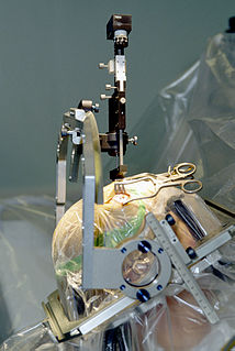
A craniotomy is a surgical operation in which a bone flap is temporarily removed from the skull to access the brain. Craniotomies are often critical operations, performed on patients who are suffering from brain lesions, such as tumors, blood clots, removal of foreign bodies such as bullets, or traumatic brain injury (TBI), and can also allow doctors to surgically implant devices, such as, deep brain stimulators for the treatment of Parkinson's disease, epilepsy, and cerebellar tremor. The procedure is also used in epilepsy surgery to remove the parts of the brain that are causing epilepsy.

Intraparenchymal hemorrhage (IPH) is one form of intracerebral bleeding in which there is bleeding within brain parenchyma. The other form is intraventricular hemorrhage (IVH).

Duret haemorrhages are small linear areas of bleeding in the midbrain and upper pons of the brainstem. They are caused by a traumatic downward displacement of the brainstem.
Hypophysectomy is the surgical removal of the hypophysis. It is most commonly performed to treat tumors, especially craniopharyngioma tumors. Sometimes it is used to treat Cushing's syndrome due to pituitary adenoma or Simmond's disease It is also applied in neurosciences to understand the functioning of hypophysis. There are various ways a hypophysectomy can be carried out. These methods include transsphenoidal hypophysectomy, open craniotomy, and stereotactic radiosurgery.

A colloid cyst is a non-malignant tumor in the brain. It consists of a gelatinous material contained within a membrane of epithelial tissue. It is almost always found just posterior to the foramen of Monro in the anterior aspect of the third ventricle, originating from the roof of the ventricle. Because of its location, it can cause obstructive hydrocephalus and increased intracranial pressure. Colloid cysts represent 0.5–1.0% of intracranial tumors.
Dysosmia is a disorder described as any qualitative alteration or distortion of the perception of smell. Qualitative alterations differ from quantitative alterations, which include anosmia and hyposmia. Dysosmia can be classified as either parosmia or phantosmia. Parosmia is a distortion in the perception of an odorant. Odorants smell different from what one remembers. Phantosmia is the perception of an odor when no odorant is present. The cause of dysosmia still remains a theory. It is typically considered a neurological disorder and clinical associations with the disorder have been made. Most cases are described as idiopathic and the main antecedents related to parosmia are URTIs, head trauma, and nasal and paranasal sinus disease. Dysosmia tends to go away on its own but there are options for treatment for patients that want immediate relief.
Kernohan's notch is a cerebral peduncle indentation associated with some forms of transtentorial herniation. It is a secondary condition caused by a primary injury on the opposite hemisphere of the brain. Kernohan's notch is an ipsilateral condition, in that a left-sided primary lesion evokes motor impairment in the left side of the body and a right-sided primary injury evokes motor impairment in the right side of the body. The seriousness of Kernohan's notch varies depending on the primary problem causing it, which may range from benign brain tumors to advanced subdural hematoma.

A central nervous system cyst is a type of cyst that presents and affects part of the central nervous system (CNS). They are usually benign and filled with either cerebrospinal fluid, blood, or tumor cells. CNS cysts are classified into two categories: cysts that originate from non-central nervous system tissue, migrate to, and form on a portion of the CNS, and cysts that originate within central nervous system tissue itself. Within these two categories, there are many types of CNS cysts that have been identified from previous studies.

A brain metastasis is a cancer that has metastasized (spread) to the brain from another location in the body and is therefore considered a secondary brain tumor. The metastasis typically shares a cancer cell type with the original site of the cancer. Metastasis is the most common cause of brain cancer, as primary tumors that originate in the brain are less common. The most common sites of primary cancer which metastasize to the brain are lung, breast, colon, kidney, and skin cancer. Brain metastases can occur in patients months or even years after their original cancer is treated. Brain metastases have a poor prognosis for cure, but modern treatments are allowing patients to live months and sometimes years after the diagnosis.
The Division of Surgical Neurooncology in the Department of Neurological Surgery and Sylvester Comprehensive Cancer Center at the University of Miami is one of the largest and most complete programs for brain tumor treatment in the United States. As the only academic medical center in the region, the University of Miami offers comprehensive approaches to these conditions with interdisciplinary discussion between neurosurgery, neurology, radiation therapy, and oncology.
Oculomotor apraxia (OMA), is the absence or defect of controlled, voluntary, and purposeful eye movement. It was first described in 1952 by the American ophthalmologist David Glendenning Cogan. People with this condition have difficulty moving their eyes horizontally and moving them quickly. The main difficulty is in saccade initiation, but there is also impaired cancellation of the vestibulo-ocular reflex. Patients have to turn their head in order to compensate for the lack of eye movement initiation in order to follow an object or see objects in their peripheral vision, but they often exceed their target. There is controversy regarding whether OMA should be considered an apraxia, since apraxia is the inability to perform a learned or skilled motor action to command, and saccade initiation is neither a learned nor a skilled action.
Awake craniotomy is a neurosurgical technique and type of craniotomy that allows a surgeon to remove a brain tumor while the patient is awake to avoid brain damage. During the surgery, the neurosurgeon performs cortical mapping to identify vital areas, called the "eloquent brain", that should not be disturbed while removing the tumor.
Günther C. Feigl is an Austrian neurosurgeon. Feigl is an internationally renowned expert in minimally invasive neurosurgery. His main areas of expertise are skull base surgery and neurooncology. He specializes in the surgery of gliomas, minimally invasive endoscopy-assisted microvascular decompression in trigeminal neuralgia and facial hemispasm as well as the surgery of acoustic neuromas, tumors of the pineal gland and meningiomas of the skull base. Furthermore, his specialties comprise treatment of pituitary adenomas, spinal cord tumours and metastases as well as the area of pediatric neurosurgery.
Brain Tumor Social Media (#BTSM) is a patient and care partner-run, grassroots Twitter community. The Twitter account @BTSMchat hosts monthly tweet chats for the #BTSM community and consistently trends among the top 15 of disease-related tweet chats. A study published in 2020 revealed the hashtag was most commonly used by brain tumor patients (33.13%), along with patient advocacy organizations (7.01%), care partners (4.63%), and clinicians (3.63%) and researchers (3.37%) specializing in brain tumors and brain cancers.
References
| | This surgery article is a stub. You can help Wikipedia by expanding it. |