Bone healing

Last updated
Bone healing of a fracture by forming a callus as shown by X-ray. Communitive midshaft humeral fracture callus.jpg
Bone healing of a fracture by forming a callus as shown by X-ray.

Bone healing, or fracture healing, is a proliferative physiological process in which the body facilitates the repair of a bone fracture.

Contents

Generally, bone fracture treatment consists of a doctor reducing (pushing) displaced bones back into place via relocation with or without anaesthetic, stabilizing their position to aid union, and then waiting for the bone's natural healing process to occur.

Adequate nutrient intake has been found to significantly affect the integrity of the fracture repair. [1] Age, bone type, drug therapy and pre-existing bone pathology are factors that affect healing. The role of bone healing is to produce new bone without a scar as seen in other tissues which would be a structural weakness or deformity. [2]

The process of the entire regeneration of the bone can depend on the angle of dislocation or fracture. While the bone formation usually spans the entire duration of the healing process, in some instances, bone marrow within the fracture has healed two or fewer weeks before the final remodelling phase.[ citation needed ]

While immobilization and surgery may facilitate healing, a fracture ultimately heals through physiological processes. The healing process is mainly determined by the periosteum (the connective tissue membrane covering the bone). The periosteum is one source of precursor cells that develop into chondroblasts and osteoblasts that are essential to the healing of bone. Other sources of precursor cells are the bone marrow (when present), endosteum, small blood vessels, and fibroblasts. [3]

Primary healing

Primary healing (also known as direct healing) requires a correct anatomical reduction which is stable, without any gap formation. Such healing requires only the remodeling of lamellar bone, the Haversian canals and the blood vessels without callus formation. This process may take a few months to a few years. [4]

Contact healing

When the gap between the bone ends is less than 0.01 mm, and interfragmentary strain is less than 2%, contact healing can occur. In this case, cutting cones, which consists of osteoclasts, form across the fracture lines, generating cavities at a rate of 50–100 μm/day. Osteoblasts fill up the cavities with the Haversian system. This causes the formation of lamellar bone that orients longitudinally along the long axis of the bone. Blood vessels form that penetrate the Haversian system. Remodelling of lamellar bone results in healing without callus formation. [4]

Gap healing

If the fracture gap is 800 μm to 1 mm, the fracture is filled by osteoblasts and then by lamellar bone oriented perpendicular to the axis of the bone. This initial process takes three to eight weeks. Perpendicular orientation of lamellar bone is weak, thus a secondary osteonal reconstruction is required to re-orient the lamellar bone longitudinally. [4]

Secondary healing

Secondary healing (also known as indirect fracture healing) is the most common form of bone healing. It usually consists of only endochondral ossification. Sometimes, intramembranous ossification occurs together with endochondral ossification. Intramembranous ossification, mediated by the periosteal layer of bone, occurs with the formation of callus. For endochondral ossification, deposition of bone only occurs after the mineralised cartilage.[ citation needed ] This process of healing occurs when the fracture is treated conservatively using orthopaedic cast or immobilisation, external fixation, or internal fixation. [4]

Reaction

After bone fracture, blood cells accumulate adjacent to the injury site. Soon after fracture, blood vessels constrict, stopping further bleeding. Within a few hours, the extravascular blood cells form a clot called a hematoma [5] that acts as a template for callus formation. These cells, including macrophages, release inflammatory mediators such as cytokines (tumor necrosis factor alpha (TNFα), interleukin-1 family (IL-1), interleukin 6 (IL-6), 11 (IL-11), and 18 (IL-18)) and increase blood capillary permeability. Inflammation peaks by 24 hours and completes by seven days. Through tumor necrosis factor receptor 1 (TNFR1) and tumor necrosis factor receptor 2, TNFα mediates the differentiation of mesenchymal stem cell (originated from the bone marrow) into osteoblast and chondrocytes. Stromal cell-derived factor 1 (SDF-1) and CXCR4 mediate recruitment of mesenchymal stem cells. IL-1 and IL-6 are the most important cytokines for bone healing. IL-1 promotes formation of callus and of blood vessels. IL-6 promotes differentiation of osteoblasts and osteoclasts. [4] All cells within the blood clot degenerate and die. Within this area, the fibroblasts replicate. Within 7–14 days, they form a loose aggregate of cells, interspersed with small blood vessels, known as granulation tissue.[ citation needed ] Osteoclasts move in to reabsorb dead bone ends, and other necrotic tissue is removed. [6]

Repair

Radiolucency around a 12-day-old scaphoid fracture that was initially barely visible. Scaphoid fracture with a radiolucent line after 12 days.jpg
Radiolucency around a 12-day-old scaphoid fracture that was initially barely visible.

Seven to nine days after fracture, the cells of the periosteum replicate and transform. The periosteal cells proximal to (on the near side of) the fracture gap develop into chondroblasts, which form hyaline cartilage. The periosteal cells distal to (at the far end of) the fracture gap develop into osteoblasts, which form woven bone [ citation needed ] through bone resorption of calcified cartilage and recruitment of bone cells and osteoclasts. [4] The fibroblasts within the granulation tissue develop into chondroblasts which also form hyaline cartilage. These two new tissues grow in size until they unite with each other. These processes culminate in a new mass of heterogeneous tissue known as a fracture callus [ citation needed ] Callus formation peaks at day 14 of fracture. [4] Eventually, the fracture gap is bridged[ citation needed ]

The next phase is the replacement of the hyaline cartilage and woven bone with lamellar bone. The replacement process is known as endochondral ossification with respect to the hyaline cartilage and bony substitution with respect to the woven bone. Substitution of woven bone happens before substitution of hyaline cartilage. The lamellar bone begins forming soon after the collagen matrix of either tissue becomes mineralized[ citation needed ] At this stage, the process is induced by IL-1 and TNFα. [4] The mineralized matrix is penetrated by microvessel and numerous osteoblasts. The osteoblasts form new lamellar bone upon the recently exposed surface of the mineralized matrix. This new lamellar bone is in the form of trabecular bone. Eventually, all of the woven bone and cartilage of the original fracture callus is replaced by trabecular bone, restoring most of the bone's original strength[ citation needed ]

Remodelling

Remodeling begins as early as three to four weeks after fracture and may take 3 to 5 years to complete. [4] The process substitutes the trabecular bone with compact bone. The trabecular bone is first resorbed by osteoclasts, creating a shallow resorption pit known as a "Howship's lacuna". Then osteoblasts deposit compact bone within the resorption pit. Eventually, the fracture callus is remodelled into a new shape which closely duplicates the bone's original shape and strength. This process can be achieved by the formation of electrical polarity during partial weight bearing a long bone;[ citation needed ] where electropositive convex surface and electronegative concave surface activates osteoclasts and osteoblasts respectively. [4] This process can be enhanced by certain synthetic injectable biomaterials, such as Cerament, which are osteoconductive and promote bone healing[ citation needed ]

Obstructions

Femur (top) healed while improperly aligned Photographs of surgical cases and specimens (1865) (14762515012).jpg
Femur (top) healed while improperly aligned
  1. Poor blood supply which leads to the death of the osteocytes. Bone cell death also depends on degree of fracture and disruption to the Haversian system.
  2. Condition of the soft tissues. Soft tissue between bone ends restricts healing.
  3. Nutrition and drug therapy. Poor general health reduces healing rate. Drugs that impair the inflammatory response impede healing also.
  4. Infection. Diverts the inflammatory response away from healing towards fighting off the infection.
  5. Age. Young bone unites more rapidly than adult bone.
  6. Pre-existing bone malignancy.
  7. Mechanical factors such as the bone not being aligned, and too much or too little movement. Excess mobility can disrupt the bridging callus, interfering with union; but slight biomechanical motion is seen to improve callus formation. [6]

Complications

Complications of fracture healing include:

  1. Infection: this is the most common complication of fractures and predominantly occurs in open fractures. Post-traumatic wound infection is the most common cause of chronic osteomyelitis in patients. Osteomyelitis can also occur following surgical fixation of a fracture. [8]
  2. Non-union: no progression of healing within six months of a fracture occurring. The fracture pieces remain separated and can be caused by infection and/or lack of blood supply (Ischaemia) to the bone. [9] There are two types of non-union, atrophic and hypertrophic. Hypertrophic involves the formation of excess callus leading to bone ends appearing sclerotic causing a radiological "Elephants Foot" appearance [6] due to excessive fracture ends mobility but adequate blood supply. [4] Atrophic non-union results in re-absorption and rounding of bone ends [6] due to inadequate blood supply and excessive mobility of the bone ends. [4]
  3. Mal-union: healing occurs but the healed bone has 'angular deformity, translation, or rotational alignment that requires surgical correction'. This is most common in long bones such as the femur. [10]
  4. Delayed union: healing times vary depending on the location of a fracture and the age of a patient. Delayed union is characterised by 'persistence of the fracture line and a scarcity or absence of callus formation' on x-ray. Healing is still occurring but at a much slower rate than normal. [9]

Radiologic timeline in young children

On medical imaging, secondary bone healing displays the following features over time in young children:

Features and when they appear (and percentage of individuals having the feature in that period or at that time point) [11]
Resolution of soft tissues7–10 days (or 2–21 days)
Gap widening4–6 weeks (56%)
Periosteal reaction7 days – 7 weeks
Marginal sclerosis4–6 weeks (85%)
First callus4–7 weeks (100%)
Radiodensity of callus > cortex13 weeks (90%)
Bridging callus2.6 – 13 weeks
Periosteal incorporation14 weeks
Remodeling9 weeks (50%)

Footnotes

  1. Karpouzos, A.; Diamantis, E.; Farmaki, P.; Savvanis, S.; Troupis, T. (2017). "Nutritional Aspects of Bone Health and Fracture Healing". Journal of Osteoporosis. 2017: 1–10. doi: 10.1155/2017/4218472 . PMC   5804294 . PMID   29464131.
  2. Gomez-Barrena E, Rosset P, Lozano D, Stanovici J, Ermthaller C, Gerbhard F. Bone fracture healing: Cell therapy in delayed unions and nonunions. Bone. 2015;70:93–101.
  3. Ferretti C, Mattioli-Belmonte M. Periosteum derived stem cells for regenerative medicine proposals: Boosting current knowledge. World Journal of Stem Cells. 2014;6(3):266-277. doi:10.4252/wjsc.v6.i3.266.
  4. 1 2 3 4 5 6 7 8 9 10 11 12 Richard, Marsell; Thomas A, Einhorn (1 June 2012). "The biology of fracture healing". Injury. 42 (6): 551–555. doi:10.1016/j.injury.2011.03.031. PMC   3105171 . PMID   21489527.
  5. "Overview of Bone Fractures". The Lecturio Medical Concept Library. Retrieved 26 August 2021.
  6. 1 2 3 4 Nyary Tamas, Scamell BE. (2015). Principles of bone and joint injuries and their healing. Surgery(Oxford). 33 (1), p 7-14.
  7. Jarraya, Mohamed; Hayashi, Daichi; Roemer, Frank W.; Crema, Michel D.; Diaz, Luis; Conlin, Jane; Marra, Monica D.; Jomaah, Nabil; Guermazi, Ali (2013). "Radiographically Occult and Subtle Fractures: A Pictorial Review". Radiology Research and Practice. 2013: 1–10. doi: 10.1155/2013/370169 . ISSN   2090-1941. PMC   3613077 . PMID   23577253. CC-BY 3.0
  8. Rowbotham, Emma; Barron, Dominic (2009). "Radiology of fracture complications". Orthopaedics and Trauma. 23 (1): 52–60. doi:10.1016/j.mporth.2008.12.008.
  9. 1 2 Jahagirdar, Rajeev; Scammell, Brigitte E (2008). "Principles of fracture healing and disorders of bone union". Surgery. 27 (2): 63–69. doi:10.1016/j.mpsur.2008.12.011.
  10. Chen, Andrew T; Vallier, Heather A (2016). "Noncontiguous and open fractures of the lower extremity: Epidemiology, complications, and unplanned procedures". Injury. 47 (3): 742–747. doi:10.1016/j.injury.2015.12.013. PMID   26776462.
  11. Unless otherwise specified in boxes, reference is:
    - Prosser, Ingrid; Lawson, Zoe; Evans, Alison; Harrison, Sara; Morris, Sue; Maguire, Sabine; Kemp, Alison M. (2012). "A Timetable for the Radiologic Features of Fracture Healing in Young Children". American Journal of Roentgenology. 198 (5): 1014–1020. doi: 10.2214/AJR.11.6734 . ISSN   0361-803X. PMID   22528890.
    - Data is taken from scientific studies, notably Islam et al. where data is contradictory to radiology textbooks:
    Islam, Omar; Soboleski, Don; Symons, S.; Davidson, L. K.; Ashworth, M. A.; Babyn, Paul (2000). "Development and Duration of Radiographic Signs of Bone Healing in Children". American Journal of Roentgenology. 175 (1): 75–78. doi:10.2214/ajr.175.1.1750075. ISSN   0361-803X. PMID   10882250.

Related Research Articles

<span class="mw-page-title-main">Bone</span> Rigid organs that constitute part of the endoskeleton of vertebrates

A bone is a rigid organ that constitutes part of the skeleton in most vertebrate animals. Bones protect the various other organs of the body, produce red and white blood cells, store minerals, provide structure and support for the body, and enable mobility. Bones come in a variety of shapes and sizes and have complex internal and external structures. They are lightweight yet strong and hard and serve multiple functions.

<span class="mw-page-title-main">Osteoblast</span> Cells secreting extracellular matrix

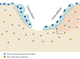
Osteoblasts are cells with a single nucleus that synthesize bone. However, in the process of bone formation, osteoblasts function in groups of connected cells. Individual cells cannot make bone. A group of organized osteoblasts together with the bone made by a unit of cells is usually called the osteon.

<span class="mw-page-title-main">Osteoclast</span> Cell that breaks down bone tissue

An osteoclast is a type of bone cell that breaks down bone tissue. This function is critical in the maintenance, repair, and remodeling of bones of the vertebral skeleton. The osteoclast disassembles and digests the composite of hydrated protein and mineral at a molecular level by secreting acid and a collagenase, a process known as bone resorption. This process also helps regulate the level of blood calcium.

<span class="mw-page-title-main">Periosteum</span> Membrane covering outer surface of bones

The periosteum is a membrane that covers the outer surface of all bones, except at the articular surfaces of long bones. Endosteum lines the inner surface of the medullary cavity of all long bones.

<span class="mw-page-title-main">Osteocyte</span> Mature osteoblasts which helps in communication between cells and also in molecular synthesis

An osteocyte, an oblate shaped type of bone cell with dendritic processes, is the most commonly found cell in mature bone. It can live as long as the organism itself. The adult human body has about 42 billion of them. Osteocytes do not divide and have an average half life of 25 years. They are derived from osteoprogenitor cells, some of which differentiate into active osteoblasts. Osteoblasts/osteocytes develop in mesenchyme.

<span class="mw-page-title-main">Chondrocyte</span> Cell that makes up cartilage

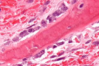
Chondrocytes are the only cells found in healthy cartilage. They produce and maintain the cartilaginous matrix, which consists mainly of collagen and proteoglycans. Although the word chondroblast is commonly used to describe an immature chondrocyte, the term is imprecise, since the progenitor of chondrocytes can differentiate into various cell types, including osteoblasts.

<span class="mw-page-title-main">Endochondral ossification</span> Cartilaginous bone development that forms the long bones

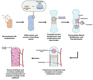
Endochondral ossification is one of the two essential pathways by which bone tissue is produced during fetal development of the mammalian skeletal system, the other pathway being intramembranous ossification. Both endochondral and intramembranous processes initiate from a precursor mesenchymal tissue, but their transformations into bone are different. In intramembranous ossification, mesenchymal tissue is directly converted into bone. On the other hand, endochondral ossification starts with mesenchymal tissue turning into an intermediate cartilage stage, which is eventually substituted by bone.

<span class="mw-page-title-main">Intramembranous ossification</span> Mesenchymal bone development that forms the non-long bones

Intramembranous ossification is one of the two essential processes during fetal development of the gnathostome skeletal system by which rudimentary bone tissue is created. Intramembranous ossification is also an essential process during the natural healing of bone fractures and the rudimentary formation of bones of the head.

<span class="mw-page-title-main">Osteon</span> Fundamental anatomical unit of compact bone

In osteology, the osteon or haversian system is the fundamental functional unit of much compact bone. Osteons are roughly cylindrical structures that are typically between 0.25 mm and 0.35 mm in diameter. Their length is often hard to define, but estimates vary from several millimeters to around 1 centimeter. They are present in many bones of most mammals and some bird, reptile, and amphibian species.

<span class="mw-page-title-main">Ossification</span> Development process in bones

Ossification in bone remodeling is the process of laying down new bone material by cells named osteoblasts. It is synonymous with bone tissue formation. There are two processes resulting in the formation of normal, healthy bone tissue: Intramembranous ossification is the direct laying down of bone into the primitive connective tissue (mesenchyme), while endochondral ossification involves cartilage as a precursor.

<span class="mw-page-title-main">Bone resorption</span> Medical condition

Bone resorption is resorption of bone tissue, that is, the process by which osteoclasts break down the tissue in bones and release the minerals, resulting in a transfer of calcium from bone tissue to the blood.

<span class="mw-page-title-main">Chondroblast</span> Mesenchymal progenitor cell that forms a chondrocyte

Chondroblasts, or perichondrial cells, is the name given to mesenchymal progenitor cells in situ which, from endochondral ossification, will form chondrocytes in the growing cartilage matrix. Another name for them is subchondral cortico-spongious progenitors. They have euchromatic nuclei and stain by basic dyes.

<span class="mw-page-title-main">Short bone</span> Bones that are as wide as they are long

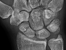
Short bones are designated as those bones that are more or less equal in length, width, and thickness. They include the tarsals in the ankle and the carpals in the wrist. They are one of five types of bones: short, long, flat, irregular and sesamoid. Most short bones are named according to their shape as they exhibit a variety of complex morphological features

<span class="mw-page-title-main">Chondroblastoma</span> Medical condition

Chondroblastoma is a rare, benign, locally aggressive bone tumor that typically affects the epiphyses or apophyses of long bones. It is thought to arise from an outgrowth of immature cartilage cells (chondroblasts) from secondary ossification centers, originating from the epiphyseal plate or some remnant of it.

<span class="mw-page-title-main">Brown tumor</span> Medical condition

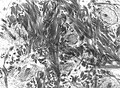
The brown tumor is a bone lesion that arises in settings of excess osteoclast activity, such as hyperparathyroidism. They are a form of osteitis fibrosa cystica. It is not a neoplasm, but rather simply a mass. It most commonly affects the maxilla and mandible, though any bone may be affected. Brown tumours are radiolucent on x-ray.

<span class="mw-page-title-main">Bone remodeling</span> Continuous turnover of bone matrix and mineral

In osteology, bone remodeling or bone metabolism is a lifelong process where mature bone tissue is removed from the skeleton and new bone tissue is formed. These processes also control the reshaping or replacement of bone following injuries like fractures but also micro-damage, which occurs during normal activity. Remodeling responds also to functional demands of the mechanical loading.

The in vivo bioreactor is a tissue engineering paradigm that uses bioreactor methodology to grow neotissue in vivo that augments or replaces malfunctioning native tissue. Tissue engineering principles are used to construct a confined, artificial bioreactor space in vivo that hosts a tissue scaffold and key biomolecules necessary for neotissue growth. Said space often requires inoculation with pluripotent or specific stem cells to encourage initial growth, and access to a blood source. A blood source allows for recruitment of stem cells from the body alongside nutrient delivery for continual growth. This delivery of cells and nutrients to the bioreactor eventually results in the formation of a neotissue product. 

<span class="mw-page-title-main">Index of trauma and orthopaedics articles</span>

Orthopedic surgery is the branch of surgery concerned with conditions involving the musculoskeletal system. Orthopedic surgeons use both surgical and nonsurgical means to treat musculoskeletal injuries, sports injuries, degenerative diseases, infections, bone tumours, and congenital limb deformities. Trauma surgery and traumatology is a sub-specialty dealing with the operative management of fractures, major trauma and the multiply-injured patient.

Craniofacial regeneration refers to the biological process by which the skull and face regrow to heal an injury. This page covers birth defects and injuries related to the craniofacial region, the mechanisms behind the regeneration, the medical application of these processes, and the scientific research conducted on this specific regeneration. This regeneration is not to be confused with tooth regeneration. Craniofacial regrowth is broadly related to the mechanisms of general bone healing.

Joints form during embryonic development in conjunction with the formation and growth of the associated bones. The joints and bones are developed from the embryonic tissue called mesenchyme.

References