Related Research Articles

Histology, also known as microscopic anatomy or microanatomy, is the branch of biology which studies the microscopic anatomy of biological tissues. Histology is the microscopic counterpart to gross anatomy, which looks at larger structures visible without a microscope. Although one may divide microscopic anatomy into organology, the study of organs, histology, the study of tissues, and cytology, the study of cells, modern usage places all of these topics under the field of histology. In medicine, histopathology is the branch of histology that includes the microscopic identification and study of diseased tissue. In the field of paleontology, the term paleohistology refers to the histology of fossil organisms.

Staining is a technique used to enhance contrast in samples, generally at the microscopic level. Stains and dyes are frequently used in histology, in cytology, and in the medical fields of histopathology, hematology, and cytopathology that focus on the study and diagnoses of diseases at the microscopic level. Stains may be used to define biological tissues, cell populations, or organelles within individual cells.

Histopathology refers to the microscopic examination of tissue in order to study the manifestations of disease. Specifically, in clinical medicine, histopathology refers to the examination of a biopsy or surgical specimen by a pathologist, after the specimen has been processed and histological sections have been placed onto glass slides. In contrast, cytopathology examines free cells or tissue micro-fragments.
Zenker's fixative is a rapid-acting fixative for animal tissues. It is employed to prepare specimens of animal or vegetable tissues for microscopic study. It provides excellent fixation of nuclear chromatin, connective tissue fibers and some cytoplasmic features, but does not preserve delicate cytoplasmic organelles such as mitochondria. Helly's fixative is preferable for traditional dye staining of mitochondria. Zenker's fixative permeabilises the plasma, but not the nuclear membrane. It can therefore be used to selectively stain mitotic cells with antibodies against chromatin
Trichrome staining is a histological staining method that uses two or more acid dyes in conjunction with a polyacid. Staining differentiates tissues by tinting them in contrasting colours. It increases the contrast of microscopic features in cells and tissues, which makes them easier to see when viewed through a microscope.

Periodic acid–Schiff (PAS) is a staining method used to detect polysaccharides such as glycogen, and mucosubstances such as glycoproteins, glycolipids and mucins in tissues. The reaction of periodic acid oxidizes the vicinal diols in these sugars, usually breaking up the bond between two adjacent carbons not involved in the glycosidic linkage or ring closure in the ring of the monosaccharide units that are parts of the long polysaccharides, and creating a pair of aldehydes at the two free tips of each broken monosaccharide ring. The oxidation condition has to be sufficiently regulated so as to not oxidize the aldehydes further. These aldehydes then react with the Schiff reagent to give a purple-magenta color. A suitable basic stain is often used as a counterstain.

Paraformaldehyde (PFA) is the smallest polyoxymethylene, the polymerization product of formaldehyde with a typical degree of polymerization of 8–100 units. Paraformaldehyde commonly has a slight odor of formaldehyde due to decomposition. Paraformaldehyde is a poly-acetal.

Masson's trichrome is a three-colour staining procedure used in histology. The recipes evolved from Claude L. Pierre Masson's (1880–1959) original formulation have different specific applications, but all are suited for distinguishing cells from surrounding connective tissue.

Papanicolaou stain is a multichromatic (multicolored) cytological staining technique developed by George Papanicolaou in 1942. The Papanicolaou stain is one of the most widely used stains in cytology, where it is used to aid pathologists in making a diagnosis. Although most notable for its use in the detection of cervical cancer in the Pap test or Pap smear, it is also used to stain non-gynecological specimen preparations from a variety of bodily secretions and from small needle biopsies of organs and tissues. Papanicolaou published three formulations of this stain in 1942, 1954, and 1960.

Orange G also called C.I. 16230, Acid Orange 10, or orange gelb is a synthetic azo dye used in histology in many staining formulations. It usually comes as a disodium salt. It has the appearance of orange crystals or powder.

Hematoxylin and eosin stain is one of the principal tissue stains used in histology. It is the most widely used stain in medical diagnosis and is often the gold standard. For example, when a pathologist looks at a biopsy of a suspected cancer, the histological section is likely to be stained with H&E.

Phosphotungstic acid haematoxylin (PTAH) is a mix of haematoxylin with phosphotungstic acid, used in histology for staining.
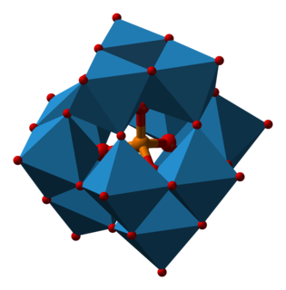
Phosphotungstic acid (PTA) or tungstophosphoric acid (TPA), is a heteropoly acid with the chemical formula H3PW12O40]. It forms hydrates H3[PW12O40]·nH2O. It is normally isolated as the n = 24 hydrate but can be desiccated to the hexahydrate. EPTA is the name of ethanolic phosphotungstic acid, its alcohol solution used in biology. It has the appearance of small, colorless-grayish or slightly yellow-green crystals, with melting point 89 °C. It is odorless and soluble in water. It is not especially toxic, but is a mild acidic irritant. The compound is known by a variety of names and acronyms.
Bone decalcification is the softening of bones due to the removal of calcium ions, and can be performed as a histological technique to study bones and extract DNA. This process also occurs naturally during bone development and growth, and when uninhibited, can cause diseases such as osteomalacia.
In the fields of histology, pathology, and cell biology, fixation is the preservation of biological tissues from decay due to autolysis or putrefaction. It terminates any ongoing biochemical reactions and may also increase the treated tissues' mechanical strength or stability. Tissue fixation is a critical step in the preparation of histological sections, its broad objective being to preserve cells and tissue components and to do this in such a way as to allow for the preparation of thin, stained sections. This allows the investigation of the tissues' structure, which is determined by the shapes and sizes of such macromolecules as proteins and nucleic acids.

Acid fuchsin or fuchsine acid, (also called Acid Violet 19 and C.I. 42685) is an acidic magenta dye with the chemical formula C20H17N3Na2O9S3. It is a sodium sulfonate derivative of fuchsine. Acid fuchsin has wide use in histology, and is one of the dyes used in Masson's trichrome stain. This method is commonly used to stain cytoplasm and nuclei of tissue sections in the histology laboratory in order to distinguish muscle from collagen. The muscle stains red with the acid fuchsin, and the collagen is stained green or blue with Light Green SF yellowish or methyl blue. It can also be used to identify growing bacteria.

Toluidine blue, also known as TBO or tolonium chloride (INN) is a blue cationic (basic) dye used in histology and sometimes clinically.
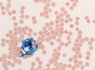
In histology, histopathology, and clinical pathology, Perls Prussian blue is a commonly used method to detect the presence of iron in tissue or cell samples. Perls Prussian Blue derives its name from the German pathologist Max Perls (1843–1881), who described the technique in 1867. The method does not involve the application of a dye, but rather causes the pigment Prussian blue to form directly within the tissue. The method stains mostly iron in the ferric state which includes ferritin and hemosiderin, rather than iron in the ferrous state.
The von Kossa histological stain is used to quantify mineralization in cell culture and histological sections.
Verhoeff's stain, also known as Verhoeff's elastic stain (VEG) or Verhoeff–Van Gieson stain (VVG), is a staining protocol used in histology, developed by American ophthalmic surgeon and pathologist Frederick Herman Verhoeff (1874–1968) in 1908. The formulation is used to demonstrate normal or pathologic elastic fibers.
References
- ↑ Carson, Freida L.; Hladik, Christa (2009). Histotechnology: A Self-Instructional Text (3 ed.). Hong Kong: American Society for Clinical Pathology Press. p. 19. ISBN 978-0-89189-581-7.
- ↑ Culling, C.F.A. 1974. Handbook of Histopathological and Histochemical Techniques (including museum techniques), 3rd ed. London: Butterworths, p.49.
- ↑ Bancroft, John D.; Gamble, Marilyn, eds. (2008). Theory and Practice of Histology Techniques (6 ed.). China: Churchill Livingstone Elsevier. p. 72. ISBN 978-0-443-10279-0.
- ↑ Baker, J.R. 1958. Principles of Biological Microtechnique. London, Methuen, pp.149-150.
- ↑ Kiernan, J.A. 2015 Histological and Histochemical Methods. Theory and Practice. Banbury, UK: Scion, p.195.
- ↑ Lincoln, Roger J.; Sheals, John Gordon, eds. (1979). Invertebrate Animals - Collection and Preservation (1 ed.). UK: British Museum (Natural History). p. 128. ISBN 0-521-296773.
- ↑ Culling, C.F.A. 1974. Handbook of Histopathological and Histochemical Techniques (including museum techniques), 3rd ed. London: Butterworths, p.49.
- ↑ Culling, C.F.A. 1974. Handbook of Histopathological and Histochemical Techniques (including museum techniques), 3rd ed. London: Butterworths, p.49.
- ↑ Dean, J.A. 1973. Lange's Handbook of Chemistry, 11th ed. New York: McGraw-Hill, p.7-383.
- ↑ Carson, Freida L.; Hladik, Christa (2009). Histotechnology: A Self-Instructional Text (3 ed.). Hong Kong: American Society for Clinical Pathology Press. p. 19. ISBN 978-0-89189-581-7.
- ↑ Gray, P. 1954. The Microtomist's Formulary and Guide. Reprinted 1975. Huntington, NY: Kreiger, p.220.