Bovine viral diarrhea

Last updated
Bovine viral diarrhea
BVDV-CP7.JPG
Immunofluorescence image of BVDV (CP7 type). Nuclei are stained blue with DAPI. The replication complexes of the viruses are marked red by NS3 protein binding antibodies
Scientific classification OOjs UI icon edit-ltr.svg
(unranked): Virus
Realm: Riboviria
Kingdom: Orthornavirae
Phylum: Kitrinoviricota
Class: Flasuviricetes
Order: Amarillovirales
Family: Flaviviridae
Genus: Pestivirus
Groups included
Cladistically included but traditionally excluded taxa
Tongue lesions on confirmed BVD/MD case (mucosal disease form) Bovine Virus Diarrhea tongue ulcers.jpg
Tongue lesions on confirmed BVD/MD case (mucosal disease form)

Bovine viral diarrhea (BVD), bovine viral diarrhoea (UK English) or mucosal disease, previously referred to as bovine virus diarrhea (BVD), is an economically significant disease of cattle that is found in the majority of countries throughout the world. [1] Worldwide reviews of the economically assessed production losses and intervention programs (e.g. eradication programs, vaccination strategies and biosecurity measures) incurred by BVD infection have been published. [2] [3] The causative agent, bovine viral diarrhea virus (BVDV), is a member of the genus Pestivirus of the family Flaviviridae. [1]

Contents

BVD infection results in a wide variety of clinical signs, due to its immunosuppressive effects, [4] as well as having a direct effect on respiratory disease and fertility. [5] In addition, BVD infection of a susceptible dam during a certain period of gestation can result in the production of a persistently infected (PI) fetus. [6]

PI animals recognise intra-cellular BVD viral particles as ‘self’ and shed virus in large quantities throughout life; they represent the cornerstone of the success of BVD as a disease.

Currently, it was shown in a worldwide review study that the PI prevalence at animal level ranged from low (≤0.8% Europe, North America, Australia), medium (>0.8% to 1.6% East Asia) to high (>1.6% West Asia). Countries that had failed to implement any BVDV control and/or eradication programmes (including vaccination) had the highest PI prevalence. [7]

Virus classification and structure

BVDVs are members of the genus Pestivirus , belonging to the family Flaviviridae . Other members of this genus cause Border disease (sheep) and classical swine fever (pigs) which cause significant financial loss to the livestock industry. [8]

Pestiviruses are small, spherical, single-stranded, enveloped RNA viruses of 40 to 60 nm in diameter. [9]

The genome consists of a single, linear, positive-sense, single-stranded RNA molecule of approximately 12.3 kb. [10] RNA synthesis is catalyzed by the BVDV RNA-dependent RNA polymerase (RdRp). This RdRp can undergo template strand switching allowing RNA-RNA copy choice recombination during elongative RNA synthesis. [11]

Two BVDV genotypes are recognised, based on the nucleotide sequence of the 5’untranslated (UTR) region; BVDV-1 and BVDV-2. [12] BVDV-1 isolates have been grouped into 16 subtypes (a –p) and BVDV-2 has currently been grouped into 3 subtypes (a – c). [13]

BVDV strains can be further divided into distinct biotypes (cytopathic or non-cytopathic) according to their effects on tissue cell culture; cytopathic (cp) biotypes, formed via mutation of non-cytopathic (ncp) biotypes, induce apoptosis in cultured cells. [14] Ncp viruses can induce persistent infection in cells and have an intact NS2/3 protein. In cp viruses the NS2/3 protein is either cleaved to NS2 and NS3 or there is a duplication of viral RNA containing an additional NS3 region. [15] The majority of BVDV infections in the field are caused by the ncp biotype. [1]

Epidemiology

BVD is considered one of the most significant infectious diseases in the livestock industry worldwide due to its high prevalence, persistence and clinical consequences. [16]

In Europe the prevalence of antibody positive animals in countries without systematic BVD control is between 60 and 80%. [17] Prevalence has been determined in individual countries and tends to be positively associated with stocking density of cattle.[ citation needed ]

BVDV-1 strains are predominant in most parts of the world, whereas BVDV-2 represents 50% of cases in North America. [16] In Europe, BVDV-2 was first isolated in the UK in 2000 and currently represents up to 11% of BVD cases in Europe. [18]

Transmission of BVDV occurs both horizontally and vertically with both persistently and transiently infected animals excreting infectious virus. Virus is transmitted via direct contact, bodily secretions and contaminated fomites, with the virus being able to persist in the environment for more than two weeks. Persistently infected animals are the most important source of the virus, continuously excreting a viral load one thousand times that shed by acutely infected animals. [19]

Pathogenesis

Turbinate cells infected with BVDV Fetal calf serum from a diffrent animal.tif
Turbinate cells infected with BVDV

Acute, transient infection

Following viral entry and contact with the mucosal lining of the mouth or nose, replication occurs in epithelial cells. BVDV replication has a predilection for the palatine tonsils, lymphoid tissues and epithelium of the oropharynx.

Phagocytes take up BVDV or virus-infected cells and transport them to peripheral lymphoid tissues; the virus can also spread systemically through the bloodstream. Viraemia occurs 2–4 days after exposure and virus isolation from serum or leukocytes is generally possible between 3–10 days post infection. [20]

During systemic spread the virus is able to gain entry into most tissues with a preference for lymphoid tissues. Neutralising antibodies can be detected from 10 to 14 days post infection with titres continuing to increase slowly for 8–10 weeks. After 2–3 weeks, antibodies effectively neutralise viral particles, promote clearance of virus and prevent seeding of target organs. [21]

Intrauterine infections

Fetal infection is of most consequence as this can result in the birth of a persistently infected neonate. The effects of fetal infection with BVDV are dependent upon the stage of gestation at which the dam suffers acute infection.

BVDV infection of the dam prior to conception, and during the first 18 days of gestation, results in delayed conception and an increased calving to conception interval. Once the embryo is attached, infection from days 29–41 can result in embryonic infection and resultant embryonic death.

Infection of the dam from approximately day 30 of gestation until day 120 can result in immunotolerance and the birth of calves persistently infected with the virus.

BVDV infection between 80 and 150 days of gestation may be teratogenic, with the type of birth defect dependent upon the stage of fetal development at infection. Abortion may occur at any time during gestation. Infection after approximately day 120 can result in the birth of a normal fetus which is BVD antigen-negative and BVD antibody-positive. This occurs because the fetal immune system has developed, by this stage of gestation, and has the ability to recognise and fight off the invading virus, producing anti-BVD antibodies.

Chronic infections

BVD virus can be maintained as a chronic infection within some immunoprivileged sites following transient infection. These sites include ovarian follicles, testicular tissues, central nervous system and white blood cells. Cattle with chronic infections elicit a significant immune response, exhibited by extremely high antibody titres.

Clinical signs

BVDV infection has a wide manifestation of clinical signs including fertility issues, milk drop, pyrexia, diarrhea, and fetal infection. [9] Occasionally, a severe acute form of BVD may occur. These outbreaks are characterized by thrombocytopenia with high morbidity and mortality. However, clinical signs are frequently mild and infection insidious, recognized only by BVDV's immunosuppressive effects perpetuating other circulating infectious diseases (particularly scours and pneumonias).

PI animals

Persistently infected animals did not have a competent immune system at the time of BVDV transplacental infection. The virus, therefore, entered the fetal cells and, during immune system development, was accepted as self. In PIs the virus remains present in a large number of the animal's body cells throughout its life and is continuously shed. PIs are often ill-thrifty and smaller than their peers, however, they can appear normal. PIs are more susceptible to disease, with only 20% of PIs surviving to two years of age. [22] If a PI dam is able to reproduce they always give birth to PI calves. [23]

Mucosal disease

The PI cattle that do survive ill-thrift are susceptible to mucosal disease. Mucosal disease only develops in PI animals and is invariably fatal. [5] Disease results when a PI animal is superinfected with a cytopathic biotype arising from mutation of the non-cytopathic strain of BVDV already circulating in that animal. [24] The cp BVDV spreads to the gastro-intestinal epithelium, and necrosis of keratinocytes results in erosion and ulceration. Fluid leaks from the epithelial surface of the gastro-intestinal tract causing diarrhoea and dehydration. In addition, bacterial infection of the damaged epithelium results in secondary septicaemia. Death occurs in the ensuing days or weeks.

Diagnosis

Various diagnostic tests are available for the detection of either active infection or evidence of historical infection. The method of diagnosis used also depends upon whether the vet is investigating at an individual or a herd level.

Virus or antigen detection

Antigen ELISA and rtPCR are currently the most frequently performed tests to detect virus or viral antigen. Individual testing of ear tissue tag samples or serum samples is performed. It is vital that repeat testing is performed on positive samples to distinguish between acute, transiently infected cattle and PIs. A second positive result, acquired at least three weeks after the primary result, indicates a PI animal. rtPCR can also be used on bulk tank milk (BTM) samples to detect any PI cows contributing to the tank. It is reported that the maximum number of contributing cows from which a PI can be detected is 300.

BVD antibody detection

Antibody (Ig) ELISAs are used to detect historical BVDV infection; these tests have been validated in serum, milk and bulk milk samples. Ig ELISAs do not diagnose active infection but detect the presence of antibodies produced by the animal in response to viral infection. Vaccination also induces an antibody response, which can result in false positive results, therefore it is important to know the vaccination status of the herd or individual when interpreting results. A standard test to assess whether virus has been circulating recently is to perform an Ig ELISA on blood from 5–10 young stock that have not been vaccinated, aged between 9 and 18 months. A positive result indicates exposure to BVDV, but also that any positive animals are very unlikely to be PI animals themselves. A positive result in a pregnant female indicates that she has previously been either vaccinated or infected with BVDV and could possibly be carrying a PI fetus, so antigen testing of the newborn is vital to rule this out. [5] A negative antibody result, at the discretion of the responsible veterinarian, may require further confirmation that the animal is not in fact a PI.

At a herd level, a positive Ig result suggests that BVD virus has been circulating or the herd is vaccinated. Negative results suggest that a PI is unlikely however this naïve herd is in danger of severe consequences should an infected animal be introduced. Antibodies from wild infection or vaccination persist for several years therefore Ig ELISA testing is more valuable when used as a surveillance tool in seronegative herds.

Eradication and control

The mainstay of eradication is the identification and removal of persistently infected animals. Re-infection is then prevented by vaccination and high levels of biosecurity, supported by continuing surveillance. PIs act as viral reservoirs and are the principal source of viral infection but transiently infected animals and contaminated fomites also play a significant role in transmission. [1]

Leading the way in BVD eradication, almost 20 years ago, were the Scandinavian countries. Despite different conditions at the start of the projects in terms of legal support, and regardless of initial prevalence of herds with PI animals, it took all countries approximately 10 years to reach their final stages. [25] [26]

Once proven that BVD eradication could be achieved in a cost efficient way, a number of regional programmes followed in Europe, some of which have developed into national schemes. [27]

Vaccination is an essential part of both control and eradication. While BVD virus is still circulating within the national herd, breeding cattle are at risk of producing PI neonates and the economic consequences of BVD are still relevant. Once eradication has been achieved, unvaccinated animals will represent a naïve and susceptible herd. Infection from imported animals or contaminated fomites brought into the farm, or via transiently infected in-contacts will have devastating consequences.

Vaccination

Modern vaccination programmes aim not only to provide a high level of protection from clinical disease for the dam, but, crucially, to protect against viraemia and prevent the production of PIs. [28] While the immune mechanisms involved are the same, the level of immune protection required for foetal protection is much higher than for prevention of clinical disease. [29]

While challenge studies indicate that killed, as well as live, vaccines prevent foetal infection under experimental conditions, the efficacy of vaccines under field conditions has been questioned. [30] The birth of PI calves into vaccinated herds suggests that killed vaccines do not stand up to the challenge presented by the viral load excreted by a PI in the field. [31]

See also

Related Research Articles

<i>Rotavirus</i> Specific genus of RNA viruses

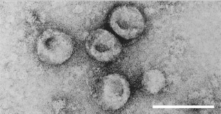
Rotavirus is a genus of double-stranded RNA viruses in the family Reoviridae. Rotaviruses are the most common cause of diarrhoeal disease among infants and young children. Nearly every child in the world is infected with a rotavirus at least once by the age of five. Immunity develops with each infection, so subsequent infections are less severe. Adults are rarely affected. There are nine species of the genus, referred to as A, B, C, D, F, G, H, I and J. Rotavirus A, the most common species, causes more than 90% of rotavirus infections in humans.

Classical swine fever (CSF) or hog cholera is a highly contagious disease of swine. It has been mentioned as a potential bioweapon.

<i>Feline leukemia virus</i> Species of virus

Feline leukemia virus (FeLV) is a retrovirus that infects cats. FeLV can be transmitted from infected cats when the transfer of saliva or nasal secretions is involved. If not defeated by the animal's immune system, the virus weakens the cat's immune system, which can lead to diseases which can be lethal. Because FeLV is cat-to-cat contagious, FeLV+ cats should only live with other FeLV+ cats.

<i>Carnivore protoparvovirus 1</i> Species of parvovirus

Carnivore protoparvovirus 1 is a species of parvovirus that infects carnivorans. It causes a highly contagious disease in both dogs and cats separately. The disease is generally divided into two major genogroups: FPV containing the classical feline panleukopenia virus (FPLV), and CPV-2 containing the canine parvovirus type 2 (CPV-2) which appeared in the 1970s.

<i>Pestivirus</i> Genus of viruses

Pestivirus is a genus of viruses, in the family Flaviviridae. Viruses in the genus Pestivirus infect mammals, including members of the family Bovidae and the family Suidae. There are 11 species in this genus. Diseases associated with this genus include: hemorrhagic syndromes, abortion, and fatal mucosal disease.

Bovine alphaherpesvirus 1 (BoHV-1) is a virus of the family Herpesviridae and the subfamily Alphaherpesvirinae, known to cause several diseases worldwide in cattle, including rhinotracheitis, vaginitis, balanoposthitis, abortion, conjunctivitis, and enteritis. BoHV-1 is also a contributing factor in shipping fever, also known as bovine respiratory disease (BRD). It is spread horizontally through sexual contact, artificial insemination, and aerosol transmission and it may also be transmitted vertically across the placenta. BoHV-1 can cause both clinical and subclinical infections, depending on the virulence of the strain. Although these symptoms are mainly non-life-threatening it is an economically important disease as infection may cause a drop in production and affect trade restrictions. Like other herpesviruses, BoHV-1 causes a lifelong latent infection and sporadic shedding of the virus. The sciatic nerve and trigeminal nerve are the sites of latency. A reactivated latent carrier is normally the source of infection in a herd. The clinical signs displayed are dependent on the virulence of the strain. There is a vaccine available which reduces the severity and incidence of disease. Some countries in Europe have successfully eradicated the disease by applying a strict culling policy.

Aujeszky's disease, usually called pseudorabies in the United States, is a viral disease in swine that is endemic in most parts of the world. It is caused by Suid herpesvirus 1 (SuHV-1). Aujeszky's disease is considered to be the most economically important viral disease of swine in areas where classical swine fever has been eradicated. Other mammals, such as cattle, sheep, goats, cats, dogs, and raccoons, are also susceptible. The disease is usually fatal in these animal species.

<span class="mw-page-title-main">Bovine malignant catarrhal fever</span> Species of virus

Bovine malignant catarrhal fever (BMCF) is a fatal lymphoproliferative disease caused by a group of ruminant gamma herpes viruses including Alcelaphine gammaherpesvirus 1 (AlHV-1) and Ovine gammaherpesvirus 2 (OvHV-2) These viruses cause unapparent infection in their reservoir hosts, but are usually fatal in cattle and other ungulates such as deer, antelope, and buffalo. In Southern Africa the disease is known as snotsiekte, from the Afrikaans.

Bovine leukemia virus (BLV) is a retrovirus which causes enzootic bovine leukosis in cattle. It is closely related to the human T‑lymphotropic virus type 1 (HTLV-I). BLV may integrate into the genomic DNA of B‑lymphocytes as a DNA intermediate, or exist as unintegrated circular or linear forms. Besides structural and enzymatic genes required for virion production, BLV expresses the Tax protein and microRNAs involved in cell proliferation and oncogenesis. In cattle, most infected animals are asymptomatic; leukemia is rare, but lymphoproliferation is more frequent (30%).

<span class="mw-page-title-main">Veterinary virology</span> Study of viruses affecting animals

Veterinary virology is the study of viruses in non-human animals. It is an important branch of veterinary medicine.

<i>Torovirus</i> Genus of viruses

Torovirus is a genus of enveloped, positive-strand RNA viruses in the order Nidovirales and family Tobaniviridae. They primarily infect vertebrates, especially cattle, pigs, and horses. Diseases associated with this genus include gastroenteritis, which commonly presents in mammals. Torovirus is the only genus in the monotypic subfamily Torovirinae. Torovirus is also a monotypic taxon, containing only one subgenus, Renitovirus.

<i>Enterovirus E</i> Species of virus

Enterovirus E is a picornavirus of the genus Enterovirus. The virus may also be referred to as enteric cytopathic bovine orphan virus (ECBO). It is endemic in cattle populations worldwide, and although normally fairly nonpathogenic, it can cause reproductive, respiratory, or enteric disease – particularly when the animal is concurrently infected with another pathogen.

<span class="mw-page-title-main">Foot-and-mouth disease</span> Infectious disease affecting cattle

Foot-and-mouth disease (FMD) or hoof-and-mouth disease (HMD) is an infectious and sometimes fatal viral disease that affects cloven-hoofed animals, including domestic and wild bovids. The virus causes a high fever lasting two to six days, followed by blisters inside the mouth and near the hoof that may rupture and cause lameness.

Bovine immunodeficiency virus (BIV) is a retrovirus belonging to the genus Lentivirus. It is similar to the human immunodeficiency virus (HIV) and infects cattle. The cells primarily infected are lymphocytes and monocytes/macrophages.

Bovine coronavirus is a coronavirus which is a member of the species Betacoronavirus 1. The infecting virus is an enveloped, positive-sense, single-stranded RNA virus which enters its host cell by binding to the N-acetyl-9-O-acetylneuraminic acid recepter. Infection causes calf enteritis and contributes to the enzootic pneumonia complex in calves. It can also cause winter dysentery in adult cattle. It can infect both domestic and wild ruminants and has a worldwide distribution. Transmission is horizontal, via oro-fecal or respiratory routes. Like other coronaviruses from genus Betacoronavirus, subgenus Embecovirus, it has a surface protein called hemagglutinin esterase (HE) in addition to the four structural proteins shared by all coronaviruses.

Porcine epidemic diarrhea is a condition caused by the porcine epidemic diarrhea virus that leads to severe gastrointestinal disease in pigs.

<span class="mw-page-title-main">Rotaviral gastroenteritis</span> Medical condition

Rotavirus gastroenteritis is a major cause of severe diarrhoea among infants and young children globally. It is caused by rotavirus, a genus of double-stranded RNA virus in the family Reoviridae. The diarrhea tends to be watery and is frequently accompanied by fever, vomiting and abdominal pain. By the age of five, nearly every child in the world has been infected with rotavirus at least once. However, with each infection, immunity develops, and subsequent infections are less severe; adults are rarely affected. There are five species of this virus, referred to as A, B, C, D, and E. Rotavirus A, the most common, causes more than 90% of infections in humans.

Bovine respiratory disease (BRD) is the most common and costly disease affecting beef cattle in the world. It is a complex, bacterial or viral infection that causes pneumonia in calves which can be fatal. The infection is usually a sum of three codependent factors: stress, an underlying viral infection, and a new bacterial infection. The diagnosis of the disease is complex since there are multiple possible causes.

Pseudocowpox is a disease caused by the Paravaccinia virus or Pseudocowpox virus, a virus of the family Poxviridae and the genus Parapoxvirus. Humans can contract the virus from contact with livestock infected with Bovine papular stomatitis and the disease is common among ranchers, milkers, and veterinarians. Infection in humans will present with fever, fatigue, and lesion on the skin.

Border disease (BD) is a viral disease of sheep and goats, primarily causing congenital diseases, but can also cause acute and persistent infections. It first appeared in the border regions of England and Wales in 1959, and has since spread world-wide. Lambs that are born with BD are commonly known as 'hairy shakers' due to the primary presentation of the disease. The disease was recognized before the virus, therefore the common name of the disease predates the understanding of the viral pathology. The virus can cause a significant reduction in the percentage of surviving lambs, thus it has a large economic impact on farmers.

References

  1. 1 2 3 4 Fray, M.D; Paton, D.J; Alenius, S.; et al. (2000). "The effects of bovine viral diarrhoea virus on cattle reproduction in relation to disease control". Animal Reproduction Science. 60–61: 615–27. doi:10.1016/s0378-4320(00)00082-8. PMID   10844229.
  2. Richter, V; Lebl, K; Baumgartner, W; Obritzhauser, W; Käsbohrer, A; Pinior, B (2017). "A systematic worldwide review of the direct monetary losses due to bovine viral diarrhea virus infection". The Veterinary Journal. 220: 80–87. doi: 10.1016/j.tvjl.2017.01.005 . PMID   28190502.
  3. Pinior, B; Firth, C; Richter, V; Lebl, K; Trauffler, M; Dzieciol, M; Hutter, S; Burgstaller, J; Obritzhauser, W; Winter, P; Käsbohrer, A (2017). "A systematic review of financial and economic assessments of bovine viral diarrhea virus (BVDV) prevention and mitigation activities worldwide". Preventive Veterinary Medicine. 137 (Pt A): 77–92. doi: 10.1016/j.prevetmed.2016.12.014 . PMID   28040270.
  4. Schaut, Robert G.; McGill, Jodi L.; Neill, John D.; Ridpath, Julia F.; Sacco, Randy E. (2015-10-02). "Bovine viral diarrhea virus type 2 in vivo infection modulates TLR4 responsiveness in differentiated myeloid cells which is associated with decreased MyD88 expression". Virus Research. 208: 44–55. doi: 10.1016/j.virusres.2015.05.017 . ISSN   1872-7492. PMID   26043978.
  5. 1 2 3 Lanyon, Sasha R.; Hill, Fraser I.; Reichel, Michael P.; Brownlie, Joe; et al. (2014). "Bovine Viral Diarrhoea: Pathogenesis and diagnosis" (PDF). Veterinary Journal. 199 (2): 201–9. doi:10.1016/j.tvjl.2013.07.024. PMID   24053990.
  6. Grooms, Daniel L. (2004). "Reproductive consequences of infection with bovine viral diarrhea virus". Veterinary Clinics of North America: Food Animal Practice. 20 (1): 5–19. doi:10.1016/j.cvfa.2003.11.006. PMID   15062471.
  7. Scharnböck, B; Roch, Franz-Ferdinand; Richter, V; Funke, C; Firth, C; Obritzhauser, W; Baumgartner, W; Käsbohrer, A; Pinior, B (2018). "A meta-analysis of bovine viral diarrhoea virus (BVDV) prevalences in the global cattle population". Scientific Reports. 8 (1): 14420. Bibcode:2018NatSR...814420S. doi:10.1038/s41598-018-32831-2. PMC   6158279 . PMID   30258185.
  8. Hornberg, Andrea; Fernández, Sandra Revilla; Vogl, Claus; Vilcek, Stefan; Matt, Monika; Fink, Maria; Köfer, Josef; Schöpf, Karl (2009). "Genetic diversity of pestivirus isolates in cattle from Western Austria" (PDF). Veterinary Microbiology. 135 (3–4): 205–213. doi:10.1016/j.vetmic.2008.09.068. PMID   19019571. S2CID   46378359.
  9. 1 2 N. James MacLachlan; Edward J. Dubovi, eds. (2011). Fenner's Veterinary Virology (4th ed.). Elsevier.
  10. Brett D. Lindenbach; Heinz-Jürgen Thiel; Charles M. Rice (2007). "Flaviviridae: The viruses and their replication" (PDF). In D. M. Knipe; P. M. Howley (eds.). Fields Virology (5th ed.). Philadelphia: Lippincott-Raven Publishers. pp. 1101–1133.
  11. Kim, M.- J.; Kao, C. (2001). "Factors regulating template switch in vitro by viral RNA-dependent RNA polymerases: Implications for RNA-RNA recombination". Proceedings of the National Academy of Sciences. 98 (9): 4972–4977. Bibcode:2001PNAS...98.4972K. doi: 10.1073/pnas.081077198 . PMC   33148 . PMID   11309487.
  12. Ridpath, J.F.; Bolin, S.R.; Dubovi, E.J. (1994). "Segregation of bovine viral diarrhoea virus into genotypes". Virology. 205 (1): 66–74. doi: 10.1006/viro.1994.1620 . PMID   7975238.
  13. Peterhans, Ernst; Bachofen, Claudia; Stalder, Hanspeter; Schweizer, Matthias (2010). "Cytopathic bovine viral diarrhea viruses (BVDV): emerging pestiviruses doomed to extinction". Veterinary Research. 41 (6): 44. doi:10.1051/vetres/2010016. PMC   2850149 . PMID   20197026.
  14. Gillespie, J. H.; Madin, S. H.; Darby, N. B. (1962). "Cellular resistance in tissue culture, induced by noncytopathogenic strains, to a cytopathogenic strain of virus diarrhea virus of cattle". Proceedings of the Society for Experimental Biology and Medicine. 110 (2): 248–50. doi:10.3181/00379727-110-27481. PMID   13898635. S2CID   12198102.
  15. Qi, Fengxia; Ridpath, Julia F.; Berry, Eugene S. (1998). "Insertion of a bovine SMT3B gene in NS4B and duplication of NS3 in a bovine viral diarrhea virus genome correlate with the cytopathogenicity of the virus". Virus Research. 57 (1): 1–9. doi:10.1016/s0168-1702(98)00073-2. PMID   9833880.
  16. 1 2 Moennig, Volker; Houe, Hans; Lindberg, Ann (2005). "BVD control in Europe: current status and perspectives". Animal Health Research Reviews. 6 (1): 63–74. doi:10.1079/ahr2005102. PMID   16164009. S2CID   10581576.
  17. Anon (2005). EU Thematic network on control of bovine viral diarrhoea virus (BVDV). Position Paper.
  18. Wolfmeyer, A.; Wolf, G.; Beer, M.; Strube, W.; Hehnen, H. R.; Schmeer, N.; Kaaden, O. R. (1997). "Genomic (50-UTR) and serological differences among German BVDV field isolates". Archives of Virology. 142 (10): 2049–2057. doi:10.1007/s007050050222. PMID   9413513. S2CID   20365815.
  19. Brownlie, J.; Clarke, M. C.; Howard, C. J.; Pocock, D. H. (1987). "Pathogenesis and epidemiology of bovine virus diarrhoea virus infection of cattle". Annales de Recherches Vétérinaires. 18 (2): 157–66. PMID   3619343.
  20. Fray, M. D.; Clarke, M. C.; Thomas, L. H.; McCauley, J. W.; Charleston, B. (1998). "Prolonged nasal shedding and viraemia of cytopathogenic bovine virus diarrhoea virus in experimental late-onset mucosal disease". Veterinary Record. 143 (22): 608–11. doi:10.1136/vr.143.22.608. PMID   9871955. S2CID   26025942.
  21. Chase, Christopher C.L; Elmowalid, Gamal; Yousif, Ausama A.A (2004). "The immune response to bovine viral diarrhea virus: a constantly changing picture". The Veterinary Clinics of North America. Food Animal Practice. 20 (1): 95–114. doi:10.1016/j.cvfa.2003.11.004. PMID   15062477.
  22. Voges, H; Young, S; Nash, M (2006). "Direct adverse effects of persistent BVDV infection in dairy heifers – A retrospective case control study". VetScript. 19 (8): 22–25.
  23. Moennig, Volker; Liess, Bernd (1995). "Pathogenesis of intrauterine infections with bovine viral diarrhoea virus". Veterinary Clinics of North America: Food Animal Practice. 11 (3): 477–488. doi:10.1016/S0749-0720(15)30462-X. PMID   8581858.
  24. Brownlie, J.; Clarke, M.; Howard, C. (1984). "Experimental production of fatal mucosal disease in cattle". The Veterinary Record. 114 (22): 535–6. doi:10.1136/vr.114.22.535. PMID   6087539. S2CID   19523700.
  25. Hult and Lindberg (2005) Prev Vet Med 72: 143–148
  26. Rikula et al. (2005) Prev Vet Med 72: 139–142
  27. Rossmanith et al. (2005) Prev Vet Med 72: 133–137
  28. Stahl and Alenius (2012) Japanese Journal of Veterinary Research 60 (Supplement) S31–39.
  29. Ridpath (2013) Biologicals 41: 14–19.
  30. O’Rourke (2002) Journal of the American Veterinary Medical Association 220(12): 1770–1772
  31. Graham et al. (2004) Revista Portuguesa de ciencias veterinarias 127: 38.