Bronchiole

Last updated
Bronchiole
Alveoli diagram.png
Diagram of the alveoli with both cross-section and external view.
Details
System Respiratory system
Identifiers
MeSH D055745
TA98 A06.5.02.026
TA2 3282
TH H3.05.02.0.00005
FMA 7410
Anatomical terminology

The bronchioles or bronchioli (pronounced bron-kee-oh-lee) are the smaller branches of the bronchial airways in the lower respiratory tract. They include the terminal bronchioles, and finally the respiratory bronchioles that mark the start of the respiratory zone delivering air to the gas exchanging units of the alveoli. The bronchioles no longer contain the cartilage that is found in the bronchi, or glands in their submucosa. [1]

Contents

Structure

A lobule of the lung enclosed in septa and supplied by a terminal bronchiole that branches into the respiratory bronchioles. Each respiratory bronchiole supplies the alveoli held in each acinus accompanied by a pulmonary artery branch. Secondary-pulmonary-lobule-illustration.jpg
A lobule of the lung enclosed in septa and supplied by a terminal bronchiole that branches into the respiratory bronchioles. Each respiratory bronchiole supplies the alveoli held in each acinus accompanied by a pulmonary artery branch.

The pulmonary lobule is the portion of the lung ventilated by one bronchiole. Bronchioles are approximately 1 mm or less in diameter and their walls consist of ciliated cuboidal epithelium and a layer of smooth muscle. Bronchioles divide into even smaller bronchioles, called terminal, which are 0.5 mm or less in diameter. Terminal bronchioles in turn divide into smaller respiratory bronchioles which divide into alveolar ducts. Terminal bronchioles mark the end of the conducting division of air flow in the respiratory system while respiratory bronchioles are the beginning of the respiratory division where gas exchange takes place.

The diameter of the bronchioles plays an important role in air flow. The bronchioles change diameter to either increase or reduce air flow. An increase in diameter is called bronchodilation and is stimulated by either epinephrine or sympathetic nerves to increase air flow. A decrease in diameter is called bronchoconstriction, which is the tightening of the smooth muscle surrounding the bronchi and bronchioles due to and stimulated by histamine, parasympathetic nerves, cold air, chemical irritants, excess mucus production, viral infections, and other factors to decrease air flow. Bronchoconstriction can result in clinical symptoms such as wheezing, chest tightness, and dyspnea, which are common features of asthma, chronic obstructive pulmonary disease (COPD), and chronic bronchitis. [2]

Bronchioles

Lungs showing bronchi and bronchioles Illu bronchi lungs.jpg
Lungs showing bronchi and bronchioles

The trachea divides into the left main bronchus which supplies the left lung, and the right main bronchus which supplies the right lung. As they enter the lungs these primary bronchi branch into secondary bronchi known as lobar bronchi which supply each lobe of the lung. These in turn give rise to tertiary bronchi (tertiary meaning "third"), known as segmental bronchi which supply each bronchopulmonary segment. [1] The segmentary bronchi subdivide into fourth order, fifth order and sixth order segmental bronchi before dividing into the bronchioles. The bronchioles are histologically distinct from the bronchi in that their walls do not have hyaline cartilage and they have club cells in their epithelial lining. The epithelium of the bronchioles starts as a simple ciliated columnar epithelium and changes to simple ciliated cuboidal epithelium as the bronchioles decreases in size. The diameter of the bronchioles is often said to be less than 1 mm, though this value can range from 5 mm to 0.3 mm. As stated, these bronchioles do not have hyaline cartilage to maintain their patency. Instead, they rely on elastic fibers attached to the surrounding lung tissue for support. The inner lining (lamina propria) of these bronchioles is thin with no glands present, and is surrounded by a layer of smooth muscle. As the bronchioles get smaller they divide into terminal bronchioles. Each bronchiole divides into between 50 and 80 terminal bronchioles. [3] These bronchioles mark the end of the conducting zone, which covers the first division through the sixteenth division of the respiratory tract. Alveoli only become present when the conducting zone changes to the respiratory zone, from the sixteenth through the twenty-third division of the tract.

Terminal bronchioles

The terminal bronchioles are the most distal segment of the conducting zone. They branch off the lesser bronchioles. Each of the terminal bronchioles divides to form respiratory bronchioles which contain a small number of alveoli. Terminal bronchioles are lined with simple ciliated cuboidal epithelium containing club cells. Club cells are non-ciliated, rounded protein-secreting cells. Their secretions are a non-sticky, proteinaceous compound to maintain the airway in the smallest bronchioles. The secretion, called pulmonary surfactant, reduces surface tension, allowing for bronchioles to expand during inspiration and keeping the bronchioles from collapsing during expiration. Club cells are a stem cell of the respiratory system, and also produce enzymes that detoxify substances dissolved in the respiratory fluid.

Respiratory bronchioles

The respiratory bronchioles are the narrowest airways of the lungs, 0.5 mm across. [4] The bronchi divide many times before evolving into the bronchioles. The respiratory bronchioles deliver air to the exchange surfaces of the lungs. [5] They are interrupted by alveoli which are thin walled evaginations. Alveolar ducts are side branches of the respiratory bronchioles. The respiratory bronchioles are lined by ciliated columnar epithelium along with some non-ciliated cells called club cells. [6]

Clinical significance

Bronchospasm, a potentially life-threatening situation, occurs when the smooth muscular tissue of the bronchioles constricts, severely narrowing their diameter. The most common cause of this is asthma. Bronchospasm is commonly treated by oxygen therapy and bronchodilators such as albuterol.

Diseases of the bronchioles include asthma, bronchiolitis obliterans, respiratory syncytial virus infections, and influenza.

Inflammation

The medical condition of inflammation of the bronchioles is termed bronchiolitis. [7]

Additional images

Related Research Articles

<span class="mw-page-title-main">Lung</span> Primary organ of the respiratory system

The lungs are the primary organs of the respiratory system in humans and most other animals, including some snails and a small number of fish. In mammals and most other vertebrates, two lungs are located near the backbone on either side of the heart. Their function in the respiratory system is to extract oxygen from the air and transfer it into the bloodstream, and to release carbon dioxide from the bloodstream into the atmosphere, in a process of gas exchange. Respiration is driven by different muscular systems in different species. Mammals, reptiles and birds use their different muscles to support and foster breathing. In earlier tetrapods, air was driven into the lungs by the pharyngeal muscles via buccal pumping, a mechanism still seen in amphibians. In humans, the main muscle of respiration that drives breathing is the diaphragm. The lungs also provide airflow that makes vocal sounds including human speech possible.

<span class="mw-page-title-main">Respiratory system</span> Biological system in animals and plants for gas exchange

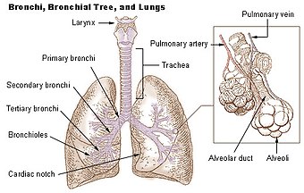
The respiratory system is a biological system consisting of specific organs and structures used for gas exchange in animals and plants. The anatomy and physiology that make this happen varies greatly, depending on the size of the organism, the environment in which it lives and its evolutionary history. In land animals the respiratory surface is internalized as linings of the lungs. Gas exchange in the lungs occurs in millions of small air sacs; in mammals and reptiles these are called alveoli, and in birds they are known as atria. These microscopic air sacs have a very rich blood supply, thus bringing the air into close contact with the blood. These air sacs communicate with the external environment via a system of airways, or hollow tubes, of which the largest is the trachea, which branches in the middle of the chest into the two main bronchi. These enter the lungs where they branch into progressively narrower secondary and tertiary bronchi that branch into numerous smaller tubes, the bronchioles. In birds the bronchioles are termed parabronchi. It is the bronchioles, or parabronchi that generally open into the microscopic alveoli in mammals and atria in birds. Air has to be pumped from the environment into the alveoli or atria by the process of breathing which involves the muscles of respiration.

<span class="mw-page-title-main">Trachea</span> Cartilaginous tube that connects the pharynx and larynx to the lungs

The trachea, also known as the windpipe, is a cartilaginous tube that connects the larynx to the bronchi of the lungs, allowing the passage of air, and so is present in almost all air-breathing animals with lungs. The trachea extends from the larynx and branches into the two primary bronchi. At the top of the trachea the cricoid cartilage attaches it to the larynx. The trachea is formed by a number of horseshoe-shaped rings, joined together vertically by overlying ligaments, and by the trachealis muscle at their ends. The epiglottis closes the opening to the larynx during swallowing.

<span class="mw-page-title-main">Pulmonary alveolus</span> Hollow cavity found in the lungs

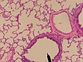
A pulmonary alveolus, also known as an air sac or air space, is one of millions of hollow, distensible cup-shaped cavities in the lungs where oxygen is exchanged for carbon dioxide. Alveoli make up the functional tissue of the mammalian lungs known as the lung parenchyma, which takes up 90 percent of the total lung volume.

<span class="mw-page-title-main">Respiratory tract</span> Organs involved in transmission of air to and from the point where gases diffuse into tissue

The respiratory tract is the subdivision of the respiratory system involved with the process of respiration in mammals. The respiratory tract is lined with respiratory epithelium as respiratory mucosa.

<span class="mw-page-title-main">Bronchus</span> Airway in the respiratory tract

A bronchus is a passage or airway in the lower respiratory tract that conducts air into the lungs. The first or primary bronchi pronounced (BRAN-KAI) to branch from the trachea at the carina are the right main bronchus and the left main bronchus. These are the widest bronchi, and enter the right lung, and the left lung at each hilum. The main bronchi branch into narrower secondary bronchi or lobar bronchi, and these branch into narrower tertiary bronchi or segmental bronchi. Further divisions of the segmental bronchi are known as 4th order, 5th order, and 6th order segmental bronchi, or grouped together as subsegmental bronchi. The bronchi, when too narrow to be supported by cartilage, are known as bronchioles. No gas exchange takes place in the bronchi.

Gas exchange is the physical process by which gases move passively by diffusion across a surface. For example, this surface might be the air/water interface of a water body, the surface of a gas bubble in a liquid, a gas-permeable membrane, or a biological membrane that forms the boundary between an organism and its extracellular environment.

<span class="mw-page-title-main">Pulmonary artery</span> Artery in pulmonary circulation carrying deoxygenated blood from heart to lungs

A pulmonary artery is an artery in the pulmonary circulation that carries deoxygenated blood from the right side of the heart to the lungs. The largest pulmonary artery is the main pulmonary artery or pulmonary trunk from the heart, and the smallest ones are the arterioles, which lead to the capillaries that surround the pulmonary alveoli.

Club cells, also known as bronchiolar exocrine cells, and formerly known as Clara cells, are low columnar/cuboidal cells with short microvilli, found in the small airways (bronchioles) of the lungs.

<span class="mw-page-title-main">Bronchoconstriction</span> Constriction of the terminal airways in the lungs

Bronchoconstriction is the constriction of the airways in the lungs due to the tightening of surrounding smooth muscle, with consequent coughing, wheezing, and shortness of breath.

<span class="mw-page-title-main">Respiratory disease</span> Disease of the respiratory system

Respiratory diseases, or lung diseases, are pathological conditions affecting the organs and tissues that make gas exchange difficult in air-breathing animals. They include conditions of the respiratory tract including the trachea, bronchi, bronchioles, alveoli, pleurae, pleural cavity, the nerves and muscles of respiration. Respiratory diseases range from mild and self-limiting, such as the common cold, influenza, and pharyngitis to life-threatening diseases such as bacterial pneumonia, pulmonary embolism, tuberculosis, acute asthma, lung cancer, and severe acute respiratory syndromes, such as COVID-19. Respiratory diseases can be classified in many different ways, including by the organ or tissue involved, by the type and pattern of associated signs and symptoms, or by the cause of the disease.

The cough reflex occurs when stimulation of cough receptors in the respiratory tract by dust or other foreign particles produces a cough, which causes rapidly moving air which usually remove the foreign material before it reaches the lungs. This typically clears particles from the bronchi and trachea, the tubes that feed air to lung tissue from the nose and mouth. The larynx and carina are especially sensitive. Cough receptors in the surface cells (epithelium) of the respiratory tract are also sensitive to chemicals. Terminal bronchioles and even the alveoli are sensitive to chemicals such as sulfur dioxide gas or chlorine gas.

<span class="mw-page-title-main">Respiratory epithelium</span> Mucosa that serves to moisten and protect the airways

Respiratory epithelium, or airway epithelium, is a type of ciliated columnar epithelium found lining most of the respiratory tract as respiratory mucosa, where it serves to moisten and protect the airways. It is not present in the vocal cords of the larynx, or the oropharynx and laryngopharynx, where instead the epithelium is stratified squamous. It also functions as a barrier to potential pathogens and foreign particles, preventing infection and tissue injury by the secretion of mucus and the action of mucociliary clearance.

<span class="mw-page-title-main">Acinus</span>

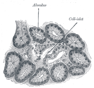
An acinus refers to any cluster of cells that resembles a many-lobed "berry," such as a raspberry. The berry-shaped termination of an exocrine gland, where the secretion is produced, is acinar in form, as is the alveolar sac containing multiple alveoli in the lungs.

<span class="mw-page-title-main">Pseudostratified columnar epithelium</span> Tissue type

A pseudostratified epithelium is a type of epithelium that, though comprising only a single layer of cells, has its cell nuclei positioned in a manner suggestive of stratified epithelia. As it rarely occurs as squamous or cuboidal epithelia, it is usually considered synonymous with the term pseudostratified columnar epithelium.

<span class="mw-page-title-main">Simple cuboidal epithelium</span> Tissue type

Simple cuboidal epithelium is a type of epithelium that consists of a single layer of cuboidal (cube-like) cells which have large, spherical and central nuclei.

<span class="mw-page-title-main">Lung bud</span>

The lung bud sometimes referred to as the respiratory bud forms from the respiratory diverticulum, an embryological endodermal structure that develops into the respiratory tract organs such as the larynx, trachea, bronchi and lungs. It arises from part of the laryngotracheal tube.

<span class="mw-page-title-main">Respiratory system of the horse</span> Biological system by which a horse circulates air for the purpose of gaseous exchange

The respiratory system of the horse is the biological system by which a horse circulates air for the purpose of gaseous exchange.

Airway basal cells are found deep in the respiratory epithelium, attached to, and lining the basement membrane.

Collateral ventilation(CV) is a back-up system of alveolar ventilation that can bypass the normal route of airflow when airways are restricted or obstructed. The pathways involved include those between adjacent alveoli, between bronchioles and alveoli, and those between bronchioles . Collateral ventilation also serves to modulate imbalances in ventilation and perfusion a feature of many diseases. The pathways are altered in lung diseases particularly asthma, and emphysema. A similar functional pattern of collateralisation is seen in the circulatory system of the heart.

References

  1. 1 2 Tortora GJ (2010). Principles of anatomy and physiology (12th ed.). Hoboken, NJ: John Wiley & Sons. pp. 883–888. ISBN   9780470233474.
  2. Bacsi A, Pan L, Ba X, Boldogh I (February 2016). "Pathophysiology of bronchoconstriction: role of oxidatively damaged DNA repair". Current Opinion in Allergy and Clinical Immunology. 16 (1): 59–67. doi:10.1097/ACI.0000000000000232. PMC   4940044 . PMID   26694039.
  3. Saladin K (2011). Human anatomy (3rd ed.). McGraw-Hill. pp. 640–641. ISBN   9780071222075.
  4. Merck Manual of Medical Information (Home ed.). Whitehouse Station, N.J.: Merck Research Laboratories. 1997. ISBN   978-0-911910-87-2.
  5. Martini FH, Timmons MJ, Tallitsch RB. Human Anatomy (6th ed.). Benjamin Cummings. p. 643. ISBN   978-0-321-49804-5.
  6. Paxton, Steve; Peckham, Michelle; Knibbs, Adele (2003). "Respiratory: Trachea, bronchioles and bronchi". University of Leeds.
  7. Friedman JN, Rieder MJ, Walton JM (November 2014). "Bronchiolitis: Recommendations for diagnosis, monitoring and management of children one to 24 months of age". Paediatrics & Child Health. 19 (9): 485–498. doi:10.1093/pch/19.9.485. PMC   4235450 . PMID   25414585.

Further reading