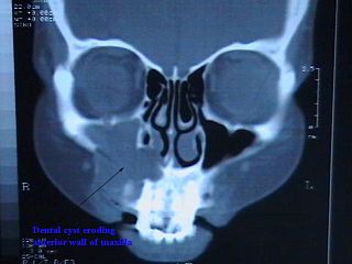
A cyst is a closed sac, having a distinct membrane and division compared with the nearby tissue. Hence, it is a cluster of cells that has grouped together to form a sac ; however, the distinguishing aspect of a cyst is that the cells forming the "shell" of such a sac are distinctly abnormal when compared with all surrounding cells for that given location. It may contain air, fluids, or semi-solid material. A collection of pus is called an abscess, not a cyst. Once formed, sometimes a cyst may resolve on its own. When a cyst fails to resolve, it may need to be removed surgically, but that would depend upon its type and location.

Oral mucocele is a clinical term for two related phenomena: mucus extravasation phenomenon and mucous retention cyst. Other names include mucous extravasation cyst, mucous cyst of the oral mucosa, and mucous retention and extravasation phenomena.

Fordyce spots are visible sebaceous glands that are present in most individuals. They appear on the genitals and/or on the face and in the mouth. They appear as small, painless, raised, pale, red or white spots or bumps 1 to 3 mm in diameter that may appear on the scrotum, shaft of the penis or on the labia, as well as the inner surface and vermilion border of the lips of the face. They are not associated with any disease or illness, nor are they infectious but rather they represent a natural occurrence on the body. No treatment is therefore required, unless the individual has cosmetic concerns. Persons with this condition sometimes consult a dermatologist because they are worried they may have a sexually transmitted disease or some form of cancer.

Pericoronitis is inflammation of the soft tissues surrounding the crown of a partially erupted tooth, including the gingiva (gums) and the dental follicle. The soft tissue covering a partially erupted tooth is known as an operculum, an area which can be difficult to access with normal oral hygiene methods. The synonym operculitis technically refers to inflammation of the operculum alone.
Oral medicine is a specialty focused on the mouth and nearby structures. It lies at the interface between medicine and dentistry.

A buccal exostosis is an exostosis on the buccal surface of the alveolar ridge of the maxilla or mandible. More commonly seen in the maxilla than the mandible, buccal exostoses are considered to be site specific. Existing as asymptomatic bony nodules, buccal exostoses don’t usually present until adult life, and some consider buccal exostoses to be a variation of normal anatomy rather than disease. Bone is thought to become hyperplastic, consisting of mature cortical and trabecular bone with a smooth outer surface. They are less common when compared with mandibular tori.
Dens evaginatus is a rare odontogenic developmental anomaly that is found in teeth where the outer surface appears to form an extra bump or cusp.

The periapical cyst is the most common odontogenic cyst. Also known as "Dental cyst". Periapical is defined as "the tissues surrounding the apex of the root of a tooth" and a cyst is "a pathological cavity lined by epithelium, having fluid or gaseous content that is not created by the accumulation of pus." Most frequently located in the maxillary anterior region, it is caused by pulpal necrosis secondary to dental caries or trauma. The cyst has lining that is derived from the epithelial cell rests of Malassez which proliferate to form the cyst. Highly common in the oral cavity, the periapical cyst is asymptomatic, but highly significant because a secondary infection can cause pain and damage. In radiographs, it appears a radiolucency around the apex of a tooth's root.
The lateral periodontal cyst is a non-inflammatory developmental cyst that arises from the epithelial post-functional dental lamina, which is a remnant from odontogenesis. It is more common in middle-aged males. Usually asymptomatic, it presents as a regular well-corticated radiolucency on the side of a mandibular canine or premolar root. Histologically, the cyst appears similar to the gingival cyst of the adult, having a non-keratinized squamous epithelial lining. The involved tooth is usually vital and has no indication for root canal treatment unless the signs of non-vital or necrotic pulpal tissue were confirmed. The cysts arise from epithelial rest cells in the periodontal ligament, although it is unknown whether from the cell rests of Malassez, reduced enamel epithelium or dental lamina remnants, and are generally treated by surgical enucleation.

Calcifying odotogenic cyst is a benign odontogenic tumor of cystic type most likely to affect the anterior areas of the jaws. It is most common in people in their second to third decades but can be seen at almost any age. On radiographs, the calcifying odontogenic cyst appears as a unilocular radiolucency. In one-third of cases, an impacted tooth is involved. Microscopically, there are many cells that are described as "ghost cells", enlarged eosinophilic epithelial cells without nuclei.

A dental abscess is a localized collection of pus associated with a tooth. The most common type of dental abscess is a periapical abscess, and the second most common is a periodontal abscess. In a periapical abscess, usually the origin is a bacterial infection that has accumulated in the soft, often dead, pulp of the tooth. This can be caused by tooth decay, broken teeth or extensive periodontal disease. A failed root canal treatment may also create a similar abscess.

Root canal treatment is a treatment sequence for the infected pulp of a tooth which results in the elimination of infection and the protection of the decontaminated tooth from future microbial invasion. Root canals, and their associated pulp chamber, are the physical hollows within a tooth that are naturally inhabited by nerve tissue, blood vessels and other cellular entities. Together, these items constitute the dental pulp.

An impacted tooth is one that fails to erupt into the dental arch within the expected developmental window. Because impacted teeth do not erupt, they are retained throughout the individual's lifetime unless extracted or exposed surgically. Teeth may become impacted because of adjacent teeth, dense overlying bone, excessive soft tissue or a genetic abnormality. Most often, the cause of impaction is inadequate arch length and space in which to erupt. That is the total length of the alveolar arch is smaller than the tooth arch. The wisdom teeth are frequently impacted because they are the last teeth to erupt in the oral cavity. Mandibular third molars are more commonly impacted than their maxillary counterparts.
Dental pertains to the teeth, including dentistry. Topics related to the dentistry, the human mouth and teeth include:

In dentistry, a furcation defect is bone loss, usually a result of periodontal disease, affecting the base of the root trunk of a tooth where two or more roots meet. The extent and configuration of the defect are factors in both diagnosis and treatment planning.
Odontogenic cyst are a group of jaw cysts that are formed from tissues involved in odontogenesis. Odontogenic cysts are closed sacs, and have a distinct membrane derived from rests of odontogenic epithelium. It may contain air, fluids, or semi-solid material. Intra-bony cysts are most common in the jaws, because the mandible and maxilla are the only bones with epithelial components. That odontogenic epithelium is critical in normal tooth development. However, epithelial rests may be the origin for the cyst lining later. Not all oral cysts are odontogenic cyst. For example, mucous cyst of the oral mucosa and nasolabial duct cyst are not of odontogenic origin.
In addition, there are several conditions with so-called (radiographic) 'pseudocystic appearance' in jaws; ranging from anatomic variants such as Stafne static bone cyst, to the aggressive aneurysmal bone cyst.
Paradental cysts constitute a family of inflammatory odontogenic cyst, that typically appear in relation to crown or root of partially erupted molar tooth. When the cyst is developed in the distal region of partially erupted third molar or in other locations in the dentition, it called simply paradental cyst, but the unique cyst that developed in the buccal bifurcation region of the mandibular first molars in the second half of the first decade of life is called buccal bifurcation cyst and has unique clinical features and management considerations in comparison to the other paradental cysts.
A cyst is a pathological epithelial lined cavity that fills with fluid or soft material and usually grows from internal pressure generated by fluid being drawn into the cavity from osmosis. The bones of the jaws, the mandible and maxilla, are the bones with the highest prevalence of cysts in the human body. This is due to the abundant amount of epithelial remnants that can be left in the bones of the jaws. The enamel of teeth is formed from ectoderm, and so remnants of epithelium can be left in the bone during odontogenesis. The bones of the jaws develop from embryologic processes which fuse together, and ectodermal tissue may be trapped along the lines of this fusion. This "resting" epithelium is usually dormant or undergoes atrophy, but, when stimulated, may form a cyst. The reasons why resting epithelium may proliferate and undergo cystic transformation are generally unknown, but inflammation is thought to be a major factor. The high prevalence of tooth impactions and dental infections that occur in the bones of the jaws is also significant to explain why cysts are more common at these sites.

Gingival cyst, also known as Epstein's pearl, is a type of cysts of the jaws that originates from the dental lamina and is found in the mouth parts. It is a superficial cyst in the alveolar mucosa. It can be seen inside the mouth as small and whitish bulge. Depending on the ages in which they develop, the cysts are classified into gingival cyst of newborn and gingival cyst of adult. Structurally, the cyst is lined by thin epithelium and shows a lumen usually filled with desquamated keratin, occasionally containing inflammatory cells. The nodes are formed as a result of cystic degeneration of epithelial rests of the dental lamina.


