
Otorhinolaryngology is a surgical subspeciality within medicine that deals with the surgical and medical management of conditions of the head and neck. Doctors who specialize in this area are called otorhinolaryngologists, otolaryngologists, head and neck surgeons, or ENT surgeons or physicians. Patients seek treatment from an otorhinolaryngologist for diseases of the ear, nose, throat, base of the skull, head, and neck. These commonly include functional diseases that affect the senses and activities of eating, drinking, speaking, breathing, swallowing, and hearing. In addition, ENT surgery encompasses the surgical management of cancers and benign tumors and reconstruction of the head and neck as well as plastic surgery of the face, scalp, and neck.
Sinusitis, also known as rhinosinusitis, is inflammation of the mucous membranes that line the sinuses resulting in symptoms that may include thick nasal mucus, a plugged nose, and facial pain. Other signs and symptoms may include fever, headaches, a poor sense of smell, sore throat, a feeling that phlegm is oozing out from the back of the nose to the throat along with a necessity to clear the throat frequently and frequent attacks of cough.

The maxilla in vertebrates is the upper fixed bone of the jaw formed from the fusion of two maxillary bones. In humans, the upper jaw includes the hard palate in the front of the mouth. The two maxillary bones are fused at the intermaxillary suture, forming the anterior nasal spine. This is similar to the mandible, which is also a fusion of two mandibular bones at the mandibular symphysis. The mandible is the movable part of the jaw.

The nasal cavity is a large, air-filled space above and behind the nose in the middle of the face. The nasal septum divides the cavity into two cavities, also known as fossae. Each cavity is the continuation of one of the two nostrils. The nasal cavity is the uppermost part of the respiratory system and provides the nasal passage for inhaled air from the nostrils to the nasopharynx and rest of the respiratory tract.

In anatomy, the orbit is the cavity or socket/hole of the skull in which the eye and its appendages are situated. "Orbit" can refer to the bony socket, or it can also be used to imply the contents. In the adult human, the volume of the orbit is 30 millilitres, of which the eye occupies 6.5 ml. The orbital contents comprise the eye, the orbital and retrobulbar fascia, extraocular muscles, cranial nerves II, III, IV, V, and VI, blood vessels, fat, the lacrimal gland with its sac and duct, the eyelids, medial and lateral palpebral ligaments, cheek ligaments, the suspensory ligament, septum, ciliary ganglion and short ciliary nerves.

The pyramid-shaped maxillary sinus is the largest of the paranasal sinuses, located in the maxilla. It drains into the middle meatus of the nose through the semilunar hiatus. It is located to the side of the nasal cavity, and below the orbit.

The ethmoid sinuses or ethmoid air cells of the ethmoid bone are one of the four paired paranasal sinuses. Unlike the other three pairs of paranasal sinuses which consist of one or two large cavities, the ethmoidal sinuses entail a number of small air-filled cavities. The cells are located within the lateral mass (labyrinth) of each ethmoid bone and are variable in both size and number. The cells are grouped into anterior, middle, and posterior groups; the groups differ in their drainage modalities, though all ultimately drain into either the superior or the middle nasal meatus of the lateral wall of the nasal cavity.

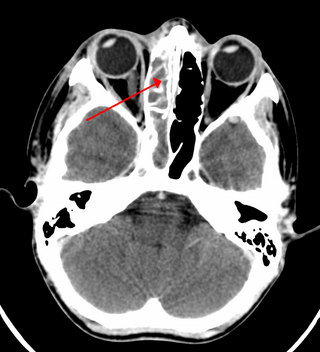
The sphenoid sinus is a paired paranasal sinus occurring within the body of the sphenoid bone. It represents one pair of the four paired paranasal sinuses. The pair of sphenoid sinuses are separated in the middle by a septum of sphenoid sinuses. Each sphenoid sinus communicates with the nasal cavity via the opening of sphenoidal sinus. The two sphenoid sinuses vary in size and shape, and are usually asymmetrical.
The anterior lacrimal crest is a bony projection on the frontal process of the maxilla. It creates the lateral margin of the lacrimal sac fossa and is continuous with the orbital margin. The medial palpebral ligament is attached to anterior lacrimal crest. It is an important structure to avoid damaging during rhinoplasty.
Silent sinus syndrome is a spontaneous, asymptomatic collapse of an air sinus associated with negative sinus pressures. It can cause painless facial asymmetry, diplopia and enophthalmos. Diagnosis is suspected based on symptoms, and can be confirmed using a CT scan. Treatment is surgical involving making an outlet for mucous drainage from the obstructed sinus, and, in some cases, paired with reconstruction of the orbital floor. It is slightly more common in middle age.
Chronic atrophic rhinitis, or simply atrophic rhinitis, is a chronic inflammation of the nose characterised by atrophy of nasal mucosa, including the glands, turbinate bones and the nerve elements supplying the nose. Chronic atrophic rhinitis may be primary and secondary. Special forms of chronic atrophic rhinitis are rhinitis sicca anterior and ozaena. It can also be described as the empty nose syndrome.

Functional endoscopic sinus surgery (FESS) is a procedure that is used to treat sinusitis and other conditions that affect the sinuses. Sinusitis is an inflammation of the sinuses that can cause symptoms such as congestion, headaches, and difficulty breathing through the nose.

An orbital blowout fracture is a traumatic deformity of the orbital floor or medial wall that typically results from the impact of a blunt object larger than the orbital aperture, or eye socket. Most commonly this results in a herniation of orbital contents through the orbital fractures. The proximity of maxillary and ethmoidal sinus increases the susceptibility of the floor and medial wall for the orbital blowout fracture in these anatomical sites. Most commonly, the inferior orbital wall, or the floor, is likely to collapse, because the bones of the roof and lateral walls are robust. Although the bone forming the medial wall is the thinnest, it is buttressed by the bone separating the ethmoidal air cells. The comparatively thin bone of the floor of the orbit and roof of the maxillary sinus has no support and so the inferior wall collapses mostly. Therefore, medial wall blowout fractures are the second-most common, and superior wall, or roof and lateral wall, blowout fractures are uncommon and rare, respectively. They are characterized by double vision, sunken ocular globes, and loss of sensation of the cheek and upper gums from infraorbital nerve injury.

The nose is the first organ of the respiratory system. It is also the principal organ in the olfactory system. The shape of the nose is determined by the nasal bones and the nasal cartilages, including the nasal septum which separates the nostrils and divides the nasal cavity into two.

The superior dental plexus is a nerve plexus that innervates the upper/maxillary teeth and as adjacent structures. It is formed by the anterior superior alveolar nerve (ASAN), middle superior alveolar nerve (MSAN), and the posterior superior alveolar nerve (PSAN). It issues dental branches and gingival branches.

The canine space, is a fascial space of the head and neck. It is a thin potential space on the face, and is paired on either side. It is located between the levator anguli oris muscle inferiorly and the levator labii superioris muscle superiorly. The term is derived from the fact that the space is in the region of the canine fossa, and that infections originating from the maxillary canine tooth may spread to involve the space. Infra-orbital is derived from infra- meaning below and orbit which refers to the eye socket.
Antral lavage is a largely obsolete surgical procedure in which a cannula is inserted into the maxillary sinus via the inferior meatus to allow irrigation and drainage of the sinus. It is also called proof puncture, as the presence of an infection can be proven during the procedure. Upon presence of infection, it can be considered as therapeutic puncture. Often, multiple repeated lavages are subsequently required to allow for full washout of infection.

Oroantral fistula (OAF) is an epithelialised oroantral communication (OAC). OAC refers to an abnormal connection between the oral cavity and antrum. The creation of an OAC is most commonly due to the extraction of a maxillary (upper) tooth closely related to the antral floor. A small OAC may heal spontaneously but a larger OAC would require surgical closure to prevent the development of persistent OAF and chronic sinusitis.
Nasal surgery is a medical procedure designed to treat various conditions that cause nasal blockages in the upper respiratory tract, for example nasal polyps, inferior turbinate hypertrophy, and chronic rhinosinusitis. It encompasses several types of techniques, including rhinoplasty, septoplasty, sinus surgery, and turbinoplasty, each with its respective postoperative treatments. Furthermore, nasal surgery is also conducted for cosmetic purposes. While there are potential risks and complications associated, the advancement of medical instruments and enhanced surgical skills have helped mitigate them.
Odontogenic sinusitis is a type of sinusitis, specifically caused by dental infections or procedures. Comprising approximately 10-12% of all chronic sinusitis cases, this condition primarily affects the maxillary sinus, which is in close proximity to the upper teeth.
