
Microscopy is the technical field of using microscopes to view objects and areas of objects that cannot be seen with the naked eye. There are three well-known branches of microscopy: optical, electron, and scanning probe microscopy, along with the emerging field of X-ray microscopy.

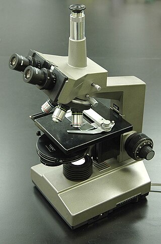
The optical microscope, also referred to as a light microscope, is a type of microscope that commonly uses visible light and a system of lenses to generate magnified images of small objects. Optical microscopes are the oldest design of microscope and were possibly invented in their present compound form in the 17th century. Basic optical microscopes can be very simple, although many complex designs aim to improve resolution and sample contrast.
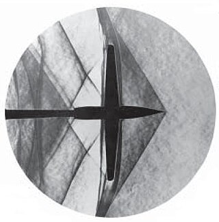
Schlieren photography is a process for photographing fluid flow. Invented by the German physicist August Toepler in 1864 to study supersonic motion, it is widely used in aeronautical engineering to photograph the flow of air around objects.
Optics is the branch of physics which involves the behavior and properties of light, including its interactions with matter and the construction of instruments that use or detect it. Optics usually describes the behavior of visible, ultraviolet, and infrared light. Because light is an electromagnetic wave, other forms of electromagnetic radiation such as X-rays, microwaves, and radio waves exhibit similar properties.

In optics, any optical instrument or system – a microscope, telescope, or camera – has a principal limit to its resolution due to the physics of diffraction. An optical instrument is said to be diffraction-limited if it has reached this limit of resolution performance. Other factors may affect an optical system's performance, such as lens imperfections or aberrations, but these are caused by errors in the manufacture or calculation of a lens, whereas the diffraction limit is the maximum resolution possible for a theoretically perfect, or ideal, optical system.
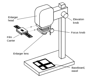
An enlarger is a specialized transparency projector used to produce photographic prints from film or glass negatives, or from transparencies.

In physics, backscatter is the reflection of waves, particles, or signals back to the direction from which they came. It is usually a diffuse reflection due to scattering, as opposed to specular reflection as from a mirror, although specular backscattering can occur at normal incidence with a surface. Backscattering has important applications in astronomy, photography, and medical ultrasonography. The opposite effect is forward scatter, e.g. when a translucent material like a cloud diffuses sunlight, giving soft light.

In photography, reciprocity is the inverse relationship between the intensity and duration of light that determines the reaction of light-sensitive material. Within a normal exposure range for film stock, for example, the reciprocity law states that the film response will be determined by the total exposure, defined as intensity × time. Therefore, the same response can result from reducing duration and increasing light intensity, and vice versa.
Medical optical imaging is the use of light as an investigational imaging technique for medical applications, pioneered by American Physical Chemist Britton Chance. Examples include optical microscopy, spectroscopy, endoscopy, scanning laser ophthalmoscopy, laser Doppler imaging, and optical coherence tomography. Because light is an electromagnetic wave, similar phenomena occur in X-rays, microwaves, and radio waves.

An optical printer is a device consisting of one or more film projectors mechanically linked to a movie camera. It allows filmmakers to re-photograph one or more strips of film. The optical printer is used for making special effects for motion pictures, or for copying and restoring film material.
In optics, a diffuser is any material that diffuses or scatters light in some manner to transmit soft light. Diffused light can be easily obtained by reflecting light from a white surface, while more compact diffusers may use translucent material, including ground glass, teflon, opal glass, and greyed glass.
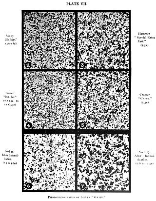
Film grain or film granularity is the random optical texture of processed photographic film due to the presence of small particles of a metallic silver, or dye clouds, developed from silver halide that have received enough photons. While film grain is a function of such particles it is not the same thing as such. It is an optical effect, the magnitude of which depends on both the film stock and the definition at which it is observed. It can be objectionably noticeable in an over-enlarged film photograph.
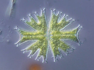
Differential interference contrast (DIC) microscopy, also known as Nomarski interference contrast (NIC) or Nomarski microscopy, is an optical microscopy technique used to enhance the contrast in unstained, transparent samples. DIC works on the principle of interferometry to gain information about the optical path length of the sample, to see otherwise invisible features. A relatively complex optical system produces an image with the object appearing black to white on a grey background. This image is similar to that obtained by phase contrast microscopy but without the bright diffraction halo. The technique was invented by Francis Hughes Smith. The "Smith DIK" was produced by Ernst Leitz Wetzlar in Germany and was difficult to manufacture. DIC was then developed further by Polish physicist Georges Nomarski in 1952.

Dark-field microscopy describes microscopy methods, in both light and electron microscopy, which exclude the unscattered beam from the image. Consequently, the field around the specimen is generally dark.

Bright-field microscopy (BF) is the simplest of all the optical microscopy illumination techniques. Sample illumination is transmitted white light, and contrast in the sample is caused by attenuation of the transmitted light in dense areas of the sample. Bright-field microscopy is the simplest of a range of techniques used for illumination of samples in light microscopes, and its simplicity makes it a popular technique. The typical appearance of a bright-field microscopy image is a dark sample on a bright background, hence the name.
Phase-contrast microscopy (PCM) is an optical microscopy technique that converts phase shifts in light passing through a transparent specimen to brightness changes in the image. Phase shifts themselves are invisible, but become visible when shown as brightness variations.
Köhler illumination is a method of specimen illumination used for transmitted and reflected light optical microscopy. Köhler illumination acts to generate an even illumination of the sample and ensures that an image of the illumination source is not visible in the resulting image. Köhler illumination is the predominant technique for sample illumination in modern scientific light microscopy. It requires additional optical elements which are more expensive and may not be present in more basic light microscopes.
The photoacoustic Doppler effect is a type of Doppler effect that occurs when an intensity modulated light wave induces a photoacoustic wave on moving particles with a specific frequency. The observed frequency shift is a good indicator of the velocity of the illuminated moving particles. A potential biomedical application is measuring blood flow.
Speckle, speckle pattern, or speckle noise is a granular noise texture degrading the quality as a consequence of interference among wavefronts in coherent imaging systems, such as radar, synthetic aperture radar (SAR), medical ultrasound and optical coherence tomography. Speckle is not external noise; rather, it is an inherent fluctuation in diffuse reflections, because the scatterers are not identical for each cell, and the coherent illumination wave is highly sensitive to small variations in phase changes.

A condenser is an optical lens that renders a divergent light beam from a point light source into a parallel or converging beam to illuminate an object to be imaged.


