Carboxypeptidase A

Last updated
Carboxypeptidase A
Carboxypeptidase A.png
Carboxypeptidase A, from bovine pancreas
Identifiers
EC no. 3.4.17.1
CAS no. 9031-98-5
Databases
IntEnz IntEnz view
BRENDA BRENDA entry
ExPASy NiceZyme view
KEGG KEGG entry
MetaCyc metabolic pathway
PRIAM profile
PDB structures RCSB PDB PDBe PDBsum
Gene Ontology AmiGO / QuickGO
Search
PMC articles
PubMed articles
NCBI proteins

Carboxypeptidase A usually refers to the pancreatic exopeptidase that hydrolyzes peptide bonds of C-terminal residues with aromatic or aliphatic side-chains. Most scientists in the field now refer to this enzyme as CPA1, and to a related pancreatic carboxypeptidase as CPA2.

Contents

Types

In addition, there are 4 other mammalian enzymes named CPA-3 through CPA-6, and none of these are expressed in the pancreas. Instead, these other CPA-like enzymes have diverse functions.

Function

CPA-1 and CPA-2 (and, it is presumed, all other CPAs) employ a zinc ion within the protein for hydrolysis of the peptide bond at the C-terminal end of an amino acid residue. Loss of the zinc leads to loss of activity, which can be replaced easily by zinc, and also by some other divalent metals (cobalt, nickel). Carboxypeptidase A is produced in the pancreas and is crucial to many processes in the human body to include digestion, post-translational modification of proteins, blood clotting, and reproduction.

Applications

This vast scope of functionality for a single protein makes it the ideal model for research regarding other zinc proteases of unknown structure. Recent biomedical research on collagenase, enkephalinase, and angiotensin-converting enzyme used carboxypeptidase A for inhibitor synthesis and kinetic testing. For example, a drug that treats high blood pressure, Captopril, was designed based on a carboxypeptidase A inhibitor. Carboxypeptidase A and the target enzyme of Captopril, angiotensin-converting enzyme, have very similar structures, as they both contain a zinc ion within the active site. This allowed for a potent carboxypeptidase A inhibitor to be used to inhibit the enzyme and, thus, lower blood pressure through the renin-angiotensin-aldosterone system. [1]

Structure

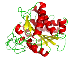
Carboxypeptidase A (CPA) contains a zinc (Zn2+) metal center in a tetrahedral geometry with amino acid residues in close proximity around zinc to facilitate catalysis and binding. Out of the 307 amino acids bonded in a peptide chain, the following amino acid residues are important for catalysis and binding; Glu-270, Arg-71, Arg-127, Asn-144, Arg-145, and Tyr-248. Figure 1 illustrates the tetrahedral zinc complex active site with the important amino acid residues that surround the complex. [2]

The zinc metal is a strong electrophilic Lewis acid catalyst which stabilizes a coordinated water molecule as well as stabilizes the negative intermediates that occur throughout the hydrolytic reaction. Stabilization of both the coordinated water molecule and negative intermediates are assisted by polar residues in the active site which are in close proximity to facilitate hydrogen bonding. [2]

Figure 1. CPA Active Site CPA Active Site.jpg
Figure 1. CPA Active Site

The active site can be characterized into two sub-sites denoted as S1’ and S1. The S1’ sub-site is the hydrophobic pocket of the enzyme, and Tyr-248 acts to ‘cap’ the hydrophobic pocket after substrate or inhibitor is bound (SITE). [2] The hydrogen bonding from the hydroxyl group in Tyr-248 facilitates this conformation due to interaction with the terminal carboxylates of substrates that bind. Substantial movement is required for this enzyme and induced fit model explains how this interaction occurs.

A triad of residues interact to the C-terminal carboxylate through hydrogen bonding:

Mechanism

Classified as a metalloexopeptidase, carboxypeptidase A consists of a single polypeptide chain bound to a zinc ion. This characteristic metal ion is located within the active site of the enzyme, along with five amino acid residues that are involved in substrate binding: Arg-71, Arg-127, Asn-144, Arg-145, Tyr-248, and Glu-270. X-ray crystallographic studies have revealed five subsites on the protein. These allosteric sites are involved in creating the ligand-enzyme specificity seen in most bioactive enzymes. One of these subsites induces a conformational change at Tyr-248 upon binding of a substrate molecule at the primary active site. The phenolic hydroxyl of tyrosine forms a hydrogen bond with the terminal carboxylate of the ligand. In addition, a second hydrogen bond is formed between the tyrosine and a peptide linkage of longer peptide substrates. These changes make the bond between the enzyme and ligand, whether it is substrate or inhibitor, much stronger. This property of carboxypeptidase A led to the first clause of Daniel E. Koshland, Jr.’s “induced fit” hypothesis.

The S1 sub-site is where catalysis occurs in CPA, and the zinc ion is coordinated by Glu-72, His-69, and His-196 enzyme residues. A plane exists that bisects the active-site groove where residues Glu-270 and Arg-127 are on opposite sides of the zinc-water coupled complex. The zinc is electron rich due to glutamine ligands coordinating the zinc because before substrate binds, Glu-72 coordinates bidentate but shifts to monodentate after substrate binds. As a result, the zinc metal is not able to deprotonate the coordinated water molecule to make a hydroxyl nucleophile. [2]

Figure 2. CPA-Catalyzed proteolysis promoted by coordinated water molecule. CPA-Catalyzed Proteolysis.jpg
Figure 2. CPA-Catalyzed proteolysis promoted by coordinated water molecule.

Glu-270 and Arg-127 play an important role in catalysis shown in Figure 2. Arg-127 acts to stabilize the carbonyl of the substrate that is bound to amino group of phenylalanine. Simultaneously, the water molecule coordinated to zinc is deprotonated by Glu-270 and interacts with the carbonyl stabilized by Arg-127. This creates an intermediate, shown in Figure 2, where the negatively charged oxygen is coordinated to zinc, and through unfavorable electrostatic interactions between Glu-270 and the ionized product facilitates the release of the product at the end of catalysis. [2]

In recent computational studies, the mechanism of catalysis is similar but the difference in mechanism is that deprotonated water molecule binds to the carbon of the carbonyl, whereas Figure 2 shows the hydroxyl group stays coordinated to zinc. Then proteolysis occurs and the water molecule is then introduced back into the active site to coordinate to zinc. [3]

Several studies have been conducted exploring the details of the bond between carboxypeptidase A and substrate and how this affects the rate of hydrolysis. In 1934, it was first discovered through kinetic experiments that, in order for substrate to bind, the peptide that is to be hydrolyzed must be adjacent to a terminal free hydroxyl group. Also, the rate of hydrolysis can be enhanced if the C-terminal residue is branched aliphatic or aromatic. However, if the substrate is a dipeptide with a free amino group, it undergoes hydrolysis slowly; this, however, can be avoided if the amino group is blocked by N-acylation. [4]

It is quite clear that the structure of the enzyme, to be specific the active site, is very important in understanding the mechanism of reaction. For this reason, Rees and colleagues studied the enzyme-ligand complex to get a clear answer for the role of the zinc ion. These studies found that, in free enzyme, the zinc coordination number is five; the metal center is coordinated with two imidazole Nδ1 nitrogens, the two carboxylate oxygens of glutamate-72, and a water molecule to form a distorted tetrahedral. However, once ligand binds at the active site of carboxypeptidase A, this coordination number can vary from five to six. When bound to dipeptide glycyl-L-tyrosine, the amino nitrogen of the dipeptide and the carbonyl oxygen replaced the water ligand. This would yield a coordination number of six for the zinc in the carboxypeptidase A- dipeptide glycyl-L-tyrosine complex. Electron density maps gave evidence that the amino nitrogen occupies a second position near glutamate-270. The closeness of these two residues would result in a steric hindrance preventing the water ligand from coordinating with zinc. This would result in a coordination number of five. Data for both are substantial, indicating that both situations occur naturally. [5]

There are two proposed mechanisms for the catalytic function of carboxypeptidase A. The first is a nucleophilic pathway involving a covalent acyl enzyme intermediate containing active site base Glu-270. Evidence for this anhydride intermediate is mixed; Suh and colleagues isolated what is assumed to by the acyl intermediate. However, confirmation of the acyl enzyme was done without trapping experiments, making the conclusions weak. [1]

The second proposed mechanism is a promoted water pathway. This mechanism involves attack of a water molecule at the scissile peptide linkage of the substrate. This process is promoted by the zinc ion and assisted by residue Glu-270. [1]

See also

Related Research Articles

<span class="mw-page-title-main">Chymotrypsin</span> Digestive enzyme

Chymotrypsin (EC 3.4.21.1, chymotrypsins A and B, alpha-chymar ophth, avazyme, chymar, chymotest, enzeon, quimar, quimotrase, alpha-chymar, alpha-chymotrypsin A, alpha-chymotrypsin) is a digestive enzyme component of pancreatic juice acting in the duodenum, where it performs proteolysis, the breakdown of proteins and polypeptides. Chymotrypsin preferentially cleaves peptide amide bonds where the side chain of the amino acid N-terminal to the scissile amide bond (the P1 position) is a large hydrophobic amino acid (tyrosine, tryptophan, and phenylalanine). These amino acids contain an aromatic ring in their side chain that fits into a hydrophobic pocket (the S1 position) of the enzyme. It is activated in the presence of trypsin. The hydrophobic and shape complementarity between the peptide substrate P1 side chain and the enzyme S1 binding cavity accounts for the substrate specificity of this enzyme. Chymotrypsin also hydrolyzes other amide bonds in peptides at slower rates, particularly those containing leucine at the P1 position.

<span class="mw-page-title-main">Urease</span> Multiprotein Nickel-containing complex which hydrolyses urea

Ureases, functionally, belong to the superfamily of amidohydrolases and phosphotriesterases. Ureases are found in numerous bacteria, fungi, algae, plants, and some invertebrates, as well as in soils, as a soil enzyme. They are nickel-containing metalloenzymes of high molecular weight.

<span class="mw-page-title-main">Active site</span> Active region of an enzyme

In biology and biochemistry, the active site is the region of an enzyme where substrate molecules bind and undergo a chemical reaction. The active site consists of amino acid residues that form temporary bonds with the substrate, the binding site, and residues that catalyse a reaction of that substrate, the catalytic site. Although the active site occupies only ~10–20% of the volume of an enzyme, it is the most important part as it directly catalyzes the chemical reaction. It usually consists of three to four amino acids, while other amino acids within the protein are required to maintain the tertiary structure of the enzymes.

DnaG is a bacterial DNA primase and is encoded by the dnaG gene. The enzyme DnaG, and any other DNA primase, synthesizes short strands of RNA known as oligonucleotides during DNA replication. These oligonucleotides are known as primers because they act as a starting point for DNA synthesis. DnaG catalyzes the synthesis of oligonucleotides that are 10 to 60 nucleotides long, however most of the oligonucleotides synthesized are 11 nucleotides. These RNA oligonucleotides serve as primers, or starting points, for DNA synthesis by bacterial DNA polymerase III. DnaG is important in bacterial DNA replication because DNA polymerase cannot initiate the synthesis of a DNA strand, but can only add nucleotides to a preexisting strand. DnaG synthesizes a single RNA primer at the origin of replication. This primer serves to prime leading strand DNA synthesis. For the other parental strand, the lagging strand, DnaG synthesizes an RNA primer every few kilobases (kb). These primers serve as substrates for the synthesis of Okazaki fragments.

Matrix metalloproteinases (MMPs), also known as matrix metallopeptidases or matrixins, are metalloproteinases that are calcium-dependent zinc-containing endopeptidases; other family members are adamalysins, serralysins, and astacins. The MMPs belong to a larger family of proteases known as the metzincin superfamily.

<span class="mw-page-title-main">Serine protease</span> Class of enzymes

Serine proteases are enzymes that cleave peptide bonds in proteins. Serine serves as the nucleophilic amino acid at the (enzyme's) active site. They are found ubiquitously in both eukaryotes and prokaryotes. Serine proteases fall into two broad categories based on their structure: chymotrypsin-like (trypsin-like) or subtilisin-like.

A metalloproteinase, or metalloprotease, is any protease enzyme whose catalytic mechanism involves a metal. An example is ADAM12 which plays a significant role in the fusion of muscle cells during embryo development, in a process known as myogenesis.

<span class="mw-page-title-main">Catalytic triad</span> Set of three coordinated amino acids

A catalytic triad is a set of three coordinated amino acids that can be found in the active site of some enzymes. Catalytic triads are most commonly found in hydrolase and transferase enzymes. An acid-base-nucleophile triad is a common motif for generating a nucleophilic residue for covalent catalysis. The residues form a charge-relay network to polarise and activate the nucleophile, which attacks the substrate, forming a covalent intermediate which is then hydrolysed to release the product and regenerate free enzyme. The nucleophile is most commonly a serine or cysteine amino acid, but occasionally threonine or even selenocysteine. The 3D structure of the enzyme brings together the triad residues in a precise orientation, even though they may be far apart in the sequence.

<span class="mw-page-title-main">Carboxypeptidase</span>

A carboxypeptidase is a protease enzyme that hydrolyzes (cleaves) a peptide bond at the carboxy-terminal (C-terminal) end of a protein or peptide. This is in contrast to an aminopeptidases, which cleave peptide bonds at the N-terminus of proteins. Humans, animals, bacteria and plants contain several types of carboxypeptidases that have diverse functions ranging from catabolism to protein maturation. At least two mechanisms have been discussed.

<span class="mw-page-title-main">Aspartoacylase</span> Hydrolytic enzyme found on human chromosome 17

Aspartoacylase is a hydrolytic enzyme that in humans is encoded by the ASPA gene. ASPA catalyzes the deacylation of N-acetyl-l-aspartate (N-acetylaspartate) into aspartate and acetate. It is a zinc-dependent hydrolase that promotes the deprotonation of water to use as a nucleophile in a mechanism analogous to many other zinc-dependent hydrolases. It is most commonly found in the brain, where it controls the levels of N-acetyl-l-aspartate. Mutations that result in loss of aspartoacylase activity are associated with Canavan disease, a rare autosomal recessive neurodegenerative disease.

<span class="mw-page-title-main">Aspartic protease</span>

Aspartic proteases are a catalytic type of protease enzymes that use an activated water molecule bound to one or more aspartate residues for catalysis of their peptide substrates. In general, they have two highly conserved aspartates in the active site and are optimally active at acidic pH. Nearly all known aspartyl proteases are inhibited by pepstatin.

<i>Bam</i>HI

BamHI is a type II restriction endonuclease, having the capacity for recognizing short sequences of DNA and specifically cleaving them at a target site. This exhibit focuses on the structure-function relations of BamHI as described by Newman, et al. (1995). BamHI binds at the recognition sequence 5'-GGATCC-3', and cleaves these sequences just after the 5'-guanine on each strand. This cleavage results in sticky ends which are 4 bp long. In its unbound form, BamHI displays a central b sheet, which resides in between α-helices.

<span class="mw-page-title-main">Mannose phosphate isomerase</span>

Mannose-6 phosphate isomerase (MPI), alternately phosphomannose isomerase (PMI) is an enzyme which facilitates the interconversion of fructose 6-phosphate (F6P) and mannose-6-phosphate (M6P). Mannose-6-phosphate isomerase may also enable the synthesis of GDP-mannose in eukaryotic organisms. M6P can be converted to F6P by mannose-6-phosphate isomerase and subsequently utilized in several metabolic pathways including glycolysis and capsular polysaccharide biosynthesis. PMI is monomeric and metallodependent on zinc as a cofactor ligand. PMI is inhibited by erythrose 4-phosphate, mannitol 1-phosphate, and to a lesser extent, the alpha anomer of M6P.

<span class="mw-page-title-main">Riboflavin synthase</span>

Riboflavin synthase is an enzyme that catalyzes the final reaction of riboflavin biosynthesis. It catalyzes the transfer of a four-carbon unit from one molecule of 6,7-dimethyl-8-ribityllumazine onto another, resulting in the synthesis of riboflavin and 5-amino-6-ribitylamino-2,4(1H,3H)-pyrimidinedione:

<span class="mw-page-title-main">Steroid Delta-isomerase</span>

In enzymology, a steroid Δ5-isomerase is an enzyme that catalyzes the chemical reaction

The discovery of an orally inactive peptide from snake venom established the unimportant role of angiotensin converting enzyme (ACE) inhibitors in regulating blood pressure. This led to the development of Captopril, the first ACE inhibitor. When the adverse effects of Captopril became apparent new derivates were designed. Then after the discovery of two active sites of ACE: N-domain and C-domain, the development of domain-specific ACE inhibitors began.

<span class="mw-page-title-main">Peptidyl-dipeptidase Dcp</span> Class of enzymes

Peptidyl-dipeptidase Dcp (EC 3.4.15.5, dipeptidyl carboxypeptidase (Dcp), dipeptidyl carboxypeptidase) is a metalloenzyme found in the cytoplasm of bacterium E. Coli responsible for the C-terminal cleavage of a variety of dipeptides and unprotected larger peptide chains. The enzyme does not hydrolyze bonds in which P1' is Proline, or both P1 and P1' are Glycine. Dcp consists of 680 amino acid residues that form into a single active monomer which aids in the intracellular degradation of peptides. Dcp coordinates to divalent zinc which sits in the pocket of the active site and is composed of four subsites: S1’, S1, S2, and S3, each subsite attracts certain amino acids at a specific position on the substrate enhancing the selectivity of the enzyme. The four subsites detect and bind different amino acid types on the substrate peptide in the P1 and P2 positions. Some metallic divalent cations such as Ni+2, Cu+2, and Zn+2 inhibit the function of the enzyme around 90%, whereas other cations such as Mn+2, Ca+2, Mg+2, and Co+2 have slight catalyzing properties, and increase the function by around 20%. Basic amino acids such as Arginine bind preferably at the S1 site, the S2 site sits deeper in the enzyme therefore is restricted to bind hydrophobic amino acids with phenylalanine in the P2 position. Dcp is divided into two subdomains (I, and II), which are the two sides of the clam shell-like structure and has a deep inner cavity where a pair of histidine residues bind to the catalytic zinc ion in the active site. Peptidyl-Dipeptidase Dcp is classified like Angiotensin-I converting enzyme (ACE) which is also a carboxypeptidase involved in blood pressure regulation, but due to structural differences and peptidase activity between these two enzymes they had to be examined separately. ACE has endopeptidase activity, whereas Dcp strictly has exopeptidase activity based on its cytoplasmic location and therefore their mechanisms of action are differentiated. Another difference between these enzymes is that the activity of Peptidyl-Dipeptidase Dcp is not enhanced in the presence of chloride anions, whereas chloride enhances ACE activity.

Lysine carboxypeptidase is an enzyme. This enzyme catalyses the following chemical reaction:

<span class="mw-page-title-main">Galactose oxidase</span>

Galactose oxidase is an enzyme that catalyzes the oxidation of D-galactose in some species of fungi.

<span class="mw-page-title-main">Aldehyde ferredoxin oxidoreductase</span>

In enzymology, an aldehyde ferredoxin oxidoreductase (EC 1.2.7.5) is an enzyme that catalyzes the chemical reaction

References

  1. 1 2 3 Christianson DW, Lipscomb WN (February 1989). "Carboxypeptidase A". Accounts of Chemical Research. 22 (2): 62–9. doi:10.1021/ar00158a003.
  2. 1 2 3 4 5 Christianson, D., W., and Lipscomb, W., N. (1989) Carboxypeptidase A.American Chemical Society, Vol (22): 62-69.
  3. Valdez CE, Morgenstern A, Eberhart ME, Alexandrova AN (November 2016). "Predictive methods for computational metalloenzyme redesign - a test case with carboxypeptidase A". Physical Chemistry Chemical Physics. 18 (46): 31744–31756. Bibcode:2016PCCP...1831744V. doi:10.1039/c6cp02247b. PMID   27841396. S2CID   3545851.
  4. Lipscomb WN (March 1970). "Structure and mechanism in the enzymic activity of carboxypeptidase A and relations to chemical sequence". Accounts of Chemical Research. 3 (3): 81–9. doi:10.1021/ar50027a001.
  5. Rees DC, Lewis M, Honzatko RB, Lipscomb WN, Hardman KD (June 1981). "Zinc environment and cis peptide bonds in carboxypeptidase A at 1.75-A resolution". Proceedings of the National Academy of Sciences of the United States of America. 78 (6): 3408–12. Bibcode:1981PNAS...78.3408R. doi: 10.1073/pnas.78.6.3408 . PMC   319577 . PMID   6943549.