Chaperone-usher fimbriae

Last updated

Chaperone-usher fimbriae (CU) are linear, unbranching, outer-membrane pili secreted by gram-negative bacteria through the chaperone-usher system rather than through type IV secretion or extracellular nucleation systems. These fimbriae are built up out of modular pilus subunits, which are transported into the periplasm in a Sec dependent manner. Chaperone-usher secreted fimbriae are important pathogenicity factors facilitating host colonisation, localisation and biofilm formation in clinically important species such as uropathogenic Escherichia coli and Pseudomonas aeruginosa .

Contents

Structure

Overall

All chaperone/usher systems are found within gene clusters consisting of at least an usher, a chaperone and one or more fimbriae subunits. [1] Overall the system includes periplasmic chaperones, periplasmic and extracellular pilus subunits, dimeric usher outer membrane pore and associated Sec machinery. The Pilus subunits polymerise via a non-covalent interaction to form the mature pilus which consists of an adhesive tip, helical body and an usher bound base. The P-pilus (pap) system is one of the best characterised and is shown below. [2]

A schematic overview of the pap chaperone-usher system showing all subunits and their organisation. PapD is the chaperone; PapC is the usher; PapA and the remaining subunits form the fimbria itself Overview of Pap system structure.png
A schematic overview of the pap chaperone-usher system showing all subunits and their organisation. PapD is the chaperone; PapC is the usher; PapA and the remaining subunits form the fimbria itself

Usher structure and function

The usher forms the outer-membrane pore and functions in vivo as a dimer, though only one of the ushers is active at any one time. [3] The usher pore (PapC) is formed by a 24 stranded beta barrel with 4 additional domains: N-terminal domain (NTD), Plug domain, and two C-terminal domains (CTD1 and CTD2 respectively). The NTD and CTDs function to bring chaperone/subunit complexes to the growing pilus and facilitate translocation across the pore. [4] The plug domain acts to stop premature formation of a pilus and can either be located inside the pore or on the periplasmic side of the membrane. In the active dimeric usher one of the usher has the plug located periplasmically and the other has it located inside the translocation pore. [5]

Chaperone structure and function

The periplasmic chaperone (PapD) has a 'boomerang' structure formed by an immunoglobulin (Ig) like fold with an essential C-terminal extension (G1). This fold is formed by 13 β strands (A1-G1) and 4 short α helices. Chaperones belong to one of two families based on the length of the loop connecting the beta strands F1 and G1. Long looped chaperones are FGL and short looped chaperones are FGS. The chaperone functions to stop aggregation and degradation of pilus subunits by binding their interfacing regions and facilitating correct protein folding before bringing them to the usher/fimbriae complex. [6]

Pilin subunit structure and function

PAP pilin major protein, PapA homo13mer, top view, E.Coli. 5flu.jpg
PAP pilin major protein, PapA homo13mer, top view, E.Coli.

The pilus subunit structure also has an Ig-like fold but with an essential N-terminal extension rather than a C-terminal extension. Because this N-terminal extension is the same β strand that normally completes the Ig fold, a large hydrophobic cleft is formed in the mature subunit (P1-P5), this cleft is both essential to pilus biogenesis but also necessitates a chaperone to stabilise the structure. [7] Particular subunits may have additional domains e.g. papG has an adhesive lectin domain. In vivo pilus subunits function to form the main body of the helical pilus (approximately 3.28 subunits per turn), and the adhesive head.

Biogenesis

Pilus biogenesis proceeds via the strand complementation mechanism, the mechanism functions as a direct consequence of the structures of both the chaperones and pilus subunits. Both the G1 extension of the chaperone and the N-terminal extension of the pilus subunits can bind to the hydrophobic P1-P5 cleft. When the G1 is bound it binds in a parallel conformation stabilising the subunit structure but doesn't form a true Ig fold (which is anti-parallel). It also only binds the cleft from sites P1-P4 leaving the P5 pocket exposed. When another subunit binds the hydrophobic cleft it binds in a P5-P2 anti-parallel fashion creating the most energetically favourable conformation of a true Ig-fold. [1]

When a chaperone/subunit complex is brought to the growing usher/pilus complex (note that a chaperone/subunit complex forms the base of this) the NTD and CTDs of the usher dimer bring and hold the chaperone/subunit complex to the pilus base. Here the N-terminal extension of the subunit interacts with the free P5 pocket of the pilus. This binding causes a residue by residue 'unzipping' of the chaperone's G1 strand with concurrent 'zipping' of the N-terminal extension of the chaperone/subunit complex, unknown as the zip in-zip out mechanism. [2]

Once the chaperone has been displaced it diffuses back to the sec machinery to bind another emerging pilus subunit. The usher using is CTDs then translocates the longer pilus across the translocation pore by one residue until the chaperone is in close association with the usher. This translocation is coupled with a slight rotation giving the pilus its helical structure. This growth proceeds until a terminating subunit binds the pilus (if one exists), terminating subunits lack the P5 pocket and thus almost irreversibly bind their chaperone. Since the usher can only translocate pilus subunits and not chaperone/subunit complexes, and the fact that the chaperone can't be energetically out-competed by another subunit (due to lack of P5) this terminating subunit anchors the pilus to the outer membrane and stop any further pilus growth. [1]

Nomenclature and Classification

Historically classification of fimbriae was made based on their appearance under the microscope creating 4 classes: Afimbrial, type 1 fimbriae, bundle forming (type IV) pilus and curli. However this type of classification doesn't give any measure of relatedness and thus has been superseded by a phylogenetic system. [8]

In chaperone/usher fimbriae there are broadly two types of classification: Based on the type of usher present, or based on the type of chaperone present. Using the usher as the base of classification forms 5 clades: alpha, beta, gamma, pi and sigma, and four sub clades: gamma 1-4. The naming of the beta and gamma clades are simply alphabetical, whereas the others are named by a defining feature: alpha for alternative chaperone/usher family; pi for P-pilus (pyelonephritis associated); and sigma for spore coat pilus from Myxococcus xanthus. [8]

As stated previously there are two types of chaperone; FGL and FGS, and this is the basis of chaperone classification. All FGL chaperones are associated with afimbrial secretion and all FGS chaperones are associated with fimbrial secretion. All FGL chaperones are found in sub-clade gamma-3, all other clades and sub-clades are FGS. [8]

Pathogenicity

Chaperone/usher fimbriae have many roles, in many stages of pathogenicity in several species. The most well understood are roles in adhesion, innate immune evasion and localisation. Adhesion is facilitated by the lectin domain at the tip of chaperone/usher fimbriae, this domain of FimH is well understood. FimH is the first subunit of the type I pilus (γ-1 clade) in species such as UPEC and binds to D-mannosylated residues in the bladder, allowing pathogen adhesion and colonisation. Immune evasion is also facilitated by the tip adhesin, for example the binding of Decay accelerating factor (DAF) by gamma3 clade fimbriae inhibit effective complement activation. Localisation could be performed by sequential expression of different chaperone/usher systems. Most species of pathogenic bacteria express more than one type of chaperone/usher system, for example in Pseudomonas aeruginosa there are five different systems. UPEC expresses both the type 1 fimbriae and the P-pilus (pap) which it expresses sequentially possibly to facilitate migration from the bladder (type I fimbriae) to the kidney (pap). [9]

This importance in pathogenicity makes the chaperone/usher system an attractive target for novel drug therapies. By targeting pathogenicity instead of the organism (in the case of conventional antibiotics) the strong selective pressure for evolution of resistance is removed. In the case of UPEC there have been some success in stopping pilus formation in clinical trials and the development of an effective vaccine in mice and cynomolgus monkeys based on FimCH complex. [9]

Related Research Articles

<span class="mw-page-title-main">Pilus</span> A proteinaceous hair-like appendage on the surface of bacteria

A pilus is a hair-like appendage found on the surface of many bacteria and archaea. The terms pilus and fimbria can be used interchangeably, although some researchers reserve the term pilus for the appendage required for bacterial conjugation. All conjugative pili are primarily composed of pilin – fibrous proteins, which are oligomeric.

<span class="mw-page-title-main">Secretion</span> Controlled release of substances by cells or tissues

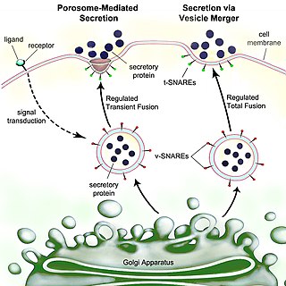
Secretion is the movement of material from one point to another, such as a secreted chemical substance from a cell or gland. In contrast, excretion is the removal of certain substances or waste products from a cell or organism. The classical mechanism of cell secretion is via secretory portals at the plasma membrane called porosomes. Porosomes are permanent cup-shaped lipoprotein structures embedded in the cell membrane, where secretory vesicles transiently dock and fuse to release intra-vesicular contents from the cell.

<span class="mw-page-title-main">RNA polymerase II</span> Protein complex that transcribes DNA

RNA polymerase II is a multiprotein complex that transcribes DNA into precursors of messenger RNA (mRNA) and most small nuclear RNA (snRNA) and microRNA. It is one of the three RNAP enzymes found in the nucleus of eukaryotic cells. A 550 kDa complex of 12 subunits, RNAP II is the most studied type of RNA polymerase. A wide range of transcription factors are required for it to bind to upstream gene promoters and begin transcription.

Adhesins are cell-surface components or appendages of bacteria that facilitate adhesion or adherence to other cells or to surfaces, usually in the host they are infecting or living in. Adhesins are a type of virulence factor.

<span class="mw-page-title-main">DNA clamp</span>

A DNA clamp, also known as a sliding clamp, is a protein complex that serves as a processivity-promoting factor in DNA replication. As a critical component of the DNA polymerase III holoenzyme, the clamp protein binds DNA polymerase and prevents this enzyme from dissociating from the template DNA strand. The clamp-polymerase protein–protein interactions are stronger and more specific than the direct interactions between the polymerase and the template DNA strand; because one of the rate-limiting steps in the DNA synthesis reaction is the association of the polymerase with the DNA template, the presence of the sliding clamp dramatically increases the number of nucleotides that the polymerase can add to the growing strand per association event. The presence of the DNA clamp can increase the rate of DNA synthesis up to 1,000-fold compared with a nonprocessive polymerase.

Pilin refers to a class of fibrous proteins that are found in pilus structures in bacteria. These structures can be used for the exchange of genetic material, or as a cell adhesion mechanism. Although not all bacteria have pili or fimbriae, bacterial pathogens often use their fimbriae to attach to host cells. In Gram-negative bacteria, where pili are more common, individual pilin molecules are linked by noncovalent protein-protein interactions, while Gram-positive bacteria often have polymerized LPXTG pilin.

<span class="mw-page-title-main">Complement component 1q</span> Protein complex

The complement component 1q is a protein complex involved in the complement system, which is part of the innate immune system. C1q together with C1r and C1s form the C1 complex.

<span class="mw-page-title-main">Mitochondrial membrane transport protein</span>

Mitochondrial membrane transport proteins, also known as mitochondrial carrier proteins, are proteins which exist in the membranes of mitochondria. They serve to transport molecules and other factors, such as ions, into or out of the organelles. Mitochondria contain both an inner and outer membrane, separated by the inter-membrane space, or inner boundary membrane. The outer membrane is porous, whereas the inner membrane restricts the movement of all molecules. The two membranes also vary in membrane potential and pH. These factors play a role in the function of mitochondrial membrane transport proteins. There are 53 discovered human mitochondrial membrane transporters, with many others that are known to still need discovered.

<span class="mw-page-title-main">LSm</span>

In molecular biology, LSm proteins are a family of RNA-binding proteins found in virtually every cellular organism. LSm is a contraction of 'like Sm', because the first identified members of the LSm protein family were the Sm proteins. LSm proteins are defined by a characteristic three-dimensional structure and their assembly into rings of six or seven individual LSm protein molecules, and play a large number of various roles in mRNA processing and regulation.

The AB5 toxins are six-component protein complexes secreted by certain pathogenic bacteria known to cause human diseases such as cholera, dysentery, and hemolytic–uremic syndrome. One component is known as the A subunit, and the remaining five components are B subunits. All of these toxins share a similar structure and mechanism for entering targeted host cells. The B subunit is responsible for binding to receptors to open up a pathway for the A subunit to enter the cell. The A subunit is then able to use its catalytic machinery to take over the host cell's regular functions.

<span class="mw-page-title-main">HSP90AB1</span>

Heat shock protein HSP 90-beta also called HSP90beta is a protein that in humans is encoded by the HSP90AB1 gene.

RNA polymerase II holoenzyme is a form of eukaryotic RNA polymerase II that is recruited to the promoters of protein-coding genes in living cells. It consists of RNA polymerase II, a subset of general transcription factors, and regulatory proteins known as SRB proteins.

<span class="mw-page-title-main">Fimbrial usher protein</span>

The fimbrial usher protein is involved in biogenesis of the pilus in Gram-negative bacteria. The biogenesis of some fimbriae requires a two-component assembly and transport system which is composed of a periplasmic chaperone and a pore-forming outer membrane protein which has been termed a molecular 'usher'; this is the chaperone-usher pathway.

Gabriel Waksman FMedSci, FRS, is Courtauld professor of biochemistry and molecular biology at University College London (UCL), and professor of structural and molecular biology at Birkbeck College, University of London. He is the director of the Institute of Structural and Molecular Biology (ISMB) at UCL and Birkbeck, head of the Department of Structural and Molecular Biology at UCL, and head of the Department of Biological Sciences at Birkbeck.

The type 2 secretion system is a type of protein secretion machinery found in various species of Gram-negative bacteria, including many human pathogens such as Pseudomonas aeruginosa and Vibrio cholerae. The type II secretion system is one of six protein secretory systems commonly found in Gram-negative bacteria, along with the type I, type III, and type IV secretion systems, as well as the chaperone/usher pathway, the autotransporter pathway/type V secretion system, and the type VI secretion system. Like these other systems, the type II secretion system enables the transport of cytoplasmic proteins across the lipid bilayers that make up the cell membranes of Gram-negative bacteria. Secretion of proteins and effector molecules out of the cell plays a critical role in signaling other cells and in the invasion and parasitism of host cells.

<span class="mw-page-title-main">Twitching motility</span> Form of crawling bacterial motility

Twitching motility is a form of crawling bacterial motility used to move over surfaces. Twitching is mediated by the activity of hair-like filaments called type IV pili which extend from the cell's exterior, bind to surrounding solid substrates and retract, pulling the cell forwards in a manner similar to the action of a grappling hook. The name twitching motility is derived from the characteristic jerky and irregular motions of individual cells when viewed under the microscope. It has been observed in many bacterial species, but is most well studied in Pseudomonas aeruginosa, Neisseria gonorrhoeae and Myxococcus xanthus. Active movement mediated by the twitching system has been shown to be an important component of the pathogenic mechanisms of several species.

<span class="mw-page-title-main">Bacterial secretion system</span> Protein complexes present on the cell membranes of bacteria for secretion of substances

Bacterial secretion systems are protein complexes present on the cell membranes of bacteria for secretion of substances. Specifically, they are the cellular devices used by pathogenic bacteria to secrete their virulence factors to invade the host cells. They can be classified into different types based on their specific structure, composition and activity. Generally, proteins can be secreted through two different processes. One process is a one-step mechanism in which proteins from the cytoplasm of bacteria are transported and delivered directly through the cell membrane into the host cell. Another involves a two-step activity in which the proteins are first transported out of the inner cell membrane, then deposited in the periplasm, and finally through the outer cell membrane into the host cell.

P fimbriae or P pili or Pap are chaperon-usher type fimbrial appendages found on the surface of many Escherichia coli bacteria. The P fimbriae is considered to be one of the most important virulence factor in uropathogenic E. coli and plays an important role in upper urinary tract infections. P fimbriae mediate adherence to host cells, a key event in the pathogenesis of urinary tract infections.

<span class="mw-page-title-main">GrpE</span>

GrpE is a bacterial nucleotide exchange factor that is important for regulation of protein folding machinery, as well as the heat shock response. It is a heat-inducible protein and during stress it prevents unfolded proteins from accumulating in the cytoplasm. Accumulation of unfolded proteins in the cytoplasm can lead to cell death.

Type VII secretion systems are bacterial secretion systems first observed in the phyla Actinomycetota and Bacillota. Bacteria use such systems to transport, or secrete, proteins into the environment. The bacterial genus Mycobacterium uses type VII secretion systems (T7SS) to secrete proteins across their cell envelope. The first T7SS system discovered was the ESX-1 System.

References

  1. 1 2 3 Busch A and Waksman G (2012). "Chaperone-usher pathways:diversity and pilus assembly mechanism". Philosophical Transactions of the Royal Society of London B. 367 (1592): 1112–1122. doi:10.1098/rstb.2011.0206. PMC   3297437 . PMID   22411982.
  2. 1 2 Hultgren S.J and Waksman G (2009). "Structural biology of the chaperone-usher pathway of pilus biogenesis". Nature Reviews Microbiology. 7 (11): 765–774. doi:10.1038/nrmicro2220. PMC   3790644 . PMID   19820722.
  3. Werneburg, Glenn T.; Henderson, Nadine S.; Portnoy, Erica B.; Sarowar, Samema; Hultgren, Scott J.; Li, Huilin; Thanassi, David G. (July 2015). "The pilus usher controls protein interactions via domain masking and is functional as an oligomer". Nature Structural & Molecular Biology. 22 (7): 540–546. doi:10.1038/nsmb.3044. ISSN   1545-9985. PMC   4496297 . PMID   26052892.
  4. Vetsh M, et al. (2006). "Mechanism of fibre assembly through the chaperone-usher pathway". EMBO Reports. 7 (7): 734–738. doi:10.1038/sj.embor.7400722. PMC   1500831 . PMID   16767077.
  5. Phan G, et al. (2011). "Crystal structure of the FimD usher bound to its cognate FimC-FimH substrate". Nature. 474 (7349): 49–53. doi:10.1038/nature10109. PMC   3162478 . PMID   21637253.
  6. Sauer F, et al. (1999). "Structural basis of chaperone function and pilus biogenesis". Science. 285 (5430): 1058–1061. doi:10.1126/science.285.5430.1058. PMID   10446050.
  7. Verger D, et al. (2007). "Crystal structure of the P-pilus rob subunit PapA". PLOS ONE. 3 (5): e73. doi:10.1371/journal.ppat.0030073. PMC   1868955 . PMID   17511517.
  8. 1 2 3 Nuccio SP, et al. (2007). "Evolution of the chaperone/usher assembly pathway: fimbrial classification goes Greek". Microbiology and Molecular Biology Reviews. 71 (4): 551–575. doi:10.1128/MMBR.00014-07. PMC   2168650 . PMID   18063717.
  9. 1 2 Beata M, et al. (2011). "Urinary tract infections of Escherichia coli strains of chaperone-usher system". Polish Journal of Microbiology. 60 (4): 279–285. doi: 10.33073/pjm-2011-040 . PMID   22390061.