Chromosome segregation

Last updated

Chromosome segregation is the process in eukaryotes by which two sister chromatids formed as a consequence of DNA replication, or paired homologous chromosomes, separate from each other and migrate to opposite poles of the nucleus. This segregation process occurs during both mitosis and meiosis. Chromosome segregation also occurs in prokaryotes. However, in contrast to eukaryotic chromosome segregation, replication and segregation are not temporally separated. Instead segregation occurs progressively following replication. [1]

Contents

Mitotic chromatid segregation

Mitosis divides the chromosomes in a cell nucleus. Mitosis Stages.svg
Mitosis divides the chromosomes in a cell nucleus.

During mitosis chromosome segregation occurs routinely as a step in cell division (see mitosis diagram). As indicated in the mitosis diagram, mitosis is preceded by a round of DNA replication, so that each chromosome forms two copies called chromatids. These chromatids separate to opposite poles, a process facilitated by a protein complex referred to as cohesin. Upon proper segregation, a complete set of chromatids ends up in each of two nuclei, and when cell division is completed, each DNA copy previously referred to as a chromatid is now called a chromosome.

Meiotic chromosome and chromatid segregation

Chromosome segregation occurs at two separate stages during meiosis called anaphase I and anaphase II (see meiosis diagram). In a diploid cell there are two sets of homologous chromosomes of different parental origin (e.g. a paternal and a maternal set). During the phase of meiosis labeled “interphase s” in the meiosis diagram there is a round of DNA replication, so that each of the chromosomes initially present is now composed of two copies called chromatids. These chromosomes (paired chromatids) then pair with the homologous chromosome (also paired chromatids) present in the same nucleus (see prophase I in the meiosis diagram). The process of alignment of paired homologous chromosomes is called synapsis (see Synapsis). During synapsis, genetic recombination usually occurs. Some of the recombination events occur by crossing over (involving physical exchange between two chromatids), but most recombination events involve information exchange but not physical exchange between two chromatids (see Synthesis-dependent strand annealing (SDSA)). Following recombination, chromosome segregation occurs as indicated by the stages metaphase I and anaphase I in the meiosis diagram.

Different pairs of chromosomes segregate independently of each other, a process termed “independent assortment of non-homologous chromosomes”. This process results in each gamete usually containing a mixture of chromosomes from both original parents.

Improper chromosome segregation (see non-disjunction, disomy) can result in aneuploid gametes having either too few or too many chromosomes.

The second stage at which segregation occurs during meiosis is prophase II (see meiosis diagram). During this stage, segregation occurs by a process similar to that during mitosis, except that in this case prophase II is not preceded by a round of DNA replication. Thus the two chromatids comprising each chromosome separate into different nuclei, so that each nucleus gets a single set of chromatids (now called chromosomes) and each nucleus becomes included in a haploid gamete (see stages following prophase II in the meiosis diagram). This segregation process is also facilitated by cohesin. Failure of proper segregation during prophase II can also lead to aneuploid gametes. Aneuploid gametes can undergo fertilization to form aneuploid zygotes and hence to serious adverse consequences for progeny.

Crossovers facilitate segregation, but are not essential

A diagram of the meiotic phases Meiosis diagram.jpg
A diagram of the meiotic phases
A current model of meiotic recombination, initiated by a double-strand break or gap, followed by pairing with an homologous chromosome and strand invasion to initiate the recombinational repair process. Repair of the gap can lead to crossover (CO) or non-crossover (NCO) of the flanking regions. CO recombination is thought to occur by the Double Holliday Junction (DHJ) model, illustrated on the right, above. NCO recombinants are thought to occur primarily by the Synthesis Dependent Strand Annealing (SDSA) model, illustrated on the left, above. Most recombination events appear to be the SDSA type. Homologous Recombination.jpg
A current model of meiotic recombination, initiated by a double-strand break or gap, followed by pairing with an homologous chromosome and strand invasion to initiate the recombinational repair process. Repair of the gap can lead to crossover (CO) or non-crossover (NCO) of the flanking regions. CO recombination is thought to occur by the Double Holliday Junction (DHJ) model, illustrated on the right, above. NCO recombinants are thought to occur primarily by the Synthesis Dependent Strand Annealing (SDSA) model, illustrated on the left, above. Most recombination events appear to be the SDSA type.

Meiotic chromosomal crossover (CO) recombination facilitates the proper segregation of homologous chromosomes. This is because, at the end of meiotic prophase I, CO recombination provides a physical link that holds homologous chromosome pairs together. These linkages are established by chiasmata, which are the cytological manifestations of CO recombination. Together with cohesion linkage between sister chromatids, CO recombination may help ensure the orderly segregation of the paired homologous chromosomes to opposite poles. In support of this, a study of aneuploidy in single spermatozoa by whole genome sequencing found that, on average, human sperm cells with aneuploid autosomes exhibit significantly fewer crossovers than normal cells. [2] After the first chromosome segregation in meiosis I is complete, there is further chromosome segregation during the second equational division of meiosis II. Both proper initial segregation of chromosomes in prophase I and the next chromosome segregation during equational division in meiosis II are required to generate gametes with the correct number of chromosomes.

CO recombinants are produced by a process involving the formation and resolution of Holliday junction intermediates. As indicated in the figure titled "A current model of meiotic recombination", the formation of meiotic crossovers can be initiated by a double-strand break (DSB). The introduction of DSBs in DNA often employs the topoisomerase-like protein SPO11. [3] CO recombination may also be initiated by external sources of DNA damage such as X-irradiation, [4] or internal sources. [5] [6]

There is evidence that CO recombination facilitates meiotic chromosome segregation. [2] Other studies, however, indicate that chiasma, while supportive, are not essential to meiotic chromosome segregation. The budding yeast Saccharomyces cerevisiae is a model organism used for studying meiotic recombination. Mutants of S. cerevisiae defective in CO recombination at the level of Holliday junction resolution were found to efficiently undergo proper chromosome segregation. The pathway that produces the majority of COs in S. cerevisiae, and possibly in mammals, involves a complex of proteins including the MLH1-MLH3 heterodimer (called MutL gamma). [7] MLH1-MLH3 binds preferentially to Holliday junctions. [8] It is an endonuclease that makes single-strand breaks in supercoiled double-stranded DNA, [8] [9] and promotes the formation of CO recombinants. [10] Double mutants deleted for both MLH3 (major pathway) and MMS4 (which is necessary for a minor Holliday junction resolution pathway) showed dramatically reduced crossing over compared to wild-type (6- to 17-fold reduction); however spore viability was reasonably high (62%) and chromosomal disjunction appeared mostly functional. [10]

The MSH4 and MSH5 proteins form a hetero-oligomeric structure (heterodimer) in S. cerevisiae and humans. [11] [12] [13] In S. cerevisiae, MSH4 and MSH5 act specifically to facilitate crossovers between homologous chromosomes during meiosis. [11] The MSH4/MSH5 complex binds and stabilizes double Holliday junctions and promotes their resolution into crossover products. An MSH4 hypomorphic (partially functional) mutant of S. cerevisiae showed a 30% genome-wide reduction in crossover numbers, and a large number of meioses with non-exchange chromosomes. [14] Nevertheless, this mutant gave rise to spore viability patterns suggesting that segregation of non-exchange chromosomes occurred efficiently. [14] Thus it appears that CO recombination facilitates proper chromosome segregation during meiosis in S. cerevisiae, but it is not essential.

The fission yeast Schizosaccharomyces pombe has the ability to segregate homologous chromosomes in the absence of meiotic recombination (achiasmate segregation). [15] This ability depends on the microtubule motor dynein that regulates the movement of chromosomes to the poles of the meiotic spindle.

See also

Related Research Articles

<span class="mw-page-title-main">Meiosis</span> Cell division producing haploid gametes

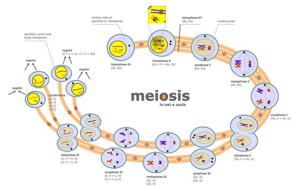
Meiosis is a special type of cell division of germ cells in sexually-reproducing organisms that produces the gametes, the sperm or egg cells. It involves two rounds of division that ultimately result in four cells, each with only one copy of each chromosome (haploid). Additionally, prior to the division, genetic material from the paternal and maternal copies of each chromosome is crossed over, creating new combinations of code on each chromosome. Later on, during fertilisation, the haploid cells produced by meiosis from a male and a female will fuse to create a zygote, a cell with two copies of each chromosome again.

<span class="mw-page-title-main">Chromosomal crossover</span> Cellular process

Chromosomal crossover, or crossing over, is the exchange of genetic material during sexual reproduction between two homologous chromosomes' non-sister chromatids that results in recombinant chromosomes. It is one of the final phases of genetic recombination, which occurs in the pachytene stage of prophase I of meiosis during a process called synapsis. Synapsis begins before the synaptonemal complex develops and is not completed until near the end of prophase I. Crossover usually occurs when matching regions on matching chromosomes break and then reconnect to the other chromosome.

<span class="mw-page-title-main">Prophase</span> First phase of cell division in both mitosis and meiosis

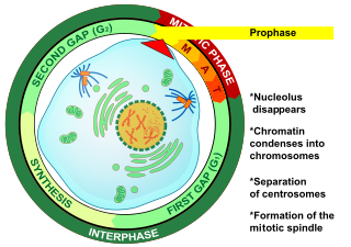
Prophase is the first stage of cell division in both mitosis and meiosis. Beginning after interphase, DNA has already been replicated when the cell enters prophase. The main occurrences in prophase are the condensation of the chromatin reticulum and the disappearance of the nucleolus.

<span class="mw-page-title-main">Genetic recombination</span> Production of offspring with combinations of traits that differ from those found in either parent

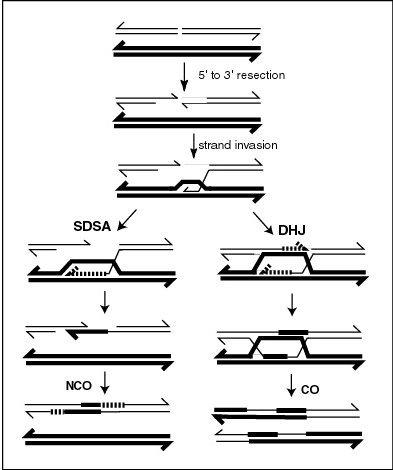
Genetic recombination is the exchange of genetic material between different organisms which leads to production of offspring with combinations of traits that differ from those found in either parent. In eukaryotes, genetic recombination during meiosis can lead to a novel set of genetic information that can be further passed on from parents to offspring. Most recombination occurs naturally and can be classified into two types: (1) interchromosomal recombination, occurring through independent assortment of alleles whose loci are on different but homologous chromosomes ; & (2) intrachromosomal recombination, occurring through crossing over.

<span class="mw-page-title-main">Homologous chromosome</span> Chromosomes that pair in fertilization

A pair of homologous chromosomes, or homologs, is a set of one maternal and one paternal chromosome that pair up with each other inside a cell during fertilization. Homologs have the same genes in the same loci, where they provide points along each chromosome that enable a pair of chromosomes to align correctly with each other before separating during meiosis. This is the basis for Mendelian inheritance, which characterizes inheritance patterns of genetic material from an organism to its offspring parent developmental cell at the given time and area.

<span class="mw-page-title-main">Nondisjunction</span> Failure to separate properly during cell division

Nondisjunction is the failure of homologous chromosomes or sister chromatids to separate properly during cell division (mitosis/meiosis). There are three forms of nondisjunction: failure of a pair of homologous chromosomes to separate in meiosis I, failure of sister chromatids to separate during meiosis II, and failure of sister chromatids to separate during mitosis. Nondisjunction results in daughter cells with abnormal chromosome numbers (aneuploidy).

<span class="mw-page-title-main">Heteroduplex</span>

A heteroduplex is a double-stranded (duplex) molecule of nucleic acid originated through the genetic recombination of single complementary strands derived from different sources, such as from different homologous chromosomes or even from different organisms.

<span class="mw-page-title-main">Synaptonemal complex</span> Protein structure

The synaptonemal complex (SC) is a protein structure that forms between homologous chromosomes during meiosis and is thought to mediate synapsis and recombination during prophase I during meiosis in eukaryotes. It is currently thought that the SC functions primarily as a scaffold to allow interacting chromatids to complete their crossover activities.

<span class="mw-page-title-main">Synapsis</span> Biological phenomenon in meiosis

Synapsis or Syzygy is the pairing of two chromosomes that occurs during meiosis. It allows matching-up of homologous pairs prior to their segregation, and possible chromosomal crossover between them. Synapsis takes place during prophase I of meiosis. When homologous chromosomes synapse, their ends are first attached to the nuclear envelope. These end-membrane complexes then migrate, assisted by the extranuclear cytoskeleton, until matching ends have been paired. Then the intervening regions of the chromosome are brought together, and may be connected by a protein-DNA complex called the synaptonemal complex. During synapsis, autosomes are held together by the synaptonemal complex along their whole length, whereas for sex chromosomes, this only takes place at one end of each chromosome.

Zygotene is the second stage of prophase I during meiosis, the specialized cell division that reduces the chromosome number by half to produce haploid gametes. It follows the Leptotene stage.

The pachytene stage, also known as pachynema, is the third stage of prophase I during meiosis, the specialized cell division that reduces chromosome number by half to produce haploid gametes. It follows the zygotene stage.

<span class="mw-page-title-main">Holliday junction</span> Branched nucleic acid structure

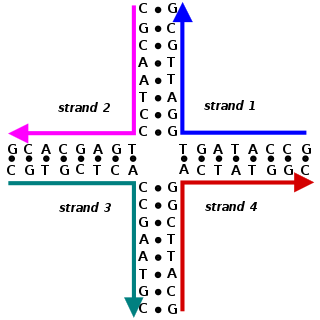
A Holliday junction is a branched nucleic acid structure that contains four double-stranded arms joined. These arms may adopt one of several conformations depending on buffer salt concentrations and the sequence of nucleobases closest to the junction. The structure is named after Robin Holliday, the molecular biologist who proposed its existence in 1964.

Mitotic recombination is a type of genetic recombination that may occur in somatic cells during their preparation for mitosis in both sexual and asexual organisms. In asexual organisms, the study of mitotic recombination is one way to understand genetic linkage because it is the only source of recombination within an individual. Additionally, mitotic recombination can result in the expression of recessive alleles in an otherwise heterozygous individual. This expression has important implications for the study of tumorigenesis and lethal recessive alleles. Mitotic homologous recombination occurs mainly between sister chromatids subsequent to replication. Inter-sister homologous recombination is ordinarily genetically silent. During mitosis the incidence of recombination between non-sister homologous chromatids is only about 1% of that between sister chromatids.

<span class="mw-page-title-main">Sister chromatid exchange</span>

Sister chromatid exchange (SCE) is the exchange of genetic material between two identical sister chromatids.

<span class="mw-page-title-main">MSH5</span> Protein-coding gene in the species Homo sapiens

MutS protein homolog 5 is a protein that in humans is encoded by the MSH5 gene.

<span class="mw-page-title-main">MSH4</span> Protein-coding gene in the species Homo sapiens

MutS protein homolog 4 is a protein that in humans is encoded by the MSH4 gene.

<span class="mw-page-title-main">MLH3</span> Protein-coding gene in the species Homo sapiens

DNA mismatch repair protein Mlh3 is a protein that in humans is encoded by the MLH3 gene.

<span class="mw-page-title-main">Meiotic recombination checkpoint</span>

The meiotic recombination checkpoint monitors meiotic recombination during meiosis, and blocks the entry into metaphase I if recombination is not efficiently processed.

The leptotene stage, also known as leptonema, is the first of five substages of prophase I during meiosis, the specialized cell division that reduces the chromosome number by half to produce haploid gametes in sexually reproducing organisms.

The origin and function of meiosis are currently not well understood scientifically, and would provide fundamental insight into the evolution of sexual reproduction in eukaryotes. There is no current consensus among biologists on the questions of how sex in eukaryotes arose in evolution, what basic function sexual reproduction serves, and why it is maintained, given the basic two-fold cost of sex. It is clear that it evolved over 1.2 billion years ago, and that almost all species which are descendants of the original sexually reproducing species are still sexual reproducers, including plants, fungi, and animals.

References

  1. Nielsen, H. J.; Youngren, B.; Hansen, F. G.; Austin, S. (2007-12-01). "Dynamics of Escherichia coli Chromosome Segregation during Multifork Replication". Journal of Bacteriology. 189 (23): 8660–8666. doi:10.1128/JB.01212-07. ISSN   0021-9193. PMC   2168957 . PMID   17905986.
  2. 1 2 Lu S, Zong C, Fan W, Yang M, Li J, Chapman AR, Zhu P, Hu X, Xu L, Yan L, Bai F, Qiao J, Tang F, Li R, Xie XS (2012). "Probing meiotic recombination and aneuploidy of single sperm cells by whole-genome sequencing". Science. 338 (6114): 1627–30. Bibcode:2012Sci...338.1627L. doi:10.1126/science.1229112. PMC   3590491 . PMID   23258895.
  3. Sansam CL, Pezza RJ (2015). "Connecting by breaking and repairing: mechanisms of DNA strand exchange in meiotic recombination". FEBS J. 282 (13): 2444–57. doi:10.1111/febs.13317. PMC   4573575 . PMID   25953379.
  4. Dernburg AF, McDonald K, Moulder G, Barstead R, Dresser M, Villeneuve AM (1998). "Meiotic recombination in C. elegans initiates by a conserved mechanism and is dispensable for homologous chromosome synapsis". Cell. 94 (3): 387–98. doi: 10.1016/s0092-8674(00)81481-6 . PMID   9708740. S2CID   10198891.
  5. Farah JA, Cromie G, Davis L, Steiner WW, Smith GR (2005). "Activation of an alternative, rec12 (spo11)-independent pathway of fission yeast meiotic recombination in the absence of a DNA flap endonuclease". Genetics. 171 (4): 1499–511. doi:10.1534/genetics.105.046821. PMC   1456079 . PMID   16118186.
  6. Pauklin S, Burkert JS, Martin J, Osman F, Weller S, Boulton SJ, Whitby MC, Petersen-Mahrt SK (2009). "Alternative induction of meiotic recombination from single-base lesions of DNA deaminases". Genetics. 182 (1): 41–54. doi:10.1534/genetics.109.101683. PMC   2674839 . PMID   19237686.
  7. Zakharyevich K, Tang S, Ma Y, Hunter N (2012). "Delineation of joint molecule resolution pathways in meiosis identifies a crossover-specific resolvase". Cell. 149 (2): 334–47. doi:10.1016/j.cell.2012.03.023. PMC   3377385 . PMID   22500800.
  8. 1 2 Ranjha L, Anand R, Cejka P (2014). "The Saccharomyces cerevisiae Mlh1-Mlh3 heterodimer is an endonuclease that preferentially binds to Holliday junctions". J. Biol. Chem. 289 (9): 5674–86. doi: 10.1074/jbc.M113.533810 . PMC   3937642 . PMID   24443562.
  9. Rogacheva MV, Manhart CM, Chen C, Guarne A, Surtees J, Alani E (2014). "Mlh1-Mlh3, a meiotic crossover and DNA mismatch repair factor, is a Msh2-Msh3-stimulated endonuclease". J. Biol. Chem. 289 (9): 5664–73. doi: 10.1074/jbc.M113.534644 . PMC   3937641 . PMID   24403070.
  10. 1 2 Sonntag Brown M, Lim E, Chen C, Nishant KT, Alani E (2013). "Genetic analysis of mlh3 mutations reveals interactions between crossover promoting factors during meiosis in baker's yeast". G3: Genes, Genomes, Genetics. 3 (1): 9–22. doi:10.1534/g3.112.004622. PMC   3538346 . PMID   23316435.
  11. 1 2 Pochart P, Woltering D, Hollingsworth NM (1997). "Conserved properties between functionally distinct MutS homologs in yeast". J. Biol. Chem. 272 (48): 30345–9. doi: 10.1074/jbc.272.48.30345 . PMID   9374523.
  12. Winand NJ, Panzer JA, Kolodner RD (1998). "Cloning and characterization of the human and Caenorhabditis elegans homologs of the Saccharomyces cerevisiae MSH5 gene". Genomics. 53 (1): 69–80. doi: 10.1006/geno.1998.5447 . PMID   9787078.
  13. Bocker T, Barusevicius A, Snowden T, Rasio D, Guerrette S, Robbins D, Schmidt C, Burczak J, Croce CM, Copeland T, Kovatich AJ, Fishel R (1999). "hMSH5: a human MutS homologue that forms a novel heterodimer with hMSH4 and is expressed during spermatogenesis". Cancer Res. 59 (4): 816–22. PMID   10029069.
  14. 1 2 Krishnaprasad GN, Anand MT, Lin G, Tekkedil MM, Steinmetz LM, Nishant KT (2015). "Variation in crossover frequencies perturb crossover assurance without affecting meiotic chromosome segregation in Saccharomyces cerevisiae". Genetics. 199 (2): 399–412. doi:10.1534/genetics.114.172320. PMC   4317650 . PMID   25467183.
  15. Davis L, Smith GR (2005). "Dynein promotes achiasmate segregation in Schizosaccharomyces pombe". Genetics. 170 (2): 581–90. doi:10.1534/genetics.104.040253. PMC   1450395 . PMID   15802518.