Arteriovenous malformation is an abnormal connection between arteries and veins, bypassing the capillary system. This vascular anomaly is widely known because of its occurrence in the central nervous system, but can appear in any location. Although many AVMs are asymptomatic, they can cause intense pain or bleeding or lead to other serious medical problems.
A fistula in anatomy is an abnormal connection between two hollow spaces, such as blood vessels, intestines, or other hollow organs. Types of fistula can be described by their location. Anal fistulas connect between the anal canal and the perianal skin. Anovaginal or rectovaginal fistulas occur when a hole develops between the anus or rectum and the vagina. Colovaginal fistulas occur between the colon and the vagina. Urinary tract fistulas are abnormal openings within the urinary tract or an abnormal connection between the urinary tract and another organ such as between the bladder and the uterus in a vesicouterine fistula, between the bladder and the vagina in a vesicovaginal fistula, and between the urethra and the vagina in urethrovaginal fistula. When occurring between two parts of the intestine, it is known as an enteroenteral fistula, between the small intestine and the skin as an enterocutaneous fistula, and between the colon and the skin as a colocutaneous fistula.

An anastomosis is a connection or opening between two things that are normally diverging or branching, such as between blood vessels, leaf veins, or streams. Such a connection may be normal or abnormal ; it may be acquired or innate ; and it may be natural or artificial. The reestablishment of an anastomosis that had become blocked is called a reanastomosis. Anastomoses that are abnormal, whether congenital or acquired, are often called fistulas.

Interventional radiology (IR) is a medical specialty that performs various minimally-invasive procedures using medical imaging guidance, such as x-ray fluoroscopy, computed tomography, magnetic resonance imaging, or ultrasound. IR performs both diagnostic and therapeutic procedures through very small incisions or body orifices. Diagnostic IR procedures are those intended to help make a diagnosis or guide further medical treatment, and include image-guided biopsy of a tumor or injection of an imaging contrast agent into a hollow structure, such as a blood vessel or a duct. By contrast, therapeutic IR procedures provide direct treatment—they include catheter-based medicine delivery, medical device placement, and angioplasty of narrowed structures.
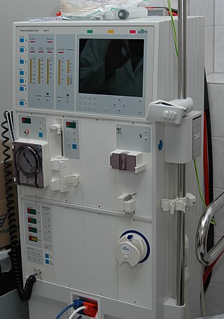
Hemodialysis, also spelled haemodialysis, or simply dialysis, is a process of purifying the blood of a person whose kidneys are not working normally. This type of dialysis achieves the extracorporeal removal of waste products such as creatinine and urea and free water from the blood when the kidneys are in a state of kidney failure. Hemodialysis is one of three renal replacement therapies. An alternative method for extracorporeal separation of blood components such as plasma or cells is apheresis.
A dialysis catheter is a catheter used for exchanging blood to and from a hemodialysis machine and a patient.

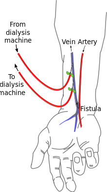
An arteriovenous fistula is an abnormal connection or passageway between an artery and a vein. It may be congenital, surgically created for hemodialysis treatments, or acquired due to pathologic process, such as trauma or erosion of an arterial aneurysm.

Hemofiltration, also haemofiltration, is a renal replacement therapy which is used in the intensive care setting. It is usually used to treat acute kidney injury (AKI), but may be of benefit in multiple organ dysfunction syndrome or sepsis. During hemofiltration, a patient's blood is passed through a set of tubing via a machine to a semipermeable membrane where waste products and water are removed by convection. Replacement fluid is added and the blood is returned to the patient.
A vascular bypass is a surgical procedure performed to redirect blood flow from one area to another by reconnecting blood vessels. Often, this is done to bypass around a diseased artery, from an area of normal blood flow to another relatively normal area. It is commonly performed due to inadequate blood flow (ischemia) caused by atherosclerosis, as a part of organ transplantation, or for vascular access in hemodialysis. In general, someone's own vein (autograft) is the preferred graft material for a vascular bypass, but other types of grafts such as polytetrafluoroethylene (Teflon), polyethylene terephthalate (Dacron), or a different person's vein (allograft) are also commonly used. Arteries can also serve as vascular grafts. A surgeon sews the graft to the source and target vessels by hand using surgical suture, creating a surgical anastomosis.
An embolus, is described as a free-floating mass, located inside blood vessels that can travel from one site in the blood stream to another. An embolus can be made up of solid, liquid, or gas. Once these masses get "stuck" in a different blood vessel, it is then known as an "embolism." An embolism can cause ischemia - or damage to an organ from lack of oxygen. A paradoxical embolism is a specific type of embolism in which the emboli travels from the right side of the heart or "venous circulation," travels to the left side of the heart or "arterial circulation," and lodges itself in a blood vessel known as an artery. Thus, it is termed "paradoxical" because the emboli lands in an artery, rather than a vein.

A dural arteriovenous fistula (DAVF) or malformation is an abnormal direct connection (fistula) between a meningeal artery and a meningeal vein or dural venous sinus.
In nephrology, vascular access steal syndrome is a syndrome caused by ischemia resulting from a vascular access device that was installed to provide access for the inflow and outflow of blood during hemodialysis.

A surgical anastomosis is a surgical technique used to make a new connection between two body structures that carry fluid, such as blood vessels or bowel. For example, an arterial anastomosis is used in vascular bypass and a colonic anastomosis is used to restore colonic continuity after the resection of colon cancer.
Vascular access refers to a rapid, direct method of introducing or removing devices or chemicals from the bloodstream. In hemodialysis, vascular access is used to remove the patient's blood so that it can be filtered through the dialyzer. Three primary methods are used to gain access to the blood: an intravenous catheter, an arteriovenous fistula (AV) or a synthetic graft. In the latter two, needles are used to puncture the graft or fistula each time dialysis is performed.

Computed tomography angiography is a computed tomography technique used for angiography—the visualization of arteries and veins—throughout the human body. Using contrast injected into the blood vessels, images are created to look for blockages, aneurysms, dissections, and stenosis. CTA can be used to visualize the vessels of the heart, the aorta and other large blood vessels, the lungs, the kidneys, the head and neck, and the arms and legs. CTA can also be used to localise arterial or venous bleed of the gastrointestinal system.
Nils Alwall was a Swedish professor at Lund University, Sweden. He was a pioneer in hemodialysis and the inventor of one of the first practical dialysis machines. Alwall pioneered the technique of ultrafiltration and introduced the principle of hemofiltration. Alwall is referred to as the "father of extracorporeal blood treatment."
Vascular myelopathy refers to an abnormality of the spinal cord in regard to its blood supply. The blood supply is complicated and supplied by two major vessel groups: the posterior spinal arteries and the anterior spinal arteries—of which the Artery of Adamkiewicz is the largest. Both the posterior and anterior spinal arteries run the entire length of the spinal cord and receive anastomotic (conjoined) vessels in many places. The anterior spinal artery has a less efficient supply of blood and is therefore more susceptible to vascular disease. Whilst atherosclerosis of spinal arteries is rare, necrosis in the anterior artery can be caused by disease in vessels originating from the segmental arteries such as atheroma or aortic dissection.
The Nicoladoni–Branham sign is named after Carl Nicoladoni, who first noticed the phenomenon of the pulse slowing in a patient with right arm phlebarteriectasia when the brachialis artery proximal to it was compressed. In modern medicine, the sign is elicited when pressure is applied to an artery proximal to an arteriovenous fistula and said to be positive if the following occurs:

Parkes Weber syndrome (PWS) is a congenital disorder of the vascular system. It is an extremely rare condition, and its exact prevalence is unknown. It is named after British dermatologist Frederick Parkes Weber, who first described the syndrome in 1907.

Vein of Galen aneurysmal malformations (VGAM) and Vein of Galen aneurysmal dilations (VGAD) are the most frequent arteriovenous malformations in infants and fetuses. VGAM consist of a tangled mass of dilated vessels supplied by an enlarged artery. The malformation increases greatly in size with age, although the mechanism of the increase is unknown. Dilation of the great cerebral vein of Galen is a secondary result of the force of arterial blood either directly from an artery via an arteriovenous fistula or by way of a tributary vein that receives the blood directly from an artery. There is usually a venous anomaly downstream from the draining vein that, together with the high blood flow into the great cerebral vein of Galen causes its dilation. The right sided cardiac chambers and pulmonary arteries also develop mild to severe dilation.