Related Research Articles
Articles related to anatomy include:
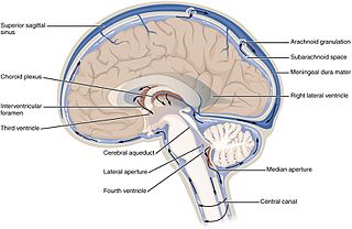
The ventricular system is a set of four interconnected cavities known as cerebral ventricles in the brain. Within each ventricle is a region of choroid plexus which produces the circulating cerebrospinal fluid (CSF). The ventricular system is continuous with the central canal of the spinal cord from the fourth ventricle, allowing for the flow of CSF to circulate.
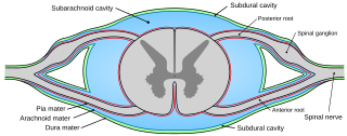
Pia mater, often referred to as simply the pia, is the delicate innermost layer of the meninges, the membranes surrounding the brain and spinal cord. Pia mater is medieval Latin meaning "tender mother". The other two meningeal membranes are the dura mater and the arachnoid mater. Both the pia and arachnoid mater are derivatives of the neural crest while the dura is derived from embryonic mesoderm. The pia mater is a thin fibrous tissue that is permeable to water and small solutes. The pia mater allows blood vessels to pass through and nourish the brain. The perivascular space between blood vessels and pia mater is proposed to be part of a pseudolymphatic system for the brain. When the pia mater becomes irritated and inflamed the result is meningitis.
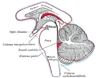
The subarachnoid cisterns are spaces formed by openings in the subarachnoid space, an anatomic space in the meninges of the brain. The space is situated between the two meninges, the arachnoid mater and the pia mater. These cisterns are filled with cerebrospinal fluid.

The cisterna magna is one of three principal openings in the subarachnoid space between the arachnoid and pia mater layers of the meninges surrounding the brain. The openings are collectively referred to as the subarachnoid cisterns. The cisterna magna is located between the cerebellum and the dorsal surface of the medulla oblongata. Cerebrospinal fluid produced in the fourth ventricle drains into the cisterna magna via the lateral apertures and median aperture.

The middle cerebral artery (MCA) is one of the three major paired arteries that supply blood to the cerebrum. The MCA arises from the internal carotid and continues into the lateral sulcus where it then branches and projects to many parts of the lateral cerebral cortex. It also supplies blood to the anterior temporal lobes and the insular cortices.

The posterior cerebral artery (PCA) is one of a pair of cerebral arteries that supply oxygenated blood to the occipital lobe, part of the back of the human brain. The two arteries originate from the distal end of the basilar artery, where it bifurcates into the left and right posterior cerebral arteries. These anastomose with the middle cerebral arteries and internal carotid arteries via the posterior communicating arteries.

The lateral aperture is a paired structure in human anatomy. It is an opening in each lateral extremity of the lateral recess of the fourth ventricle of the human brain, which also has a single median aperture. The two lateral apertures provide a conduit for cerebrospinal fluid to flow from the brain's ventricular system into the subarachnoid space; specifically into the pontocerebellar cistern at the cerebellopontine angle. The structure is also called the lateral aperture of the fourth ventricle or the foramen of Luschka after anatomist Hubert von Luschka.
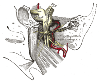
The trigeminal cave is a dura mater pouch containing cerebrospinal fluid.

The lateral corticospinal tract is the largest part of the corticospinal tract. It extends throughout the entire length of the spinal cord, and on transverse section appears as an oval area in front of the posterior column and medial to the posterior spinocerebellar tract.

The anterior corticospinal tract is a small bundle of descending fibers that connect the cerebral cortex to the spinal cord. Descending tracts are pathways by which motor signals are sent from upper motor neurons in the brain to lower motor neurons which then directly innervate muscle to produce movement. The anterior corticospinal tract is usually small, varying inversely in size with the lateral corticospinal tract, which is the main part of the corticospinal tract.
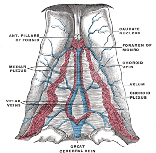
The basal vein is a vein in the brain. It is formed at the anterior perforated substance by the union of

Weber's syndrome, also known as midbrain stroke syndrome or superior alternating hemiplegia, is a form of stroke that affects the medial portion of the midbrain. It involves oculomotor fascicles in the interpeduncular cisterns and cerebral peduncle so it characterizes the presence of an ipsilateral lower motor neuron type oculomotor nerve palsy and contralateral hemiparesis or hemiplegia.

The interpeduncular cistern of Sweeney is the subarachnoid cistern that encloses the cerebral peduncles and the structures contained in the interpeduncular fossa and contains the arterial circle of Willis as well as the oculomotor nerve (CN3).
The chiasmatic cistern is formed as the interpeduncular cistern extends forward across the optic chiasm and onto the upper surface of the corpus callosum – the arachnoid stretches across from one cerebral hemisphere to the other immediately beneath the free border of the falx cerebri, and thus leaves a space in which the anterior cerebral arteries are contained. The "leaf" or extension of the chiasmatic cistern above the chiasma, which is separated from the optic recess of the third ventricle by the thin lamina terminalis, has been called the suprachiasmatic cistern. As spaces filled with freely circulating cerebrospinal fluid, cisterns receive little direct study, but are mentioned in pathological conditions. Cysts and tumors of the lamina terminalis extend into the suprachiasmatic cistern, as can pituitary tumors, or the cistern can be partially or completely effaced by injury and hematoma or by blockage of the cerebral aqueduct.
The superior cistern is a dilation as a subarachnoid cistern of the subarachnoid space around the brain. It lies between the splenium of the corpus callosum and the superior surface of the cerebellum. It extends between the layers of the tela choroidea of the third ventricle. It contains the great cerebral vein, posterior cerebral artery, quadrigeminal artery, glossopharyngeal nerve, and the pineal gland.
The lenticulostriate arteries, anterolateral central arteries, or antero-lateral ganglionic branches are a group of small arteries arising from the initial part M1 of the middle cerebral artery that supply the basal ganglia.

The middle cerebral veins are the superficial middle cerebral vein and the deep middle cerebral vein.
The cistern of lamina terminalis is one of the a subarachnoid cisterns in the subarachnoid space in the brain. It lies in front of the lamina terminalis and anterior commissure between the two frontal lobes of the cerebrum. The cistern contains cerebrospinal fluid, and connects the chiasmatic cistern to the pericallosal cistern. The anterior cerebral artery and the anterior communicating artery travel within this cistern.
References
![]() This article incorporates text in the public domain from page 877 of the 20th edition of Gray's Anatomy (1918)
This article incorporates text in the public domain from page 877 of the 20th edition of Gray's Anatomy (1918)