Coincidence detection in neurobiology

Last updated

Coincidence detection is a neuronal process in which a neural circuit encodes information by detecting the occurrence of temporally close but spatially distributed input signals. Coincidence detectors influence neuronal information processing by reducing temporal jitter and spontaneous activity, allowing the creation of variable associations between separate neural events in memory. [1] The study of coincidence detectors has been crucial in neuroscience with regards to understanding the formation of computational maps in the brain.

Contents

Principles of coincidence detection

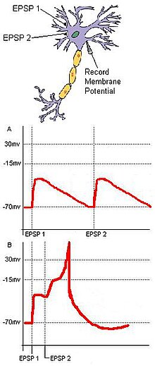
Fig. 1: Spatial and temporal summation. Two EPSPs innervated in rapid succession sum to produce a larger EPSP, or an action potential in the postsynaptic cell. Spatial summation.JPG
Fig. 1: Spatial and temporal summation. Two EPSPs innervated in rapid succession sum to produce a larger EPSP, or an action potential in the postsynaptic cell.

Coincidence detection relies on separate inputs converging on a common target. For example (Fig. 1), in a basic neural circuit with two input neurons—A and B—that have excitatory synaptic terminals converging on a single output neuron (C), if each input neuron's EPSP is sub-threshold for an action potential at C, then C cannot fire unless the two inputs from A and B are temporally close. The synchronous arrival of these two inputs may push the membrane potential of a target neuron over the threshold required to create an action potential. Conversely, if the two inputs temporally arrive too far apart, the depolarization of the first input may have time to drop significantly, preventing the membrane potential of the target neuron from reaching the action potential threshold. Hence, the function of coincidence detection is to reduce the jitter caused by spontaneous neuronal activity, and while random sub-threshold stimulations from cells may not often fire coincidentally, coincident synaptic inputs derived from a unitary external stimulus ensure that a target neuron will fire as a result of the stimulus.

Distal coincidence detection

The above description applies well to feedforward inputs to neurons, which provide inputs from either sensory nerves or lower-level regions in the brain. About 90% of interneural connections are, however, not feedforward but predictive (or modulatory, or attentional) in nature. These connections receive inputs mainly from nearby cells in the same layer as the receiving cell, and also from distant connections which are fed through Layer 1. The dendrites which receive these inputs are quite distant from the cell body, and therefore they exhibit different electrical and signal-processing behaviour compared with the proximal (or feedforward) dendrites described above.

In a short section (perhaps 40  μm long) of distal dendrite, the reaction to activations coming in on synapses to the dendritic spines acts to raise the overall local potential with each incoming signal. This rising potential acts against a background of decay in the potential back to the resting level. If sufficient signals are received within a short period of time (i.e. before the overall voltage decays to background), the voltage of the segment will rise above a threshold, giving rise to a non-linear dendritic spike, which travels, effectively undiminished, all the way to the cell body, and which causes it to become partially depolarised.

This is perhaps the most important form of dendritic coincidence detection in the brain. The more easily understood proximal activation acts over much longer time periods, and is thus much less sensitive to the time factor in coincidence detection.

Sound localization

Fig. 2: If a sound arrives at the left ear before the right ear, the impulse in the left auditory tract will reach X sooner than the impulse in the right auditory tract reaches Y. Neurons 4 or 5 may therefore receive coincident inputs. Jeffress Model2.png
Fig. 2: If a sound arrives at the left ear before the right ear, the impulse in the left auditory tract will reach X sooner than the impulse in the right auditory tract reaches Y. Neurons 4 or 5 may therefore receive coincident inputs.

Coincidence detection has been shown to be a major factor in sound localization along the azimuth plane in several organisms. In 1948, Lloyd A. Jeffress proposed that some organisms may have a collection of neurons that receive auditory input from each ear. [2] The neural pathways to these neurons are called delay lines. Jeffress claimed that the neurons that the delay lines link act as coincidence detectors by firing maximally when receiving simultaneous inputs from both ears. When a sound is heard, sound waves may reach the ears at different times. This is referred to as the interaural time difference (ITD). Due to differing lengths and a finite conduction speed within the axons of the delay lines, different coincidence detector neurons will fire when sound comes from different positions along the azimuth. Jeffress' model proposes that two signals even from an asynchronous arrival of sound in the cochlea of each ear will converge synchronously on a coincidence detector in the auditory cortex based on the magnitude of the ITD (Fig. 2). Therefore, the ITD should correspond to an anatomical map that can be found within the brain. Masakazu Konishi's study on barn owls shows that this is true. [3] Sensory information from the hair cells of the ears travels to the ipsilateral nucleus magnocellularis. From here, the signals project ipsilaterally and contralaterally to two nucleus laminari. Each nucleus laminaris contains coincidence detectors that receive auditory input from the left and the right ear. Since the ipsilateral axons enter the nucleus laminaris dorsally while the contralateral axons enter ventrally, sounds from various positions along the azimuth correspond directly to stimulation of different depths of the nucleus laminaris. From this information, a neural map of auditory space was formed. The function of the nucleus laminaris parallels that of the medial superior olive in mammals. [4]

Synaptic plasticity and associativity

In 1949, Donald Hebb postulated that synaptic efficiency will increase through repeated and persistent stimulation of a postsynaptic cell by a presynaptic cell. This is often informally summarized as "cells that fire together, wire together". The theory was validated in part by the discovery of long-term potentiation. Studies of LTP on multiple presynaptic cells stimulating a postsynaptic cell uncovered the property of associativity. A weak neuronal stimulation onto a pyramidal neuron may not induce long-term potentiation. However, this same stimulation paired with a simultaneous strong stimulation from another neuron will strengthen both synapses. [5] This process suggests that two neuronal pathways converging on the same cell may both strengthen if stimulated coincidentally.

Molecular mechanism of long-term potentiation

LTP in the hippocampus requires a prolonged depolarization that can expel the Mg2+ block of postsynaptic NMDA receptors. The removal of the Mg2+ block allows the flow of Ca2+ into the cell. A large elevation of calcium levels activate protein kinases that ultimately increase the number of postsynaptic AMPA receptors. This increases the sensitivity of the postsynaptic cell to glutamate. As a result, both synapses strengthen. The prolonged depolarization needed for the expulsion of Mg2+ from NMDA receptors requires a high frequency stimulation. [6] Associativity becomes a factor because this can be achieved through two simultaneous inputs that may not be strong enough to activate LTP by themselves.

Besides the NMDA-receptor based processes, further cellular mechanisms allow of the association between two different input signals converging on the same neuron, in a defined timeframe. Upon a simultaneous increase in the intracellular concentrations of cAMP and Ca2+, a transcriptional coactivator called TORC1 (CRTC1) becomes activated, that converts the temporal coincidence of the two second messengers into long term changes such as LTP. [7] This cellular mechanism, through calcium-dependent adenylate cyclase activation, might also account for the detection of the repetitive stimulation of a given synapse.

Adenylyl cyclase (also commonly known as adenyl cyclase and adenylate cyclase) has been implicated in memory formation as a coincidence detector. [8] [9] [10] [11]

Molecular mechanism of long-term depression

Long-term depression also works through associative properties although it is not always the reverse process of LTP. LTD in the cerebellum requires a coincident stimulation of parallel fibers and climbing fibers. Glutamate released from the parallel fibers activates AMPA receptors which depolarize the postsynaptic cell. The parallel fibers also activate metabotropic glutamate receptors that release the second messengers IP3 and DAG. The climbing fibers stimulate a large increase in postsynaptic Ca2+ levels when activated. The Ca2+, IP3, and DAG work together in a signal transduction pathway to internalize AMPA receptors and decrease the sensitivity of the postsynaptic cell to glutamate. [6]

See also

Related Research Articles

<span class="mw-page-title-main">Long-term potentiation</span> Persistent strengthening of synapses based on recent patterns of activity

In neuroscience, long-term potentiation (LTP) is a persistent strengthening of synapses based on recent patterns of activity. These are patterns of synaptic activity that produce a long-lasting increase in signal transmission between two neurons. The opposite of LTP is long-term depression, which produces a long-lasting decrease in synaptic strength.

In neuroscience, synaptic plasticity is the ability of synapses to strengthen or weaken over time, in response to increases or decreases in their activity. Since memories are postulated to be represented by vastly interconnected neural circuits in the brain, synaptic plasticity is one of the important neurochemical foundations of learning and memory.

An inhibitory postsynaptic potential (IPSP) is a kind of synaptic potential that makes a postsynaptic neuron less likely to generate an action potential. The opposite of an inhibitory postsynaptic potential is an excitatory postsynaptic potential (EPSP), which is a synaptic potential that makes a postsynaptic neuron more likely to generate an action potential. IPSPs can take place at all chemical synapses, which use the secretion of neurotransmitters to create cell-to-cell signalling. EPSPs and IPSPs compete with each other at numerous synapses of a neuron. This determines whether an action potential occurring at the presynaptic terminal produces an action potential at the postsynaptic membrane. Some common neurotransmitters involved in IPSPs are GABA and glycine.

<span class="mw-page-title-main">Excitatory postsynaptic potential</span> Process causing temporary increase in postsynaptic potential

In neuroscience, an excitatory postsynaptic potential (EPSP) is a postsynaptic potential that makes the postsynaptic neuron more likely to fire an action potential. This temporary depolarization of postsynaptic membrane potential, caused by the flow of positively charged ions into the postsynaptic cell, is a result of opening ligand-gated ion channels. These are the opposite of inhibitory postsynaptic potentials (IPSPs), which usually result from the flow of negative ions into the cell or positive ions out of the cell. EPSPs can also result from a decrease in outgoing positive charges, while IPSPs are sometimes caused by an increase in positive charge outflow. The flow of ions that causes an EPSP is an excitatory postsynaptic current (EPSC).

In neurophysiology, long-term depression (LTD) is an activity-dependent reduction in the efficacy of neuronal synapses lasting hours or longer following a long patterned stimulus. LTD occurs in many areas of the CNS with varying mechanisms depending upon brain region and developmental progress.

In neuroscience, a silent synapse is an excitatory glutamatergic synapse whose postsynaptic membrane contains NMDA-type glutamate receptors but no AMPA-type glutamate receptors. These synapses are named "silent" because normal AMPA receptor-mediated signaling is not present, rendering the synapse inactive under typical conditions. Silent synapses are typically considered to be immature glutamatergic synapses. As the brain matures, the relative number of silent synapses decreases. However, recent research on hippocampal silent synapses shows that while they may indeed be a developmental landmark in the formation of a synapse, that synapses can be "silenced" by activity, even once they have acquired AMPA receptors. Thus, silence may be a state that synapses can visit many times during their lifetimes.

Spike-timing-dependent plasticity (STDP) is a biological process that adjusts the strength of connections between neurons in the brain. The process adjusts the connection strengths based on the relative timing of a particular neuron's output and input action potentials. The STDP process partially explains the activity-dependent development of nervous systems, especially with regard to long-term potentiation and long-term depression.

Synaptogenesis is the formation of synapses between neurons in the nervous system. Although it occurs throughout a healthy person's lifespan, an explosion of synapse formation occurs during early brain development, known as exuberant synaptogenesis. Synaptogenesis is particularly important during an individual's critical period, during which there is a certain degree of synaptic pruning due to competition for neural growth factors by neurons and synapses. Processes that are not used, or inhibited during their critical period will fail to develop normally later on in life.

Schaffer collaterals are axon collaterals given off by CA3 pyramidal cells in the hippocampus. These collaterals project to area CA1 of the hippocampus and are an integral part of memory formation and the emotional network of the Papez circuit, and of the hippocampal trisynaptic loop. It is one of the most studied synapses in the world and named after the Hungarian anatomist-neurologist Károly Schaffer.

<span class="mw-page-title-main">Glutamate receptor</span> Cell-surface proteins that bind glutamate and trigger changes which influence the behavior of cells

Glutamate receptors are synaptic and non synaptic receptors located primarily on the membranes of neuronal and glial cells. Glutamate is abundant in the human body, but particularly in the nervous system and especially prominent in the human brain where it is the body's most prominent neurotransmitter, the brain's main excitatory neurotransmitter, and also the precursor for GABA, the brain's main inhibitory neurotransmitter. Glutamate receptors are responsible for the glutamate-mediated postsynaptic excitation of neural cells, and are important for neural communication, memory formation, learning, and regulation.

Metaplasticity is a term originally coined by W.C. Abraham and M.F. Bear to refer to the plasticity of synaptic plasticity. Until that time synaptic plasticity had referred to the plastic nature of individual synapses. However this new form referred to the plasticity of the plasticity itself, thus the term meta-plasticity. The idea is that the synapse's previous history of activity determines its current plasticity. This may play a role in some of the underlying mechanisms thought to be important in memory and learning such as long-term potentiation (LTP), long-term depression (LTD) and so forth. These mechanisms depend on current synaptic "state", as set by ongoing extrinsic influences such as the level of synaptic inhibition, the activity of modulatory afferents such as catecholamines, and the pool of hormones affecting the synapses under study. Recently, it has become clear that the prior history of synaptic activity is an additional variable that influences the synaptic state, and thereby the degree, of LTP or LTD produced by a given experimental protocol. In a sense, then, synaptic plasticity is governed by an activity-dependent plasticity of the synaptic state; such plasticity of synaptic plasticity has been termed metaplasticity. There is little known about metaplasticity, and there is much research currently underway on the subject, despite its difficulty of study, because of its theoretical importance in brain and cognitive science. Most research of this type is done via cultured hippocampus cells or hippocampal slices.

<span class="mw-page-title-main">Synapse</span> Structure connecting neurons in the nervous system

In the nervous system, a synapse is a structure that permits a neuron to pass an electrical or chemical signal to another neuron or to the target effector cell.
Ca<sup>2+</sup>/calmodulin-dependent protein kinase II Class of enzymes

Ca2+
/calmodulin-dependent protein kinase II
is a serine/threonine-specific protein kinase that is regulated by the Ca2+
/calmodulin complex. CaMKII is involved in many signaling cascades and is thought to be an important mediator of learning and memory. CaMKII is also necessary for Ca2+
homeostasis and reuptake in cardiomyocytes, chloride transport in epithelia, positive T-cell selection, and CD8 T-cell activation.

<span class="mw-page-title-main">Calyx of Held</span>

The Calyx of Held is a particularly large synapse in the mammalian auditory central nervous system, so named after Hans Held who first described it in his 1893 article Die centrale Gehörleitung because of its resemblance to the calyx of a flower. Globular bushy cells in the anteroventral cochlear nucleus (AVCN) send axons to the contralateral medial nucleus of the trapezoid body (MNTB), where they synapse via these calyces on MNTB principal cells. These principal cells then project to the ipsilateral lateral superior olive (LSO), where they inhibit postsynaptic neurons and provide a basis for interaural level detection (ILD), required for high frequency sound localization. This synapse has been described as the largest in the brain.

Gliotransmitters are chemicals released from glial cells that facilitate neuronal communication between neurons and other glial cells. They are usually induced from Ca2+ signaling, although recent research has questioned the role of Ca2+ in gliotransmitters and may require a revision of the relevance of gliotransmitters in neuronal signalling in general.

Neural backpropagation is the phenomenon in which, after the action potential of a neuron creates a voltage spike down the axon, another impulse is generated from the soma and propagates towards the apical portions of the dendritic arbor or dendrites. In addition to active backpropagation of the action potential, there is also passive electrotonic spread. While there is ample evidence to prove the existence of backpropagating action potentials, the function of such action potentials and the extent to which they invade the most distal dendrites remain highly controversial.

<span class="mw-page-title-main">Nonsynaptic plasticity</span> Form of neuroplasticity

Nonsynaptic plasticity is a form of neuroplasticity that involves modification of ion channel function in the axon, dendrites, and cell body that results in specific changes in the integration of excitatory postsynaptic potentials and inhibitory postsynaptic potentials. Nonsynaptic plasticity is a modification of the intrinsic excitability of the neuron. It interacts with synaptic plasticity, but it is considered a separate entity from synaptic plasticity. Intrinsic modification of the electrical properties of neurons plays a role in many aspects of plasticity from homeostatic plasticity to learning and memory itself. Nonsynaptic plasticity affects synaptic integration, subthreshold propagation, spike generation, and other fundamental mechanisms of neurons at the cellular level. These individual neuronal alterations can result in changes in higher brain function, especially learning and memory. However, as an emerging field in neuroscience, much of the knowledge about nonsynaptic plasticity is uncertain and still requires further investigation to better define its role in brain function and behavior.

Many experiments have been done to find out how the brain interprets stimuli and how animals develop fear responses. The emotion, fear, has been hard-wired into almost every individual, due to its vital role in the survival of the individual. Researchers have found that fear is established unconsciously and that the amygdala is involved with fear conditioning.

<span class="mw-page-title-main">Homosynaptic plasticity</span> Type of synaptic plasticity.

Homosynaptic plasticity is one type of synaptic plasticity. Homosynaptic plasticity is input-specific, meaning changes in synapse strength occur only at post-synaptic targets specifically stimulated by a pre-synaptic target. Therefore, the spread of the signal from the pre-synaptic cell is localized.

<span class="mw-page-title-main">Heterosynaptic plasticity</span>

Synaptic plasticity refers to a chemical synapse's ability to undergo changes in strength. Synaptic plasticity is typically input-specific, meaning that the activity in a particular neuron alters the efficacy of a synaptic connection between that neuron and its target. However, in the case of heterosynaptic plasticity, the activity of a particular neuron leads to input unspecific changes in the strength of synaptic connections from other unactivated neurons. A number of distinct forms of heterosynaptic plasticity have been found in a variety of brain regions and organisms. These different forms of heterosynaptic plasticity contribute to a variety of neural processes including associative learning, the development of neural circuits, and homeostasis of synaptic input.
References

  1. Marsálek, P.; Koch, C.; Maunsell, J. (1997). "On the relationship between synaptic input and spike output jitter in individual neurons". Proceedings of the National Academy of Sciences of the United States of America. 94 (2): 735–740. Bibcode:1997PNAS...94..735M. doi: 10.1073/pnas.94.2.735 . PMC   19583 . PMID   9012854.
  2. Jeffress, L. A. (1948). "A place theory of sound localization". Journal of Comparative and Physiological Psychology. 41 (1): 35–39. doi:10.1037/h0061495. PMID   18904764.
  3. Carr, C. E.; Konishi, M. (1988). "Axonal delay lines for time measurement in the owl's brainstem". Proceedings of the National Academy of Sciences of the United States of America. 85 (21): 8311–8315. Bibcode:1988PNAS...85.8311C. doi: 10.1073/pnas.85.21.8311 . PMC   282419 . PMID   3186725.
  4. Zupanc, G.K.H. 2004. Behavioral Neurobiology: An Integrative Approach. Oxford University Press: Oxford, UK. pp. 133-150
  5. Frey, Uwe; Morris, Richard G. M. (February 1997). "Synaptic tagging and long-term potentiation". Nature. 385 (6616): 533–536. Bibcode:1997Natur.385..533F. doi:10.1038/385533a0. ISSN   0028-0836. PMID   9020359. S2CID   4339789.
  6. 1 2 Purves, Dale (2004). Neuroscience (3 ed.). Sunderland, MA: Sinauer Associates. pp. 575–608. ISBN   9780878937257.
  7. Kovacs, K. A.; Steullet, P.; Steinmann, M.; Do, K. Q.; Magistretti, P. J.; Halfon, O.; Cardinaux, J. -R. (2007). "TORC1 is a calcium- and cAMP-sensitive coincidence detector involved in hippocampal long-term synaptic plasticity". Proceedings of the National Academy of Sciences. 104 (11): 4700–4705. Bibcode:2007PNAS..104.4700K. doi: 10.1073/pnas.0607524104 . PMC   1838663 . PMID   17360587.
  8. Willoughby, Debbie; Cooper, Dermot M. F. (July 2007). "Organization and Ca2+ regulation of adenylyl cyclases in cAMP microdomains". Physiological Reviews. 87 (3): 965–1010. CiteSeerX   10.1.1.336.3746 . doi:10.1152/physrev.00049.2006. ISSN   0031-9333. PMID   17615394.
  9. Mons, N.; Guillou, J.-L.; Jaffard, R. (1999-04-01). "The role of Ca2+/calmodulin-stimulable adenylyl cyclases as molecular coincidence detectors in memory formation". Cellular and Molecular Life Sciences. 55 (4): 525–533. doi:10.1007/s000180050311. ISSN   1420-682X. PMID   10357223. S2CID   10849274.
  10. Hanoune, J.; Defer, N. (2001). "Regulation and role of adenylyl cyclase isoforms". Annual Review of Pharmacology and Toxicology. 41: 145–174. doi:10.1146/annurev.pharmtox.41.1.145. ISSN   0362-1642. PMID   11264454.
  11. Neve, Kim A.; Seamans, Jeremy K.; Trantham-Davidson, Heather (August 2004). "Dopamine receptor signaling". Journal of Receptor and Signal Transduction Research. 24 (3): 165–205. CiteSeerX   10.1.1.465.5011 . doi:10.1081/RRS-200029981. ISSN   1079-9893. PMID   15521361. S2CID   12407397.

Further reading