Continuous noninvasive arterial pressure

Last updated

Continuous noninvasive arterial pressure (CNAP) is the method of measuring beat-to-beat arterial blood pressure in real-time without any interruptions (continuously) and without cannulating the human body (noninvasive).

Contents

Benefit of CNAP technology

Continuous noninvasive arterial blood pressure measurement (CNAP) combines the advantages of the following two clinical “gold standards”: it measures blood pressure (BP) continuously in real-time like the invasive arterial catheter system (IBP) and it is non-invasive like the standard upper arm sphygmomanometer (NBP). Latest developments in this field show promising results in terms of accuracy, ease of use and clinical acceptance.[ citation needed ]

Clinical requirements

For the use in clinical environment a CNAP system must provide the following blood pressure information:

  1. Absolute blood pressure obtained from a proximal artery (e.g. arteria brachialis)
  2. Blood pressure changes in order to detect hemodynamic instabilities
  3. Physiological rhythms that deliver insight into the hemodynamic control function and/or fluid management
  4. Blood pressure pulse waves for quality control – further pulse wave analysis provides additional cardiovascular parameters such as stroke volume, cardiac output and arterial stiffness.
Different blood pressure information according to time resolution Blood Pressure Information.jpg
Different blood pressure information according to time resolution

A high demand for easily applicable and accurate CNAP-systems is proven. This is why researchers, practitioners and the medical device industry focus on such devices. Like in other fields of innovation, the use of small but powerful microcomputers and digital signal processors facilitates the development of efficient blood pressure measurement instruments. These processors enable complex and computationally intensive mathematical functions in small inexpensive devices, which are necessary for this purpose.[ citation needed ]

Medical need and outcome

Recent literature,[ when? ] [1] a nationally representative survey among 200 German and Austrian physicians [2] and additional expert interviews provide strong evidence that in only 15% to 18% of inpatient surgeries blood pressure is measured continuously with invasive catheters (IBP). In all other inpatient and outpatient surgeries intermittent, noninvasive blood pressure (NBP) monitoring is the standard of care. Due to the discontinuous character of NBP, dangerous hypotensive episodes might be missed: In women undergoing Caesarean section, CNAP detected hypotensive phases in 39% of the cases, whereas only 9% were detected by the standard NBP. [3] Dangerous fetal acidosis did not occur when systolic blood pressure measured with CNAP was above 100mmHg. [3] Another study showed more than 22% of missed hypotensive episodes leading to delayed or no treatment. [4]

Hemodynamic optimization

A further advantage of CNAP is hemodynamic optimization using continuous blood pressure and its parameters derived from physiological rhythms and pulse wave analysis. The concept has quickly found wide acceptance in anesthesia and critical care: The evaluation of Pulse Pressure Variation (PPV) allows for goal-directed fluid management in sedated and ventilated patients. [5] [6]

Respiratory changes of blood pressure - the change of pulse pressure maximum (PPmax) to minimum (PPmin) is called PPV (in %). Pulse pressure variation.jpg
Respiratory changes of blood pressure – the change of pulse pressure maximum (PPmax) to minimum (PPmin) is called PPV (in %).

Further, the mathematical analysis of CNAP pulse waves enables the noninvasive estimation of stroke volume and cardiac output. [7] A meta-analysis of 29 clinical trials evidences that goal-directed therapy using these hemodynamic parameters leads to lower rates of morbidity and mortality in moderate and high-risk surgical procedures. [8]

Current noninvasive blood pressure technologies

Detecting pressure changes inside an artery from the outside is difficult, whereas volume and flow changes of the artery can well be determined by using e.g. light, echography, impedance, etc. But unfortunately these volume changes are not linearly correlated with the arterial pressure– especially when measured in the periphery, where the access to the arteries is easy. Thus, noninvasive devices have to find a way to transform the peripheral volume signal to arterial pressure.

Vascular unloading technique

Pulse oximeters can measure finger blood volume changes using light. These volume changes must be transformed into pressure, because of the non-linearity of the elastic components of the arterial wall as well as the non-elastic parts of the smooth muscles of the finger artery.[ citation needed ]

The method is to unload the arterial wall in order to linearize this phenomenon with a counter pressure as high as the pressure inside the artery. Blood volume is kept constant by applying this corresponding pressure from the outside. The continuously changing outside pressure that is needed to keep the arterial blood volume constant directly corresponds to the arterial pressure. This is the basic principle of the so-called “Vascular Unloading Technique”.[ citation needed ]

For the realization, a cuff is placed over the finger. Inside the cuff, the blood volume in the finger arteries is measured using a light source and a light detector. The resulting light signal is kept constant by controlling the alterable cuff pressure. During systole, when blood volume increases in the finger, the control system increases cuff pressure, too, until the excess blood volume is squeezed out. On the other hand, during diastole, the blood volume in the finger is decreased; as a result, cuff pressure is lowered and again the overall blood volume remains constant. As blood volume and, thus, the light signal is held constant over time, intra-arterial pressure is equal to the cuff pressure. This pressure can easily be measured with a manometer.[ citation needed ]

Principle of the Vascular Unloading Technique Vascaular unloading.png
Principle of the Vascular Unloading Technique

As the volume of the finger artery is clamped on a constant diameter, the method is also known as “Volume Clamped Method”.

The Czech physiologist Jan Peňáz introduced this type of measurement of continuous noninvasive arterial blood pressure in 1973 by means of an electro-pneumatic control loop. [9] Two research groups have improved this method:

Getinge incorporates the vascular unloading technique in the NICCI Technology. Utilizing a dual finger cuff, which automatically alternates between fingers, the NICCI sensor performs a continuous measurement of blood pressure and analyzes the pressure curve to derive blood flow, preload, afterload and contractility parameters. The three different sensor cuff sizes allow noninvasive hemodynamic monitoring even in pediatrics.[ citation needed ]

Tonometry

The non-linear effect of the vascular wall decreases in bigger arteries. It is well known that good access to a “big” artery is at the wrist by palpating. Different mechanisms have been developed for the automatic noninvasive palpation on the arteria radialis. [15] In order to obtain a stable blood pressure signal, the tonometric sensor must be protected against movement and other mechanical artifacts.

Pulse transit time

When the heart ejects stroke volume to the arteries, it takes a certain transit time until the blood pressure wave arrives in the periphery. This pulse transit time (PTT) indirectly depends on blood pressure – the higher the pressure, the faster PTT. This circumstance can be used for the noninvasive detection of blood pressure changes. [16] [17] For absolute values, this method needs calibration.

Pulse Decomposition Analysis

Pulse Decomposition Analysis (PDA), which is a pulse contour analysis approach, [18] is based on the concept that five individual component pulses constitute the peripheral arterial pressure pulse of the upper body. These component pulses are due to the left ventricular ejection and the reflections and re-reflections of the first component pulse from two central arteries reflection sites. [19] [20] PDA is the operational principle of the Caretaker physiological monitor, which has demonstrated compliance with the ANSI/AAMI/ISO 81060-2:2013 standard and received FDA clearances (K151499, K163255) for the non-invasive and continuous monitoring of blood pressure, heart rate and respiration rate.

Calibration and correction to proximal arteries

All methods measure peripheral arterial pressure, which is inherently different from the blood pressure detected from proximal arteries. Even the comparison between the two clinical “gold standards” invasive continuous blood pressure at the arteria radialis and noninvasive, but intermittent, upper arm cuff shows large differences. [21]

Blood pressure changes along the arterial pathway Arterial Pathway.jpg
Blood pressure changes along the arterial pathway

Physicians are trained to derive treatment decisions from proximal arteries – e.g. noninvasively from the arteria brachialis. Calibration to the noninvasive “gold standard” NBP is performed in most of the devices that are currently marketed, although the calibration methods differ:

Changes in arterial tone

The pitfall of all noninvasive technologies is change in vascular tone. Small arteries beginning from arteria radialis downwards to the periphery have smooth muscles in order to open (vasodilation) and close (vasoconstriction). This human mechanism is activated by sympathetic tone and is further influenced by vasoactive drugs. Especially in critical care, vasoactive drugs are needed to control and maintain sedation and blood pressure. Mathematically advanced correction methods have to be developed for these noninvasive technologies in order to fulfill accuracy and clinical acceptance:

VERIFI

The VERIFI-algorithm corrects vasomotor tone by means of a fast pulse wave analysis. It establishes correct mean arterial blood pressure in the finger cuff by checking typical characteristics of the pulse wave. VERIFI-correction is performed after every heart beat, as vasomotor changes can occur immediately. This allows for a true continuous CNAP-signal without interruption during hemodynamic instable situations. VERIFI is implemented in the Task Force Monitor, CNAP Monitor 500, CNAP Smart Pod and in the LiDCOrapid. [10]

PhysioCal

PhysioCal is used in Finapres and its successor devices. The so-called PhysioCal algorithm eliminates the changes in tone of smooth muscles in the arterial wall, the hematocrit and other finger volume changes during measurement periods of constant pressure. Physiocal is achieved by opening the Vascular Unloading feedback loop. A new pressure ramp search is then performed before the measurement starts again. This algorithm needs to interrupt the blood pressure tracings for recalibration purposes, which results in short data loss during that time. [24]

Repeated calibration

For other methods like PTT, a closed-mashed re-calibration to NBP can overcome vasomotor changes.

Accuracy

The overall accuracy of CNAP devices has been demonstrated in comparison with the current gold standard invasive blood pressure (IBP) monitoring in numerous studies during the last few years.[ when? ] As examples, investigators came to the following conclusions:

See also

Related Research Articles

<span class="mw-page-title-main">Blood pressure</span> Pressure exerted by circulating blood upon the walls of arteries

Blood pressure (BP) is the pressure of circulating blood against the walls of blood vessels. Most of this pressure results from the heart pumping blood through the circulatory system. When used without qualification, the term "blood pressure" refers to the pressure in the large arteries. Blood pressure is usually expressed in terms of the systolic pressure over diastolic pressure in the cardiac cycle. It is measured in millimeters of mercury (mmHg) above the surrounding atmospheric pressure.

<span class="mw-page-title-main">Cardiac output</span> Measurement of blood pumped by the heart

In cardiac physiology, cardiac output (CO), also known as heart output and often denoted by the symbols , , or , is the volumetric flow rate of the heart's pumping output: that is, the volume of blood being pumped by a single ventricle of the heart, per unit time. Cardiac output (CO) is the product of the heart rate (HR), i.e. the number of heartbeats per minute (bpm), and the stroke volume (SV), which is the volume of blood pumped from the left ventricle per beat; thus giving the formula:

Hemodynamics or haemodynamics are the dynamics of blood flow. The circulatory system is controlled by homeostatic mechanisms of autoregulation, just as hydraulic circuits are controlled by control systems. The hemodynamic response continuously monitors and adjusts to conditions in the body and its environment. Hemodynamics explains the physical laws that govern the flow of blood in the blood vessels.

<span class="mw-page-title-main">Sphygmomanometer</span> Instrument for measuring blood pressure

A sphygmomanometer, a.k.a. a blood pressure monitor, or blood pressure gauge, is a device used to measure blood pressure, composed of an inflatable cuff to collapse and then release the artery under the cuff in a controlled manner, and a mercury or aneroid manometer to measure the pressure. Manual sphygmomanometers are used with a stethoscope when using the auscultatory technique.
<span class="mw-page-title-main">Photoplethysmogram</span> Chart of tissue blood volume changes

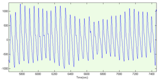
A photoplethysmogram (PPG) is an optically obtained plethysmogram that can be used to detect blood volume changes in the microvascular bed of tissue. A PPG is often obtained by using a pulse oximeter which illuminates the skin and measures changes in light absorption. A conventional pulse oximeter monitors the perfusion of blood to the dermis and subcutaneous tissue of the skin.

<span class="mw-page-title-main">Pulse oximetry</span> Measurement of blood oxygen saturation

Pulse oximetry is a noninvasive method for monitoring a person's blood oxygen saturation. Peripheral oxygen saturation (SpO2) readings are typically within 2% accuracy of the more accurate reading of arterial oxygen saturation (SaO2) from arterial blood gas analysis. But the two are correlated well enough that the safe, convenient, noninvasive, inexpensive pulse oximetry method is valuable for measuring oxygen saturation in clinical use.

<span class="mw-page-title-main">Mean arterial pressure</span> Average blood pressure in an individual during a single cardiac cycle

In medicine, the mean arterial pressure (MAP) is an average blood pressure in an individual during a single cardiac cycle. MAP is altered by cardiac output and systemic vascular resistance.

Compliance is the ability of a hollow organ (vessel) to distend and increase volume with increasing transmural pressure or the tendency of a hollow organ to resist recoil toward its original dimensions on application of a distending or compressing force. It is the reciprocal of "elastance", hence elastance is a measure of the tendency of a hollow organ to recoil toward its original dimensions upon removal of a distending or compressing force.

<span class="mw-page-title-main">Cardiac catheterization</span> Insertion of a catheter into a chamber or vessel of the heart

Cardiac catheterization is the insertion of a catheter into a chamber or vessel of the heart. This is done both for diagnostic and interventional purposes.

<span class="mw-page-title-main">Ankle–brachial pressure index</span> The ratio of the blood pressure at the ankle to the blood pressure in the upper arm

The ankle-brachial pressure index (ABPI) or ankle-brachial index (ABI) is the ratio of the blood pressure at the ankle to the blood pressure in the upper arm (brachium). Compared to the arm, lower blood pressure in the leg suggests blocked arteries due to peripheral artery disease (PAD). The ABPI is calculated by dividing the systolic blood pressure at the ankle by the systolic blood pressure in the arm.

<span class="mw-page-title-main">Transcranial Doppler</span>

Transcranial Doppler (TCD) and transcranial color Doppler (TCCD) are types of Doppler ultrasonography that measure the velocity of blood flow through the brain's blood vessels by measuring the echoes of ultrasound waves moving transcranially. These modes of medical imaging conduct a spectral analysis of the acoustic signals they receive and can therefore be classified as methods of active acoustocerebrography. They are used as tests to help diagnose emboli, stenosis, vasospasm from a subarachnoid hemorrhage, and other problems. These relatively quick and inexpensive tests are growing in popularity. The tests are effective for detecting sickle cell disease, ischemic cerebrovascular disease, subarachnoid hemorrhage, arteriovenous malformations, and cerebral circulatory arrest. The tests are possibly useful for perioperative monitoring and meningeal infection. The equipment used for these tests is becoming increasingly portable, making it possible for a clinician to travel to a hospital, to a doctor's office, or to a nursing home for both inpatient and outpatient studies. The tests are often used in conjunction with other tests such as MRI, MRA, carotid duplex ultrasound and CT scans. The tests are also used for research in cognitive neuroscience.

Impedance cardiography (ICG) is a non-invasive technology measuring total electrical conductivity of the thorax and its changes in time to process continuously a number of cardiodynamic parameters, such as stroke volume (SV), heart rate (HR), cardiac output (CO), ventricular ejection time (VET), pre-ejection period and used to detect the impedance changes caused by a high-frequency, low magnitude current flowing through the thorax between additional two pairs of electrodes located outside of the measured segment. The sensing electrodes also detect the ECG signal, which is used as a timing clock of the system.

Arterial stiffness occurs as a consequence of biological aging and arteriosclerosis. Inflammation plays a major role in arteriosclerosis development, and consequently it is a major contributor in large arteries stiffening. Increased arterial stiffness is associated with an increased risk of cardiovascular events such as myocardial infarction, hypertension, heart failure and stroke, the two leading causes of death in the developed world. The World Health Organization predicts that in 2010, cardiovascular disease will also be the leading killer in the developing world and represents a major global health problem.

Pulse wave velocity (PWV) is the velocity at which the blood pressure pulse propagates through the circulatory system, usually an artery or a combined length of arteries. PWV is used clinically as a measure of arterial stiffness and can be readily measured non-invasively in humans, with measurement of carotid to femoral PWV (cfPWV) being the recommended method. cfPWV is highly reproducible, and predicts future cardiovascular events and all-cause mortality independent of conventional cardiovascular risk factors. It has been recognized by the European Society of Hypertension as an indicator of target organ damage and a useful additional test in the investigation of hypertension.

Increased intracranial pressure (ICP) is one of the major causes of secondary brain ischemia that accompanies a variety of pathological conditions, most notably traumatic brain injury (TBI), strokes, and intracranial hemorrhages. It can cause complications such as vision impairment due to intracranial pressure (VIIP), permanent neurological problems, reversible neurological problems, seizures, stroke, and death. However, aside from a few Level I trauma centers, ICP monitoring is rarely a part of the clinical management of patients with these conditions. The infrequency of ICP can be attributed to the invasive nature of the standard monitoring methods. Additional risks presented to patients can include high costs associated with an ICP sensor's implantation procedure, and the limited access to trained personnel, e.g. a neurosurgeon. Alternative, non-invasive measurement of intracranial pressure, non-invasive methods for estimating ICP have, as a result, been sought.

Cerebral autoregulation is a process in mammals, which aims to maintain adequate and stable cerebral blood flow. While most systems of the body show some degree of autoregulation, the brain is very sensitive to over- and underperfusion. Cerebral autoregulation plays an important role in maintaining an appropriate blood flow to that region. Brain perfusion is essential for life since the brain has a high metabolic demand. By means of cerebral autoregulation the body is able to deliver sufficient blood containing oxygen and nutrients to the brain tissue for this metabolic need, and remove CO2 and other waste products.

The article reviews the evolution of continuous noninvasive arterial pressure measurement (CNAP). The historical gap between ease of use, but intermittent upper arm instruments and bulky, but continuous “pulse writers” (sphygmographs) is discussed starting with the first efforts to measure pulse, published by Jules Harrison in 1835. Such sphygmographs led a shadowy existence in the past, while Riva Rocci's upper arm blood pressure measurement started its triumphant success over 100 years ago. In recent times, CNAP measurement introduced by Jan Penáz in 1973 enabled the first recording of noninvasive beat-to-beat blood pressure resulting in marketed products such as the Finapres™ device and its successors. Recently, a novel method for CNAP monitoring has been designed for patient monitoring in perioperative, critical and emergency care, where blood pressure needs to be measured repeatedly or even continuously to facilitate the best care for patients.

<span class="mw-page-title-main">Blood pressure measurement</span> Techniques for determining blood pressure

Arterial blood pressure is most commonly measured via a sphygmomanometer, which historically used the height of a column of mercury to reflect the circulating pressure. Blood pressure values are generally reported in millimetres of mercury (mmHg), though aneroid and electronic devices do not contain mercury.

quantium Medical Cardiac Output (qCO) uses impedance cardiography in a simple, continuous, and non-invasive way to estimate the cardiac output (CO) and other hemodynamic parameters such as the stroke volume (SV) and cardiac index (CI). The CO estimated by the qCO monitor is referred to as the "qCO". The impedance plethysmography allows determining changes in volume of the body tissues based on the measurement of the electric impedance at the body surface.

Peripheral Arterial Tone (PAT) is a noninvasive measure designed to track pulsatile volume changes in peripheral arterial beds. The collected information gives specialists important insight into the autonomic nervous system and the cardiovascular system. PAT technology is mostly used to detect heart disease, erectile dysfunction and obstructive sleep apnea.

References

  1. Maguire, S., Rinehart, J., Vakharia, S., & Cannesson, M. (2011). Technical communication: respiratory variation in pulse pressure and plethysmographic waveforms: intraoperative applicability in a North American academic center. Anesthesia & Analgesia, 112(1), 94–6.
  2. von Skerst B: Market survey, N=198 physicians in Germany and Austria, Dec.2007 - Mar 2008, InnoTech Consult GmbH, Germany
  3. 1 2 Ilies, C., Kiskalt, H., Siedenhans, D., Meybohm, P., Steinfath, M., Bein, B., & Hanss, R. (2012). Detection of hypotension during Caesarean section with continuous non-invasive arterial pressure device or intermittent oscillometric arterial pressure measurement. British Journal of Anaesthesia, 3–9.
  4. Dueck R, Jameson LC. Reliability of hypotension detection with noninvasive radial artery beat-to-beat versus upper arm cuff BP monitoring. Anesth Analg 2006, 102 Suppl:S10
  5. Michard, F., Chemla, D., Richard, C., Wysocki, M., Pinsky, M. R., Lecarpentier, Y., & Teboul, J. L. (1999). Clinical use of respiratory changes in arterial pulse pressure to monitor the hemodynamic effects of PEEP. American journal of respiratory and critical care medicine, 159(3), 935–9.
  6. Michard, F., Warltier, D. C., & Ph, D. (2005). Changes in arterial pressure during mechanical ventilation. Anesthesiology, 103(2), 419–28; quiz 449–5.
  7. Wesseling, K. H., Jansen, J. R., Settels, J. J., & Schreuder, J. J. (1993). Computation of aortic flow from pressure in humans using a nonlinear, three-element model Computation of aortic flow from pressure in humans using a nonlinear, three-element. Journal of Applied Physiology (1993), 74(5), 2566–2573.
  8. Hamilton, M. a, Cecconi, M., & Rhodes, A. (2010). A Systematic Review and Meta-Analysis on the Use of Preemptive Hemodynamic Intervention to Improve Postoperative Outcomes in Moderate and High-Risk Surgical Patients. Anesthesia & Analgesia, 112(6), 1392–402.
  9. Peňáz J: Photoelectric Measurement of blood pressure, volume and flow in the finger. Digest of the 10th international conference on medical and biological engineering – Dresden (1973).
  10. 1 2 3 Fortin, J., Marte, W., Grüllenberger, R., Hacker, A., Habenbacher, W., Heller, A., Wagner, C., et al. (2006). Continuous non-invasive blood pressure monitoring using concentrically interlocking control loops. Computers in biology and medicine, 36(9), 941–57.
  11. "Home". cnsystems.at.
  12. Imholz, B. P., Wieling, W., van Montfrans, G. A., Wesseling, K. H. (1998). Fifteen years experience with finger arterial pressure monitoring: assessment of the technology. Cardiovascular research, 38(3), 605–16.
  13. "Cakp.ru - срок регистрации домена истёк". Archived from the original on 2015-10-11. Retrieved 2014-09-15.
  14. Pivovarov V. V. A Spiroarteriocardiorhythmograph.\\ Biomedical Engineering January 2006, Volume 40, Issue 1, pp 45-47.
  15. http://www.tensysmedical.com, http://www.atcor.com. http://www.hdii.com
  16. "Home". soterawireless.com.
  17. Solà, Josep (2011). Continuous non-invasive blood pressure estimation (PDF). Zurich: ETHZ PhD dissertation.
  18. Baruch MC, Warburton DE, Bredin SS, Cote A, Gerdt DW, Adkins CM. Pulse decomposition analysis of the digital arterial pulse during hemorrhage simulation. Nonlinear Biomed Phys. 2011;5(1):1.
  19. Latham RD, Westerhof N, Sipkema P, Rubal BJ, Reuderink P, Murgo JP. Regional wave travel and reflections along the human aorta: a study with six simultaneous micromanometric pressures. Circulation. 1985;72:1257–69.
  20. Kríz J, Seba P. Force plate monitoring of human hemodynamics. Nonlinear Biomed Phys. 2008;2(1):1.
  21. Wax, D. B., Lin, H.-M., & Leibowitz, A. B. (2011). Invasive and concomitant noninvasive intraoperative blood pressure monitoring: observed differences in measurements and associated therapeutic interventions. Anesthesiology, 115(5), 973–8
  22. Bos, W. J. W., van Goudoever, J., van Montfrans, G. A., Van Den Meiracker, a H., & Wesseling, K. H. (1996). Reconstruction of brachial artery pressure from noninvasive finger pressure measurements. Circulation, 94(8), 1870–5.
  23. "Home". finapres.com.
  24. Wesseling KH., de Wit B., van der Hoeven G. M. A., van Goudoever J., Settels J. J.: Physiocal, calibrating finger vascular physiology for Finapres. Homeostasis. 36(2-3):76-82, 1995.
  25. Jeleazcov, C., Krajinovic, L., Münster, T., Birkholz, T., Fried, R., Schüttler, J., & Fechner, J. (2010). Precision and accuracy of a new device (CNAPTM) for continuous non-invasive arterial pressure monitoring: assessment during general anaesthesia. British Journal of Anaesthesia, 105(3), 264–72.
  26. Biais, M., Vidil, L., Roullet, S., Masson, F., Quinart, A., Revel, P., & Sztark, F. (2010). Continuous non-invasive arterial pressure measurement: evaluation of CNAP device during vascular surgery. Annales françaises d’anesthèsie et de rèanimation, 29(7-8), 530–5.
  27. Jagadeesh, a M., Singh, N. G., & Mahankali, S. (2012). A comparison of a continuous noninvasive arterial pressure (CNAPTM) monitor with an invasive arterial blood pressure (IBP) monitor in the cardiac surgical ICU. Annals of cardiac anaesthesia, 15(3), 180–4.
  28. Jerson R. Martina, Hollmann, M. W., Ph, D., & Lahpor, J. R. (2012). Noninvasive Continuous Arterial Blood Pressure Monitoring with Nexfin ®. Anesthesiology, 116, :1092–103.