Coronary arteries

Last updated
Coronary arteries
Coronary arteries.svg
Coronary arteries (labeled in red text) and other major landmarks (in blue text)
Identifiers
FMA 49893
Anatomical terminology

The coronary arteries are the arterial blood vessels of coronary circulation, which transport oxygenated blood to the heart muscle. The heart requires a continuous supply of oxygen to function and survive, much like any other tissue or organ of the body. [1]

Contents

The coronary arteries wrap around the entire heart and both lungs . The two main branches are the left coronary artery and right coronary artery. The arteries can additionally be categorized based on the area of the heart for which they provide circulation. These categories are called epicardial (above the epicardium, or the outermost tissue of the heart) and microvascular (close to the endocardium, or the innermost tissue of the heart). [2]

Reduced function of the coronary arteries can lead to decreased flow of oxygen and nutrients to the heart. Not only does this affect supply to the heart muscle itself, but it also can affect the ability of the heart to pump blood throughout the body. Therefore, any disorder or disease of the coronary arteries can have a serious impact on health, possibly leading to angina, a heart attack, and even death. [3]

Structure

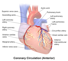
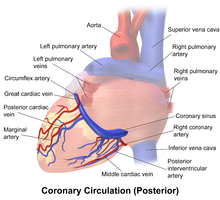
The coronary arteries are mainly composed of the left and right coronary arteries, both of which give off several branches, as shown in the 'coronary artery flow' figure.

The left coronary artery arises from the aorta within the left cusp of the aortic valve and feeds blood to the left side of the heart. It branches into two arteries, the left anterior descending and the left circumflex. The left anterior descending artery perfuses the interventricular septum and anterior wall of the left ventricle. The left circumflex artery perfuses the left ventricular free wall. In approximately 33% of individuals, the left coronary artery gives rise to the posterior descending artery [4] which perfuses the posterior and inferior walls of the left ventricle. Sometimes a third branch is formed at the fork between left anterior descending and left circumflex arteries, known as a ramus or intermediate artery. [5]

The right coronary artery (RCA) originates within the right cusp of the aortic valve. It travels down the right coronary sulcus, towards the crux of the heart. The RCA primarily branches into the right marginal arteries, and, in 67% of individuals, gives place to the posterior descending artery. [4] The right marginal arteries perfuse the right ventricle and the posterior descending artery perfuses the left ventricular posterior and inferior walls.

There is also the conus artery, which is only present in about 45 percent of the human population, and which provides collateral blood flow to the heart when the left anterior descending artery is occluded. [6] [7]

Clinical significance

Atherosclerosis Atherosclerosis 2011.jpg
Atherosclerosis

Narrowing of the arteries can be caused by a process known as atherosclerosis (most common), arteriosclerosis, or arteriolosclerosis. This occurs when plaques (made up of deposits of cholesterol and other substances) build up over time in the walls of the arteries. Coronary artery disease (CAD) or ischemic heart disease are the terms used to describe narrowing of the coronary arteries. [8]

As the disease progresses, plaque buildup can partially block blood flow to the heart muscle. Without enough blood supply (ischemia), the heart is unable to work properly, especially under increased stress. Stable angina is chest pain on exertion that improves with rest. Unstable angina is chest pain that can occur at rest, feels more severe, and/or last longer than stable angina. It is caused by more severe narrowing of the arteries. [9]

Heart attack Heart attack-NIH.gif
Heart attack

A heart attack results from a sudden plaque rupture and formation of a thrombus (blood clot) that completely blocks blood flow to a portion of the heart, leading to tissue death (infarct).

CAD can also result in heart failure or arrhythmias. Heart failure is caused by chronic oxygen deprivation due to reduced blood flow, which weakens the heart over time. Arrhythmias are caused by inadequate blood supply to the heart that interferes with the heart's electric impulse.

The coronary arteries can constrict as a response to various stimuli, mostly chemical. This is known as a coronary reflex.

There is also a rare condition known as spontaneous coronary artery dissection, in which the wall of one of the coronary arteries tears, causing severe pain. [10] Unlike CAD, spontaneous coronary artery dissection is not due to plaque buildup in arteries, and tends to occur in younger individuals, including women who have recently given birth or men who do intense exercise. [11]

Coronary artery dominance is described as the coronary artery that give branches to supply the right posterior descending artery and supplies the inferior wall of the heart. In 80 to 85% of the population, the right coronary artery supplies the posterior descending artery, making it right heart dominant while in 7 to 13% of the population, the left coronary artery supplies the posterior descending artery, making it left heart dominant. In 7 to 8% of the population, both right and left coronary arteries supplies the posterior descending artery, making it right and left co-dominance. Narrowing of coronary arteries is more frequent in those who are left dominant when compared to those who have right dominant or co-dominant hearts. [12]

Name etymology

Heart with ventricles and arteries.jpg
Model of human heart

The word corona is a Latin word meaning "crown", from the Ancient Greek κορώνη (korōnè, "garland, wreath"). It was applied to the coronary arteries because of a notional resemblance (compare the photos).

The word arterie in Anglo-French (artaire in Old French, and artērium in Latin) means "windpipe" and "an artery". It was applied to the coronary arteries because the arteries do not contain blood after death.

See also

Additional images

Blausen 0260 CoronaryVessels Anterior.png
Blausen 0261 CoronaryVessels Posterior.png

Related Research Articles

<span class="mw-page-title-main">Aorta</span> Largest artery in the human body

The aorta is the main and largest artery in the human body, originating from the left ventricle of the heart, branching upwards immediately after, and extending down to the abdomen, where it splits at the aortic bifurcation into two smaller arteries. The aorta distributes oxygenated blood to all parts of the body through the systemic circulation.

<span class="mw-page-title-main">Heart</span> Organ found inside most animals

The heart is a muscular organ in most animals. This organ pumps blood through the blood vessels of the circulatory system. The pumped blood carries oxygen and nutrients to the body, while carrying metabolic waste such as carbon dioxide to the lungs. In humans, the heart is approximately the size of a closed fist and is located between the lungs, in the middle compartment of the chest, called the mediastinum.

<span class="mw-page-title-main">Circulatory system</span> Organ system for circulating blood in animals

The circulatory system is a system of organs that includes the heart, blood vessels, and blood which is circulated throughout the entire body of a human or other vertebrate. It includes the cardiovascular system, or vascular system, that consists of the heart and blood vessels. The circulatory system has two divisions, a systemic circulation or circuit, and a pulmonary circulation or circuit. Some sources use the terms cardiovascular system and vascular system interchangeably with circulatory system.

<span class="mw-page-title-main">Coronary circulation</span> Circulation of blood in the blood vessels of the heart muscle (myocardium)

Coronary circulation is the circulation of blood in the arteries and veins that supply the heart muscle (myocardium). Coronary arteries supply oxygenated blood to the heart muscle. Cardiac veins then drain away the blood after it has been deoxygenated. Because the rest of the body, and most especially the brain, needs a steady supply of oxygenated blood that is free of all but the slightest interruptions, the heart is required to function continuously. Therefore its circulation is of major importance not only to its own tissues but to the entire body and even the level of consciousness of the brain from moment to moment. Interruptions of coronary circulation quickly cause heart attacks, in which the heart muscle is damaged by oxygen starvation. Such interruptions are usually caused by coronary ischemia linked to coronary artery disease, and sometimes to embolism from other causes like obstruction in blood flow through vessels.

<span class="mw-page-title-main">Coronary artery bypass surgery</span> Surgical procedure to restore normal blood flow to an obstructed coronary artery

Coronary artery bypass surgery, also known as coronary artery bypass graft, is a surgical procedure to treat coronary artery disease (CAD), the buildup of plaques in the arteries of the heart. It can relieve chest pain caused by CAD, slow the progression of CAD, and increase life expectancy. It aims to bypass narrowings in heart arteries by using arteries or veins harvested from other parts of the body, thus restoring adequate blood supply to the previously ischemic heart.

<span class="mw-page-title-main">Aortic valve</span> Valve in the human heart between the left ventricle and the aorta

The aortic valve is a valve in the heart of humans and most other animals, located between the left ventricle and the aorta. It is one of the four valves of the heart and one of the two semilunar valves, the other being the pulmonary valve. The aortic valve normally has three cusps or leaflets, although in 1–2% of the population it is found to congenitally have two leaflets. The aortic valve is the last structure in the heart the blood travels through before stopping the flow through the systemic circulation.

<span class="mw-page-title-main">Papillary muscle</span> Heart ventricle muscles

The papillary muscles are muscles located in the ventricles of the heart. They attach to the cusps of the atrioventricular valves via the chordae tendineae and contract to prevent inversion or prolapse of these valves on systole.

<span class="mw-page-title-main">Pulmonary artery</span> Artery in pulmonary circulation carrying deoxygenated blood from heart to lungs

A pulmonary artery is an artery in the pulmonary circulation that carries deoxygenated blood from the right side of the heart to the lungs. The largest pulmonary artery is the main pulmonary artery or pulmonary trunk from the heart, and the smallest ones are the arterioles, which lead to the capillaries that surround the pulmonary alveoli.

<span class="mw-page-title-main">Atrioventricular node</span> Part of the electrical conduction system of the heart

The atrioventricular node or AV node electrically connects the heart's atria and ventricles to coordinate beating in the top of the heart; it is part of the electrical conduction system of the heart. The AV node lies at the lower back section of the interatrial septum near the opening of the coronary sinus, and conducts the normal electrical impulse from the atria to the ventricles. The AV node is quite compact.

<span class="mw-page-title-main">Right coronary artery</span> Blood vessel supplying the human heart

In the blood supply of the heart, the right coronary artery (RCA) is an artery originating above the right cusp of the aortic valve, at the right aortic sinus in the heart. It travels down the right coronary sulcus, towards the crux of the heart. It gives off many branches, including the sinoatrial nodal artery, right marginal artery, posterior interventricular artery, conus artery, and atrioventricular nodal branch. It contributes the right side of the heart, and parts of the interventricular septum.

<span class="mw-page-title-main">Interventricular septum</span> Wall of tissue separating ventricles of human heart

The interventricular septum is the stout wall separating the ventricles, the lower chambers of the heart, from one another.

<span class="mw-page-title-main">Coronary sulcus</span> Groove on the surface of the heart that separates the atria from the ventricles

The coronary sulcus is a groove on the surface of the heart at the base of right auricle that separates the atria from the ventricles. The structure contains the trunks of the nutrient vessels of the heart, and is deficient in front, where it is crossed by the root of the pulmonary trunk. On the posterior surface of the heart, the coronary sulcus contains the coronary sinus. The right coronary artery, circumflex branch of left coronary artery, and small cardiac vein all travel along parts of the coronary sulcus.

<span class="mw-page-title-main">Posterior descending artery</span> Artery supplying the heart

In the coronary circulation, the posterior descending artery (PDA), also called the posterior interventricular artery, is an artery running in the posterior interventricular sulcus to the apex of the heart where it meets with the left anterior descending artery also known as the anterior interventricular artery. The PDA supplies the posterior third of the interventricular septum. The remaining anterior two-thirds is supplied by the left anterior descending artery, which is a branch of left coronary artery.

<span class="mw-page-title-main">Circumflex branch of left coronary artery</span> Artery of heart

The circumflex branch of left coronary artery is a branch of the left coronary artery. It winds around the left side of the heart along the atrioventricular groove. It supplies the posterolateral portion of the left ventricle.

A circulatory anastomosis is a connection between two blood vessels, such as between arteries, between veins or between an artery and a vein. Anastomoses between arteries and between veins result in a multitude of arteries and veins, respectively, serving the same volume of tissue. Such anastomoses occur normally in the body in the circulatory system, serving as back-up routes in a collateral circulation that allow blood to flow if one link is blocked or otherwise compromised, but may also occur pathologically.

<span class="mw-page-title-main">Left anterior descending artery</span> Artery of the heart

The left anterior descending artery is a branch of the left coronary artery. It supplies the anterior portion of the left ventricle. It provides about half of the arterial supply to the left ventricle and is thus considered the most important vessel supplying the left ventricle. Blockage of this artery is often called the widow-maker infarction due to a high risk of death.

<span class="mw-page-title-main">Atrioventricular nodal branch</span>

The atrioventricular nodal branch is a coronary artery that supplies arterial blood to the atrioventricular node, which is responsible for initiating muscular contraction of the ventricles. The AV nodal branch is most often a branch of the right coronary artery.

The following outline is provided as an overview of and topical guide to cardiology, the branch of medicine dealing with disorders of the human heart. The field includes medical diagnosis and treatment of congenital heart defects, coronary artery disease, heart failure, valvular heart disease and electrophysiology. Physicians who specialize in cardiology are called cardiologists.

<span class="mw-page-title-main">Coronary artery ectasia</span> Medical condition

Coronary artery ectasia is a rare disease that occurs in only 0.3-4.9% of people in North America. Coronary artery ectasia is characterized by the enlargement of a coronary artery to 1.5 times or more than its normal diameter. The disease is commonly asymptomatic and is normally discovered when performing tests for other conditions such as coronary artery disease, stable angina and other acute coronary syndromes. Coronary artery ectasia occurs 4 times more frequently in males than in females and in people who have risk factors for heart disease such as smokers. While the disease is commonly found in patients with atherosclerosis and coronary artery disease, it can occur by itself and in both cases, it can cause health problems. The disease can cause the heart tissue to be deprived of blood and die due to decreased blood flow, and blockages due to blood clots or spasms of the blood vessel. This blood flow disruption can cause permanent damage to the muscle if the deprivation is prolonged. Coronary artery ectasia also increases the chance of developing large weak spots in the affected coronary arteries, or aneurysms that can rupture and result in death. The damage can result in angina which is pain in the chest and is a common complaint in these patients.

The heart is a muscular organ situated in the mediastinum. It consists of four chambers, four valves, two main arteries, and the conduction system. The left and right sides of the heart have different functions: the right side receives de-oxygenated blood through the superior and inferior venae cavae and pumps blood to the lungs through the pulmonary artery, and the left side receives saturated blood from the lungs.

References

  1. "Coronary Arteries". Texas Heart Institute. Retrieved 2019-09-01.
  2. Petersen, J. W.; Pepine, C. J. (2014). "Microvascular Coronary Dysfunction and Ischemic Heart Disease – Where Are We in 2014?". Trends in Cardiovascular Medicine. 25 (2): 98–103. doi:10.1016/j.tcm.2014.09.013. PMC   4336803 . PMID   25454903.
  3. "Anatomy and Function of the Coronary Arteries". www.hopkinsmedicine.org. Retrieved 2019-09-01.
  4. 1 2 Costanzo, Linda S. (2018). Physiology (6th ed.). Philadelphia, PA: Elsevier. ISBN   9780323511896. OCLC   965761862.
  5. Fuster, V; Alexander RW; O'Rourke RA (2001). Hurst's The Heart (10th ed.). McGraw-Hill. p. 53. ISBN   978-0-07-135694-7.
  6. Wynn GJ, Noronha B, Burgess MI (2008). "Functional significance of the conus artery as a collateral to an occluded left anterior descending artery demonstrated by stress echocardiography". International Journal of Cardiology. 140 (1): e14–e15. doi:10.1016/j.ijcard.2008.11.039. PMID   19108914.
  7. Schlesinger MJ, Zoll PM, Wessler S (1949). "The conus artery: a third coronary artery". American Heart Journal. 38 (6): 823–838. doi:10.1016/0002-8703(49)90884-4. PMID   15395916.
  8. "Coronary Artery Disease: Causes, Diagonosis & Prevention| cdc.gov". www.cdc.gov. 2018-10-09. Retrieved 2019-09-01.
  9. "Coronary Artery Disease". Cleveland Clinic. Retrieved 2019-09-01.
  10. Franke, Kyle B; Wong, Dennis TL; Baumann, Angus; Nicholls, Stephen J; Gulati, Rajiv; Psaltis, Peter J (April 4, 2019). "Current State-of-play in Spontaneous Coronary Artery Dissection". Cardiovascular Diagnosis and Therapy. 9 (3): 281–298. doi: 10.21037/cdt.2019.04.03 . PMC   6603494 . PMID   31275818.
  11. "Coronary Artery Dissection: Not Just a Heart Attack". www.heart.org. Retrieved 2019-09-02.
  12. Aricatt DP, Prabhu A, Avadhani R, Subramanyam K, Manzil AS, Ezhilan J, Das R (January 2022). "A study of coronary dominance and its clinical significance". Folia Morphologica. 82 (1): 102–107. doi: 10.5603/FM.a2022.0005 . PMID   35099044. S2CID   246428206.