The larynx, commonly called the voice box, is an organ in the top of the neck involved in breathing, producing sound and protecting the trachea against food aspiration. The opening of larynx into pharynx known as the laryngeal inlet is about 4–5 centimeters in diameter. The larynx houses the vocal cords, and manipulates pitch and volume, which is essential for phonation. It is situated just below where the tract of the pharynx splits into the trachea and the esophagus. The word 'larynx' comes from the Ancient Greek word lárunx ʻlarynx, gullet, throat.ʼ

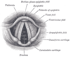
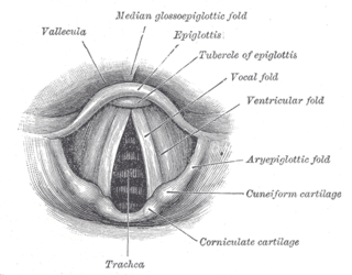
The epiglottis is a leaf-shaped flap in the throat that prevents food and water from entering the trachea and the lungs. It stays open during breathing, allowing air into the larynx. During swallowing, it closes to prevent aspiration of food into the lungs, forcing the swallowed liquids or food to go along the esophagus toward the stomach instead. It is thus the valve that diverts passage to either the trachea or the esophagus.

Elastic cartilage, fibroelastic cartilage or yellow fibrocartilage is a type of cartilage present in the pinnae (auricles) of the ear giving it shape, provides shape for the lateral region of the external auditory meatus, medial part of the auditory canal Eustachian tube, corniculate and cuneiform laryneal cartilages, and the epiglottis. It contains elastic fiber networks and collagen type II fibers. The principal protein is elastin.

The rima glottidis is the opening between the two true vocal cords anteriorly, and the two arytenoid cartilages posteriorly. It is part of the larynx.

The posterior cricoarytenoid muscle is a intrinsic muscle of the larynx. It arises from the cricoid cartilage; it inserts onto the arytenoid cartilage of the same side. It is innervated by the recurrent laryngeal nerve. Each acts to open the vocal folds by pulling the vocal fold of the same side laterally. It participates in the production of sounds.

The cricoid cartilage, or simply cricoid or cricoid ring, is the only complete ring of cartilage around the trachea. It forms the back part of the voice box and functions as an attachment site for muscles, cartilages, and ligaments involved in opening and closing the airway and in producing speech.

The arytenoid cartilages are a pair of small three-sided pyramids which form part of the larynx. They are the site of attachment of the vocal cords. Each is pyramidal or ladle-shaped and has three surfaces, a base, and an apex. The arytenoid cartilages allow for movement of the vocal cords by articulating with the cricoid cartilage. They may be affected by arthritis, dislocations, or sclerosis.
The laryngeal inlet is the opening that connects the pharynx and the larynx.

The stylopharyngeus muscle is a muscle in the head. It originates from the temporal styloid process. Some of its fibres insert onto the thyroid cartilage, while others end by intermingling with proximal structures. It is innervated by the glossopharyngeal nerve. It acts to elevate the larynx and pharynx, and dilate the pharynx, thus facilitating swallowing.

The thyroarytenoid muscle is a broad, thin muscle that forms the body of the vocal fold and that supports the wall of the ventricle and its appendix. It functions to shorten the vocal folds.
The thyrohyoid membrane is a broad, fibro-elastic sheet of the larynx. It connects the upper border of the thyroid cartilage to the hyoid bone.

The oblique arytenoid is bilaterally paired intrinsic muscle of the larynx. It is superficial to the transverse arytenoid; the oblique and transverse arytenoids are often considered two parts of a single muscle - the interarytenoid muscle.

The superior laryngeal nerve is a branch of the vagus nerve. It arises from the middle of the inferior ganglion of vagus nerve and additionally also receives a sympathetic branch from the superior cervical ganglion.

The portion of the cavity of the larynx above the vestibular fold is called the laryngeal vestibule; it is wide and triangular in shape, its base or anterior wall presenting, however, about its center the backward projection of the tubercle of the epiglottis. It contains the vestibular folds, and between these and the vocal folds are the laryngeal ventricles.

The corniculate cartilages are two small conical nodules consisting of elastic cartilage, which articulate with the summits of the arytenoid cartilages and serve to prolong them posteriorly and medially.
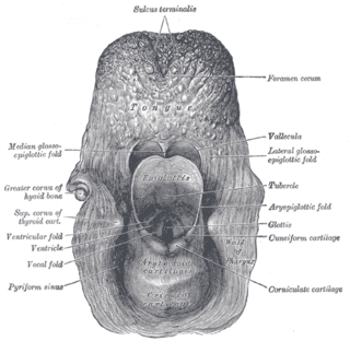
The aryepiglottic folds are triangular folds of mucous membrane of the larynx. They enclose ligamentous and muscular fibres. They extend from the lateral borders of the epiglottis to the arytenoid cartilages, hence the name 'aryepiglottic'. They contain the aryepiglottic muscles and form the upper borders of the quadrangular membrane. They have a role in growling as a form of phonation. They may be narrowed and cause stridor, or be shortened and cause laryngomalacia.

The vestibular fold is one of two thick folds of mucous membrane, each enclosing a narrow band of fibrous tissue, the vestibular ligament, which is attached in front to the angle of the thyroid cartilage immediately below the attachment of the epiglottis, and behind to the antero-lateral surface of the arytenoid cartilage, a short distance above the vocal process.
The quadrangular membrane is a layer of submucosa. It extends between the lateral margin of the epiglottis, and the apex and fovea triangularis of the ipsilateral arytenoid cartilage. It has free superior and inferior borders.
In the human larynx, the vocal process is the anterior angle of the base of the arytenoid cartilage, as it projects horizontally forward and gives attachment to the vocal ligament.

The pharynx is the part of the throat behind the mouth and nasal cavity, and above the esophagus and trachea. It is found in vertebrates and invertebrates, though its structure varies across species. The pharynx carries food to the esophagus and air to the larynx. The flap of cartilage called the epiglottis stops food from entering the larynx.
 Posterior view of the cartilages of the larynx
 Posterior view of the muscles of the larynx