Cutis marmorata telangiectatica congenita

Last updated
Cutis marmorata telangiectatica congenita
Other namesCMTC [1]
Autosomal recessive - en.svg
Cutis marmorata telangiectatica congenital is inherited via autosomal recessive manner [2]
Specialty Dermatology   OOjs UI icon edit-ltr-progressive.svg

Cutis marmorata telangiectatica congenita is a rare congenital vascular disorder that usually manifests in affecting the blood vessels of the skin. The condition was first recognised and described in 1922 by Cato van Lohuizen, [3] a Dutch pediatrician whose name was later adopted in the other common name used to describe the condition – Van Lohuizen syndrome. CMTC is also used synonymously with congenital generalized phlebectasia, nevus vascularis reticularis, congenital phlebectasia, livedo telangiectatica, congenital livedo reticularis and Van Lohuizen syndrome. [4]

Contents

It should not be confused with the more general term "cutis marmorata", which refers to livedo reticularis caused by cold. [5]

For a full and up-to-date description visit the CMTC webpages of the global non-profit patient organisation for people with CMTC and other vascular malformation CMTC-OVM: www.cmtc.nl/en

Signs and symptoms

People with visible marks generally feel fine (physically) and can act normally, but when it is mentioned, they may become withdrawn and self-conscious. Some children may have low self-esteem due to the condition. CMTC is an uncommon, sporadic congenital vascular malformation characterized by a generalized or localized reticulated cutaneous vascular network.[ citation needed ]

Cutaneous lesions described in patients with CMTC include nevus flammeus, hemangioma, nevus anemicus, café-au-lait spots, melanocytic nevus, aplasia cutis and acral cyanosis. [6]

It has a marbled bluish to deep-purple appearance. The dark skin lesions often show a palpable loss of dermal substance. The reticulated mottling frequently appears more prominent in a cold environment (physiologic cutis marmorata), but tends not to disappear with warming. Hence, the erythema may be worsened by cooling, physical activity, or crying. CMTC frequently involves the extremities, with the lower extremities involved most commonly, followed by the upper extremities, and then the trunk and face. The lower extremities often show atrophy and seldom show hypertrophy resulting in limb circumference discrepancy.[ citation needed ]

When located on the trunk, the lesions of CMTC tend to show mosaic distribution in streaks with a sharp midline demarcation seen across the abdomen. [6] The lesions are primarily localized, but can be segmental or generalized, often unilateral in appearance. [6] Diffuse involvement of the skin is usually not observed.

Although its course is variable, the majority of lesions in mild cases fade by adolescence. Ulceration and secondary infection are complications in severe cases and can be fatal if present in the neonatal period. [7]

Causes

Fewer than 100 cases of CMTC have been published worldwide. Petrozzi reported the first case of CMTC in the United States in 1970. [8] CMTC is believed to be more common than suspected, as studies have shown that milder forms of the disease are not being recognized as CMTC. [6]

The pathophysiology is still unclear, with most cases occurring sporadically, although rare cases were reported in families. Studies indicated the primary involvement of capillaries, venules and veins, and possibly also that of arterioles and lymphatics.

Hypotheses that have been proposed include: environmental/external factors; [9] peripheral neural dysfunction; [10] failure of the development of mesodermic vessels in an early embryonic stage; autosomal dominant inheritance with incomplete penetrance and, finally, the theory of Happle. [10]

Diagnosis

Differential diagnosis

During the first few weeks after birth, when the lesions are not very reticulated, CMTC may look very similar to vascular lesions such as port-wine stains. However, during follow-up, CMTC lesions become characteristic in their appearance. They must be differentiated from other causes of persistent reticulated vascular lesions, such as those in the following table:

DiseasesCharacteristics
Diffuse phlebectasiarare progressive harmartomatous malformation involving the deeper veins
Livedo reticularis associated with collagen-vascular diseaselace pattern of cyanotic skin discoloration secondary to dilation of subpapillary veinous plexi and occlusion of small vessels feeding the upper cutis
Neonatal lupus erythematosuswell-demarcated erythematous, mild-scaling plaque that is often annular and appears predominantly on the scalp, neck, or face
Nevus anemicuscongenital single patch manifested by skin pallor, most commonly seen on the trunk
Nevus flammeus (port-wine stain)pale pink to red-purple, usually unilateral macules of the face or extremities
Physiologic cutis marmoratareticulated mottling appearance of the skin that physiologically responds to cold environments
Primary antiphospholipid syndrome (APS)increased tendency to form venous and/or arterial thromboses, often accompanied by thrombocytopenia in the presence of the antiphospholipid antibodies

Histology

Some patients have a few or no histopathologic abnormalities. Histological examination of a biopsy may show an increase in the number and size of capillaries and veins (rarely lymphatics), dilated capillaries located in the deeper dermis, and hyperplasia and swollen endothelial cells with occasional dilated veins and venous lakes.[ citation needed ]

Associated abnormalities

Associated abnormalities include the following:

Treatment

In general, there is no treatment available for CMTC, although associated abnormalities can be treated. In the case of limb asymmetry, when no functional problems are noted, treatment is not warranted, except for an elevation device for the shorter leg.

Laser therapy has not been successful in the treatment of CMTC, possibly due to the presence of many large and deep capillaries and dilated veins. Pulsed-dye laser and long-pulsed-dye laser have not yet been evaluated in CMTC, but neither argon laser therapy nor YAG laser therapy has been helpful. [12]

When ulcers develop secondary to the congenital disease, antibiotic treatment such as oxacillin and gentamicin administered for 10 days has been prescribed. In one study, the wound grew Escherichia coli while blood cultures were negative. [7]

Prognosis

The prognosis is favorable in most patients with an isolated cutaneous abnormality. In the majority of cases, both the vivid red marking and the difference in circumference of the extremities regress spontaneously during the first year of life. It is theorized that this may be due to the normal maturation process, with thickening of the epidermis and dermis. Improvements for some patients can continue for up to 10 years, while in other cases, the marbled skin may persist for the patient's lifetime.

One study reported an improvement in lesions in 46% of patients within three years. [9] If CMTC persists into adulthood, it can result in complaints due to paresthesia, increased sensitivity to cold and pain, and the formation of ulcers. [7]

Few reports included long-term follow up of CMTC into adolescence and adulthood. While about 50% of patients seem to show definite improvement in the reticular vascular pattern, [12] the exact incidence and cause of persistent cases are unknown.

Epidemiology

Usually observed at birth or shortly thereafter in 94% of patients, [9] in other reports, patients did not develop skin lesions until three months or even two years after birth. [13] [14] Females are typically affected more often than males (64%). [15]

Eponym

It is named for Dr. Cato van Lohuizen. [16]

Related Research Articles

<span class="mw-page-title-main">Infantile hemangioma</span> Raised red skin lesion that affects infants caused by benign vascular tumor

An infantile hemangioma (IH), sometimes called a strawberry mark due to appearance, is a type of benign vascular tumor or anomaly that affects babies. Other names include capillary hemangioma, "strawberry hemangioma", strawberry birthmark and strawberry nevus. and formerly known as a cavernous hemangioma. They appear as a red or blue raised lesion on the skin. Typically, they begin during the first four weeks of life, growing until about five months of life, and then shrinking in size and disappearing over the next few years. Often skin changes remain after they shrink. Complications may include pain, bleeding, ulcer formation, disfigurement, or heart failure. It is the most common tumor of orbit and periorbital areas in childhood. It may occur in the skin, subcutaneous tissues and mucous membranes of oral cavities and lips as well as in extracutaneous locations including the liver and gastrointestinal tract.

<span class="mw-page-title-main">Livedo reticularis</span> Medical condition

Livedo reticularis is a common skin finding consisting of a mottled reticulated vascular pattern that appears as a lace-like purplish discoloration of the skin. The discoloration is caused by reduction in blood flow through the arterioles that supply the cutaneous capillaries, resulting in deoxygenated blood showing as blue discoloration. This can be a secondary effect of a condition that increases a person's risk of forming blood clots, including a wide array of pathological and nonpathological conditions. Examples include hyperlipidemia, microvascular hematological or anemia states, nutritional deficiencies, hyper- and autoimmune diseases, and drugs/toxins.

<span class="mw-page-title-main">Becker's nevus</span> Medical condition

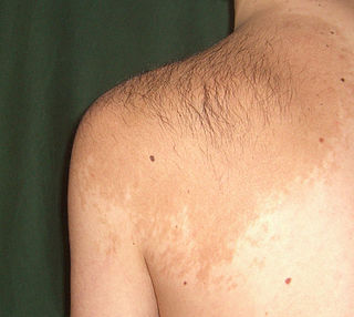
Becker's nevus is a benign skin disorder predominantly affecting males. The nevus can be present at birth, but more often shows up around puberty. It generally first appears as an irregular pigmentation on the torso or upper arm, and gradually enlarges irregularly, becoming thickened and often hairy (hypertrichosis). The nevus is due to an overgrowth of the epidermis, pigment cells (melanocytes), and hair follicles. This form of nevus was first documented in 1948 by American dermatologist Samuel William Becker (1894–1964).

Adams–Oliver syndrome (AOS) is a rare congenital disorder characterized by defects of the scalp and cranium, transverse defects of the limbs, and mottling of the skin.

CMTC may refer to:

<span class="mw-page-title-main">Angiokeratoma</span> Medical condition

Angiokeratoma is a benign cutaneous lesion of capillaries, resulting in small marks of red to blue color and characterized by hyperkeratosis. Angiokeratoma corporis diffusum refers to Fabry's disease, but this is usually considered a distinct condition.

<span class="mw-page-title-main">Sneddon's syndrome</span> Medical condition

Sneddon's syndrome is a form of arteriopathy characterized by several symptoms, including:

Phakomatosis pigmentovascularis is a rare neurocutanous condition where there is coexistence of a capillary malformation with various melanocytic lesions, including dermal melanocytosis, nevus spilus, and nevus of Ota.

Eccrine angiomatous hamartoma (EAH), first described by Lotzbeck in 1859, is a rare benign vascular hamartoma characterized histologically by a proliferation of eccrine and vascular components. EAH exists on a spectrum of cutaneous tumors that include eccrine nevus, mucinous eccrine nevus and EAH. Each diagnostic subtype is characterized by an increase in the number as well as size of mature eccrine glands or ducts, with EAH being distinguished by the added vascular component.

<span class="mw-page-title-main">Nevus anemicus</span> Medical condition

Nevus anemicus is a congenital disorder characterized by macules of varying size and shape that are paler than the surrounding skin and cannot be made red by trauma, cold, or heat. The paler area is due to the blood vessels within the area which are more sensitive to the body’s normal vasoconstricting chemicals.

<span class="mw-page-title-main">Aplasia cutis congenita</span> Medical condition

Aplasia cutis congenita is a rare disorder characterized by congenital absence of skin. Ilona J. Frieden classified ACC in 1986 into 9 groups on the basis of location of the lesions and associated congenital anomalies. The scalp is the most commonly involved area with lesser involvement of trunk and extremities. Frieden classified ACC with fetus papyraceus as type 5. This type presents as truncal ACC with symmetrical absence of skin in stellate or butterfly pattern with or without involvement of proximal limbs. It is the most common congenital cicatricial alopecia, and is a congenital focal absence of epidermis with or without evidence of other layers of the skin.

<span class="mw-page-title-main">Livedoid vasculopathy</span> Medical condition

Livedoid vasculopathy(LV) is an uncommon thrombotic dermal vasculopathy that is characterized by excruciating, recurrent ulcers on the lower limbs. Livedo racemosa, a painful ulceration in the distal regions of the lower extremities, is the characteristic clinical appearance. It heals to form porcelain-white, atrophic scars, also known as Atrophie blanche.

<span class="mw-page-title-main">Blue rubber bleb nevus syndrome</span> Medical condition

Blue rubber bleb nevus syndrome is a rare disorder that consists mainly of abnormal blood vessels affecting the skin or internal organs – usually the gastrointestinal tract. The disease is characterized by the presence of fluid-filled blisters (blebs) as visible, circumscribed, chronic lesions (nevi).

<span class="mw-page-title-main">Nevus depigmentosus</span> Medical condition

Nevus depigmentosus is a loss of pigment in the skin which can be easily differentiated from vitiligo. Although age factor has not much involvement in the nevus depigmentosus but in about 19% of the cases these are noted at birth. Their size may however grow in proportion to growth of the body. The distribution is also fairly stable and are nonprogressive hypopigmented patches. The exact cause of nevus depigmentosus is still not clearly understood. A sporadic defect in the embryonic development has been suggested to be a causative factor. It has been described as "localised albinism", though this is incorrect.

A vascular anomaly is any of a range of lesions from a simple birthmark to a large tumor that may be disfiguring. They are caused by a disorder of the vascular system. A vascular anomaly is a localized defect in blood or lymph vessels. These defects are characterized by an increased number of vessels, and vessels that are both enlarged and sinuous. Some vascular anomalies are congenital, others appear within weeks to years after birth, and others are acquired by trauma or during pregnancy. Inherited vascular anomalies are also described and often present with a number of lesions that increase with age. Vascular anomalies can also be a part of a syndrome.

<span class="mw-page-title-main">Macrocephaly-capillary malformation</span> Medical condition

Macrocephaly-capillary malformation (M-CM) is a multiple malformation syndrome causing abnormal body and head overgrowth and cutaneous, vascular, neurologic, and limb abnormalities. Though not every patient has all features, commonly found signs include macrocephaly, congenital macrosomia, extensive cutaneous capillary malformation, body asymmetry, polydactyly or syndactyly of the hands and feet, lax joints, doughy skin, variable developmental delay and other neurologic problems such as seizures and low muscle tone.

<span class="mw-page-title-main">Cutis marmorata</span> Human skin condition

Cutis marmorata is a benign skin condition which, if persistent, occurs in Cornelia de Lange syndrome, trisomy 13 and trisomy 18 syndromes. When a newborn infant is exposed to low environmental temperatures, an evanescent, lacy, reticulated red and/or blue cutaneous vascular pattern appears over most of the body surface. This vascular change represents an accentuated physiologic vasomotor response that disappears with increasing age, although it is sometimes discernible even in older children. It is also seen in cardiogenic shock.

Diffuse capillary malformation with overgrowth (DCMO) is a subset of capillary malformations (CM) associated with hypertrophy, i.e. increased size of body structures. CM can be considered an umbrella term for various vascular anomalies caused by increased diameter or number of capillary blood vessels. It is commonly referred to as "port-wine stain", and is thought to affect approximately 0.5% of the population. Typically capillaries in the papillary dermis are involved, and this gives rise to pink or violaceous colored lesions. The majority of DCMO lesions are diffuse, reticulated pale-colored stains.

Cutaneous manifestations of COVID-19 are characteristic signs or symptoms of the Coronavirus disease 2019 that occur in the skin. The American Academy of Dermatology reports that skin lesions such as morbilliform, pernio, urticaria, macular erythema, vesicular purpura, papulosquamous purpura and retiform purpura are seen in people with COVID-19. Pernio-like lesions were more common in mild disease while retiform purpura was seen only in critically ill patients. The major dermatologic patterns identified in individuals with COVID-19 are urticarial rash, confluent erythematous/morbilliform rash, papulovesicular exanthem, chilbain-like acral pattern, livedo reticularis and purpuric "vasculitic" pattern. Chilblains and Multisystem inflammatory syndrome in children are also cutaneous manifestations of COVID-19.

References

  1. RESERVED, INSERM US14-- ALL RIGHTS. "Orphanet: Cutis marmorata telangiectatica congenita". www.orpha.net. Retrieved 28 April 2019.{{cite web}}: CS1 maint: numeric names: authors list (link)
  2. "OMIM Entry – 219250 – Cutis Marmorata Telangiectatica Congenita; CMTC". omim.org. Retrieved 10 July 2017.
  3. Van Lohuizen CHJ. Cutis marmorata telangiectatica congenita,. Acta Derm Venereol. (Stockh). 1922:3:202-11.
  4. Gerritsen MJ, et al. Cutis marmorata telangiectatica congenita: report of 18 cases. Br J Dermatol. 2000 Feb;142(2):366-9.
  5. "cutis marmorata" "at Dorland's Medical Dictionary
  6. 1 2 3 4 Torrelo A, Zambrano A, Happle R. Cutis marmorata telangiectatica congenita and extensive mongolian spots: type 5 phacomatosis pigmentovascularis. Br J Dermatol. 2003 Feb;148(2):342-5.
  7. 1 2 3 Hu IJ, Chen MT, Tai HC, et al. Cutis marmorata telangiectatica congenita with gangrenous ulceration and hypovolaemic shock. Eur J Pediatr. 2005 Jul;164(7):411-3.
  8. Petrozzi JW, Rahn EK, Mofenson H, et al. Cutis marmorata telangiectatica congenita. Arch Dermatol. 1970 Jan;101(1):74-7.
  9. 1 2 3 Amitai DB, Fichman S, Merlob P, et al. Cutis marmorata telangiectatica congenita: clinical findings in 85 patients. Ped Dermatol. 2000 Mar–Apr;17(2):100-4.
  10. 1 2 Bormann G, Wohlrab J, Fischer M, et al. Cutis marmorata telangiectatica congenita: laser doppler fluxmetry evidence for a functional nervous defect. Ped Dermatol. 2001Mar-Apr;18(2):110-3.
  11. Melani L, Antiga E, Torchia D, et al. Cutis marmorata telangiectatica congenita and chronic autoimmune urticaria in a young man. J Dermatol. 2007 Mar;34(3):210-3.
  12. 1 2 Mazereeuw-Hautier J, Carel-Caneppele S, Bonafe JL. Cutis marmorata telangiectatica congenita: report of two persistent cases. Ped Dermatol. 2002 Nov–Dec;19(6): 506–9.
  13. Powel ST, Su WP. Cutis marmorata telangiectatica congenita: report of nine cases and review of the literature. Cutis. 1984 Sep;34(3):305-12. Review.
  14. Lee S, Lee JB, Kim JH, et al. Cutis marmorata telangiectatica congenita with multiple congenital anomalies (van Lohuizen’s syndrome). Dermatologica. 1981;163(5):408-12.
  15. Vascular Lesions and Congenital Nevi in the Newborn. UpToDate viewed on 03-28-2008 [ permanent dead link ].
  16. doctor/3295 at Who Named It?