The cytoskeleton is a complex, dynamic network of interlinking protein filaments present in the cytoplasm of all cells, including those of bacteria and archaea. In eukaryotes, it extends from the cell nucleus to the cell membrane and is composed of similar proteins in the various organisms. It is composed of three main components:microfilaments, intermediate filaments, and microtubules, and these are all capable of rapid growth or disassembly depending on the cell's requirements.

Actin is a family of globular multi-functional proteins that form microfilaments in the cytoskeleton, and the thin filaments in muscle fibrils. It is found in essentially all eukaryotic cells, where it may be present at a concentration of over 100 μM; its mass is roughly 42 kDa, with a diameter of 4 to 7 nm.

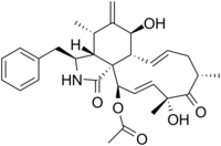
Phalloidin belongs to a class of toxins called phallotoxins, which are found in the death cap mushroom (Amanita phalloides). It is a rigid bicyclic heptapeptide that is lethal after a few days when injected into the bloodstream. The major symptom of phalloidin poisoning is acute hunger due to the destruction of liver cells. It functions by binding and stabilizing filamentous actin (F-actin) and effectively prevents the depolymerization of actin fibers. Due to its tight and selective binding to F-actin, derivatives of phalloidin containing fluorescent tags are used widely in microscopy to visualize F-actin in biomedical research.

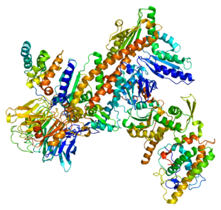
The Wiskott–Aldrich Syndrome protein (WASp) is a 502-amino acid protein expressed in cells of the hematopoietic system that in humans is encoded by the WAS gene. In the inactive state, WASp exists in an autoinhibited conformation with sequences near its C-terminus binding to a region near its N-terminus. Its activation is dependent upon CDC42 and PIP2 acting to disrupt this interaction, causing the WASp protein to 'open'. This exposes a domain near the WASp C-terminus that binds to and activates the Arp2/3 complex. Activated Arp2/3 nucleates new F-actin.

Cytochalasin B, the name of which comes from the Greek cytos (cell) and chalasis (relaxation), is a cell-permeable mycotoxin. It was found that substoichiometric concentrations of cytochalasin B (CB) strongly inhibit network formation by actin filaments. Due to this, it is often used in cytological research. It inhibits cytoplasmic division by blocking the formation of contractile microfilaments. It inhibits cell movement and induces nuclear extrusion. Cytochalasin B shortens actin filaments by blocking monomer addition at the fast-growing end of polymers. Cytochalasin B inhibits glucose transport and platelet aggregation. It blocks adenosine-induced apoptotic body formation without affecting activation of endogenous ADP-ribosylation in leukemia HL-60 cells. It is also used in cloning through nuclear transfer. Here enucleated recipient cells are treated with cytochalasin B. Cytochalasin B makes the cytoplasm of the oocytes more fluid and makes it possible to aspirate the nuclear genome of the oocyte within a small vesicle of plasma membrane into a micro-needle. Thereby, the oocyte genome is removed from the oocyte, while preventing rupture of the plasma membrane.
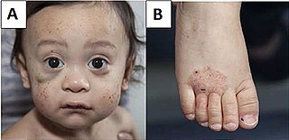
Wiskott–Aldrich syndrome (WAS) is a rare X-linked recessive disease characterized by eczema, thrombocytopenia, immune deficiency, and bloody diarrhea. It is also sometimes called the eczema-thrombocytopenia-immunodeficiency syndrome in keeping with Aldrich's original description in 1954. The WAS-related disorders of X-linked thrombocytopenia (XLT) and X-linked congenital neutropenia (XLN) may present with similar but less severe symptoms and are caused by mutations of the same gene.
Tropomyosin is a two-stranded alpha-helical, coiled coil protein found in many animal and fungal cells. In animals, it is an important component of the muscular system which works in conjunction with troponin to regulate muscle contraction. It is present in smooth and striated muscle tissues, which can be found in various organs and body systems, including the heart, blood vessels, respiratory system, and digestive system. In fungi, tropomyosin is found in cell walls and helps maintain the structural integrity of cells.
Filopodia are slender cytoplasmic projections that extend beyond the leading edge of lamellipodia in migrating cells. Within the lamellipodium, actin ribs are known as microspikes, and when they extend beyond the lamellipodia, they're known as filopodia. They contain microfilaments cross-linked into bundles by actin-bundling proteins, such as fascin and fimbrin. Filopodia form focal adhesions with the substratum, linking them to the cell surface. Many types of migrating cells display filopodia, which are thought to be involved in both sensation of chemotropic cues, and resulting changes in directed locomotion.
Cytochalasins are fungal metabolites that have the ability to bind to actin filaments and block polymerization and the elongation of actin. As a result of the inhibition of actin polymerization, cytochalasins can change cellular morphology, inhibit cellular processes such as cell division, and even cause cells to undergo apoptosis. Cytochalasins have the ability to permeate cell membranes, prevent cellular translocation and cause cells to enucleate. Cytochalasins can also have an effect on other aspects of biological processes unrelated to actin polymerization. For example, cytochalasin A and cytochalasin B can also inhibit the transport of monosaccharides across the cell membrane, cytochalasin H has been found to regulate plant growth, cytochalasin D inhibits protein synthesis and cytochalasin E prevents angiogenesis.
The latrunculins are a family of natural products and toxins produced by certain sponges, including genus Latrunculia and Negombata, whence the name is derived. It binds actin monomers near the nucleotide binding cleft with 1:1 stoichiometry and prevents them from polymerizing. Administered in vivo, this effect results in disruption of the actin filaments of the cytoskeleton, and allows visualization of the corresponding changes made to the cellular processes. This property is similar to that of cytochalasin, but has a narrow effective concentration range. Latrunculin has been used to great effect in the discovery of cadherin distribution regulation and has potential medical applications. Latrunculin A, a type of the toxin, was found to be able to make reversible morphological changes to mammalian cells by disrupting the actin network.

Cytochalasin E, a member of the cytochalasin group, is an inhibitor of actin polymerization in blood platelets. It inhibits angiogenesis and tumor growth. Unlike cytochalasin A and cytochalasin B, it does not inhibit glucose transport. Cytochalasin E, however, was noted to decrease glucose absorption in mice around the intestinal tissues by increasing the Km needed for glucose to reach the Vmax, which meant that a higher concentration of glucose was required in its presence to attain Vmax. Since Vmax remained the same according to another study, it is evident that CE is indeed a competitive inhibitor at the intestinal receptor sites for glucose.

In biology, a protein filament is a long chain of protein monomers, such as those found in hair, muscle, or in flagella. Protein filaments form together to make the cytoskeleton of the cell. They are often bundled together to provide support, strength, and rigidity to the cell. When the filaments are packed up together, they are able to form three different cellular parts. The three major classes of protein filaments that make up the cytoskeleton include: actin filaments, microtubules and intermediate filaments.
Actin-related protein 2 is a protein that in humans is encoded by the ACTR2 gene.

ACTC1 encodes cardiac muscle alpha actin. This isoform differs from the alpha actin that is expressed in skeletal muscle, ACTA1. Alpha cardiac actin is the major protein of the thin filament in cardiac sarcomeres, which are responsible for muscle contraction and generation of force to support the pump function of the heart.

Vasodilator-stimulated phosphoprotein is a protein that in humans is encoded by the VASP gene.

Invadopodia are actin-rich protrusions of the plasma membrane that are associated with degradation of the extracellular matrix in cancer invasiveness and metastasis. Very similar to podosomes, invadopodia are found in invasive cancer cells and are important for their ability to invade through the extracellular matrix, especially in cancer cell extravasation. Invadopodia are generally visualized by the holes they create in ECM -coated plates, in combination with immunohistochemistry for the invadopodia localizing proteins such as cortactin, actin, Tks5 etc. Invadopodia can also be used as a marker to quantify the invasiveness of cancer cell lines in vitro using a hyaluronic acid hydrogel assay.
Thromboelastometry (TEM), previously named rotational thromboelastography (ROTEG) or rotational thromboelastometry (ROTEM), is an established viscoelastic method for hemostasis testing in whole blood. It is a modification of traditional thromboelastography (TEG).
Actin remodeling is a biochemical process in cells. In the actin remodeling of neurons, the protein actin is part of the process to change the shape and structure of dendritic spines. G-actin is the monomer form of actin, and is uniformly distributed throughout the axon and the dendrite. F-actin is the polymer form of actin, and its presence in dendritic spines is associated with their change in shape and structure. Actin plays a role in the formation of new spines as well as stabilizing spine volume increase. The changes that actin brings about lead to the formation of new synapses as well as increased cell communication.
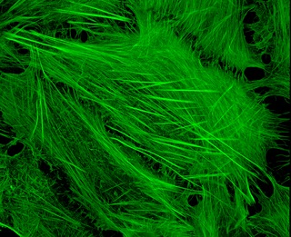
Cytoskeletal drugs are small molecules that interact with actin or tubulin. These drugs can act on the cytoskeletal components within a cell in three main ways. Some cytoskeletal drugs stabilize a component of the cytoskeleton, such as taxol, which stabilizes microtubules, or Phalloidin, which stabilizes actin filaments. Others, such as Cytochalasin D, bind to actin monomers and prevent them from polymerizing into filaments. Drugs such as demecolcine act by enhancing the depolymerisation of already formed microtubules. Some of these drugs have multiple effects on the cytoskeleton: for example, Latrunculin both prevents actin polymerization as well as enhancing its rate of depolymerization. Typically the microtubule targeting drugs can be found in the clinic where they are used therapeutically in the treatment of some forms of cancer. As a result of the lack of specificity for specific type of actin, the use of these drugs in animals results in unacceptable off-target effects. Despite this, the actin targeting compounds are still useful tools that can be used on a cellular level to help further our understanding of how this complex part of the cells' internal machinery operates. For example, Phalloidin that has been conjugated with a fluorescent probe can be used for visualizing the filamentous actin in fixed samples.

Thomas P. Stossel was an American hematologist, inventor, medical researcher, and writer that discovered gelsolin, and invented the BioAegis technology estate. He was also a professor emeritus of medicine at Harvard Medical School and professor emeritus of clinical research at the American Cancer Society. He was Chief Scientific Advisor to BioAegis Therapeutics Inc., a clinical stage biotech company developing a non-immunosuppressive, anti-inflammatory with potential to address a wide range of infectious, inflammatory and degenerative diseases. He is the holder of more than 50 patents and had authored more than 300 papers, studies, and reviews. He was also a member of the National Academy of Sciences, the American Academy of Arts and Sciences, and the National Academy of Medicine, past editor-in-chief of Current Opinion in Hematology and past editor of the Journal of Clinical Investigation, past president of the American Society of Hematology and the American Society for Clinical Investigation.