Collagen is the main structural protein in the extracellular matrix found in the body's various connective tissues. As the main component of connective tissue, it is the most abundant protein in mammals, making up from 25% to 35% of the whole-body protein content. Collagen consists of amino acids bound together to form a triple helix of elongated fibril known as a collagen helix. It is mostly found in connective tissue such as cartilage, bones, tendons, ligaments, and skin.
A ligament is the fibrous connective tissue that connects bones to other bones. It is also known as articular ligament, articular larua, fibrous ligament, or true ligament. Other ligaments in the body include the:
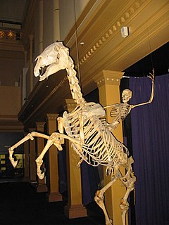
A skeleton is a structural frame that supports an animal body. There are several different skeletal types: the exoskeleton, which is the stable outer shell of an organism, the endoskeleton, which forms the support structure inside the body, and the hydroskeleton, a flexible skeleton supported by fluid pressure. The term comes from Greek σκελετός (skeletós) 'dried up'.

In biology, tissue is a biological organizational level between cells and a complete organ. A tissue is an ensemble of similar cells and their extracellular matrix from the same origin that together carry out a specific function. Organs are then formed by the functional grouping together of multiple tissues.

A tendon or sinew is a tough, high-tensile-strength band of dense fibrous connective tissue that connects muscle to bone. It is able to efficiently transmit the mechanical forces of muscle contraction to the skeletal system without sacrificing its ability to withstand significant amounts of tension.

Connective tissue is one of the many basic types of animal tissue, along with epithelial tissue, muscle tissue, and nervous tissue. In embryology it develops from the mesoderm. Connective tissue is found in between other tissues everywhere in the body, including the nervous system. The three outer membranes that envelop the brain and spinal cord are composed of dense inert connective tissue. All connective tissue consists of three main components: fibers, ground substance and cells. Not all authorities include blood or lymph as connective tissue because they lack the fiber component. All are immersed in the body water. The cells of connective tissue include fibroblasts, adipocytes, macrophages, mast cells and leucocytes.

The pubic symphysis is a secondary cartilaginous joint between the left and right superior rami of the pubis of the hip bones. It is in front of and below the urinary bladder. In males, the suspensory ligament of the penis attaches to the pubic symphysis. In females, the pubic symphysis is close to the clitoris. In most adults it can be moved roughly 2 mm and with 1 degree rotation. This increases for women at the time of childbirth.
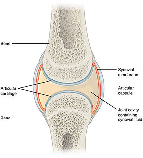
A synovial joint, also known as diarthrosis, joins bones or cartilage with a fibrous joint capsule that is continuous with the periosteum of the joined bones, constitutes the outer boundary of a synovial cavity, and surrounds the bones' articulating surfaces. This joint unites long bones and permits free bone movement and greater mobility. The synovial cavity/joint is filled with synovial fluid. The joint capsule is made up of an outer layer, the articular capsule, which keeps the bones together structurally, and an inner layer, the synovial membrane, which seals in the synovial fluid.
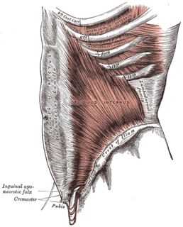
A fascia is a band or sheet of connective tissue, primarily collagen, beneath the skin that attaches to, stabilizes, encloses, and separates muscles and other internal organs. Fascia is classified by layer, as superficial fascia, deep fascia, and visceral or parietal fascia, or by its function and anatomical location.

The human musculoskeletal system is an organ system that gives humans the ability to move using their muscular and skeletal systems. The musculoskeletal system provides form, support, stability, and movement to the body.

Loose connective tissue is a category of connective tissue which includes areolar tissue, reticular tissue, and adipose tissue. Loose connective tissue is the most common type of connective tissue in vertebrates. It holds organs in place and attaches epithelial tissue to other underlying tissues. For example, it forms telae, such as the tela submucosa and tela subserosa, which connect mucous and serous membranes to the muscular layer. It also surrounds the blood vessels and nerves. Cells called fibroblasts are widely dispersed in this tissue; they are irregular branching cells that secrete strong fibrous proteins and proteoglycans as an extracellular matrix. The cells of this type of tissue are generally quite separated by a gelatinous substance primarily made up of collagenous and elastic fibers.

Reticular fibers, reticular fibres or reticulin is a type of fiber in connective tissue composed of type III collagen secreted by reticular cells. Reticular fibers crosslink to form a fine meshwork (reticulin). This network acts as a supporting mesh in soft tissues such as liver, bone marrow, and the tissues and organs of the lymphatic system.

Dense connective tissue, also called dense fibrous tissue, is a type of connective tissue with fibers as its main matrix element. The fibers are mainly composed of type I collagen. Crowded between the collagen fibers are rows of fibroblasts, fiber-forming cells, that generate the fibers. Dense connective tissue forms strong, rope-like structures such as tendons and ligaments. Tendons attach skeletal muscles to bones; ligaments connect bones to bones at joints. Ligaments are more stretchy and contain more elastic fibers than tendons. Dense connective tissue also make up the lower layers of the skin (dermis), where it is arranged in sheets.
The cardiac skeleton, also known as the fibrous skeleton of the heart, is a high density single/homogeneous structure of connective tissue that forms and anchors the valves and influences the forces exerted by and through them. The cardiac skeleton separates and partitions the atria from the ventricles. The unique matrix of connective tissue within the cardiac skeleton isolates electrical influence within these defined chambers. In normal anatomy, there is only one conduit for electrical conduction from the upper chambers to the lower chambers known as the atrioventricular (AV) node. The physiologic cardiac skeleton manages to form a firewall governing autonomic/electrical influence until bordering the His Bundle which further governs autonomic flow to the bundle branches of the ventricles. Understood as such, the cardiac skeleton efficiently centers and robustly funnels electrical energy from the atria to the ventricles. This is why atrial fibrillation almost never degrades to ventricular fibrillation.
Deep fascia is a fascia, a layer of dense connective tissue that can surround individual muscles and groups of muscles to separate into fascial compartments.

The palmar aponeurosis invests the muscles of the palm, and consists of central, lateral, and medial portions.
Sharpey's fibres are a matrix of connective tissue consisting of bundles of strong predominantly type I collagen fibres connecting periosteum to bone. They are part of the outer fibrous layer of periosteum, entering into the outer circumferential and interstitial lamellae of bone tissue.
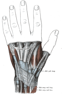
The extensor retinaculum is an anatomical term for the thickened part of the antebrachial fascia that holds the tendons of the extensor muscles in place. It is located on the back of the forearm, just proximal to the hand. It is continuous with the palmar carpal ligament, which is located on the anterior side of the forearm.

The endoneurium is a layer of delicate connective tissue around the myelin sheath of each myelinated nerve fiber in the peripheral nervous system. Its component cells are called endoneurial cells. The endoneuria with their enclosed nerve fibers are bundled into groups called nerve fascicles, each fascicle within its own protective sheath called a perineurium. In sufficiently large nerves multiple fascicles, each with its blood supply and fatty tissue, may be bundled within yet another sheath, the epineurium.
Role of skin in locomotion describes how the integumentary system is involved in locomotion. Typically the integumentary system can be thought of as skin, however the integumentary system also includes the segmented exoskeleton in arthropods and feathers of birds. The primary role of the integumentary system is to provide protection for the body. However, the structure of the skin has evolved to aid animals in their different modes of locomotion. Soft bodied animals such as starfish rely on the arrangement of the fibers in their tube feet for movement. Eels, snakes, and fish use their skin like an external tendon to generate the propulsive forces need for undulatory locomotion. Vertebrates that fly, glide, and parachute also have a characteristic fiber arrangements of their flight membranes that allows for the skin to maintain its structural integrity during the stress and strain experienced during flight.