Histology, also known as microscopic anatomy or microanatomy, is the branch of biology that studies the microscopic anatomy of biological tissues. Histology is the microscopic counterpart to gross anatomy, which looks at larger structures visible without a microscope. Although one may divide microscopic anatomy into organology, the study of organs, histology, the study of tissues, and cytology, the study of cells, modern usage places all of these topics under the field of histology. In medicine, histopathology is the branch of histology that includes the microscopic identification and study of diseased tissue. In the field of paleontology, the term paleohistology refers to the histology of fossil organisms.

Paul Ehrlich was a Nobel Prize-winning German physician and scientist who worked in the fields of hematology, immunology, and antimicrobial chemotherapy. Among his foremost achievements were finding a cure for syphilis in 1909 and inventing the precursor technique to Gram staining bacteria. The methods he developed for staining tissue made it possible to distinguish between different types of blood cells, which led to the ability to diagnose numerous blood diseases.

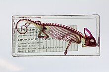
A skeleton is the structural frame that supports the body of most animals. There are several types of skeletons, including the exoskeleton, which is the stable outer shell of an organism, the endoskeleton, which forms the support structure inside the body, and the hydroskeleton, a flexible internal skeleton supported by fluid pressure. Vertebrates are animals with a vertebral column, and their skeletons are typically composed of bone and cartilage. Invertebrates are animals that lack a vertebral column. The skeletons of invertebrates vary, including hard exoskeleton shells, plated endoskeletons, or spicules. Cartilage is a rigid connective tissue that is found in the skeletal systems of vertebrates and invertebrates.

A pigment is a colored substance that is completely or nearly insoluble in water. In contrast, dyes are typically soluble, at least at some stage in their use. Generally dyes are often organic compounds whereas pigments are often inorganic compounds. Pigments of prehistoric and historic value include ochre, charcoal, and lapis lazuli.

Alizarin is an organic compound with formula C
14H
8O
4 that has been used throughout history as a prominent red dye, principally for dyeing textile fabrics. Historically it was derived from the roots of plants of the madder genus. In 1869, it became the first natural dye to be produced synthetically.

A microscope slide is a thin flat piece of glass, typically 75 by 26 mm and about 1 mm thick, used to hold objects for examination under a microscope. Typically the object is mounted (secured) on the slide, and then both are inserted together in the microscope for viewing. This arrangement allows several slide-mounted objects to be quickly inserted and removed from the microscope, labeled, transported, and stored in appropriate slide cases or folders etc.

Staining is a technique used to enhance contrast in samples, generally at the microscopic level. Stains and dyes are frequently used in histology, in cytology, and in the medical fields of histopathology, hematology, and cytopathology that focus on the study and diagnoses of diseases at the microscopic level. Stains may be used to define biological tissues, cell populations, or organelles within individual cells.

An acid dye is a dye that is typically applied to a textile at low pH. They are mainly used to dye wool, not cotton fabrics. Some acid dyes are used as food colorants, and some can also be used to stain organelles in the medical field.

Periodic acid–Schiff (PAS) is a staining method used to detect polysaccharides such as glycogen, and mucosubstances such as glycoproteins, glycolipids and mucins in tissues. The reaction of periodic acid oxidizes the vicinal diols in these sugars, usually breaking up the bond between two adjacent carbons not involved in the glycosidic linkage or ring closure in the ring of the monosaccharide units that are parts of the long polysaccharides, and creating a pair of aldehydes at the two free tips of each broken monosaccharide ring. The oxidation condition has to be sufficiently regulated so as to not oxidize the aldehydes further. These aldehydes then react with the Schiff reagent to give a purple-magenta color. A suitable basic stain is often used as a counterstain.
Metachromasia is a characteristical change in the color of staining carried out in biological tissues, exhibited by certain dyes when they bind to particular substances present in these tissues, called chromotropes. For example, toluidine blue becomes dark blue when bound to cartilage. Other widely used metachromatic stains are the haematological Giemsa and May-Grunwald stains that also contain thiazine dyes. The white cell nucleus stains purple, basophil granules intense magenta, whilst the cytoplasms stains blue. The absence of color change in staining is named orthochromasia.
Bismarck brown Y also called C.I. 21000 and C.I. Basic Brown 1, is a diazo dye with the idealized formula [(H2N)2C6H3N2]2C6H4. The dye is a mixture of closely related compounds. It was one of the earliest azo dyes, being described in 1863 by German chemist Carl Alexander von Martius. It is used in histology for staining tissues.

Papanicolaou stain is a multichromatic (multicolored) cytological staining technique developed by George Papanicolaou in 1942. The Papanicolaou stain is one of the most widely used stains in cytology, where it is used to aid pathologists in making a diagnosis. Although most notable for its use in the detection of cervical cancer in the Pap test or Pap smear, it is also used to stain non-gynecological specimen preparations from a variety of bodily secretions and from small needle biopsies of organs and tissues. Papanicolaou published three formulations of this stain in 1942, 1954, and 1960.
Henry Edward Schunck, also known as Edward von Schunck, was a British chemist who did much work with dyes.

Rose madder is a red paint made from the pigment madder lake, a traditional lake pigment extracted from the common madder plant Rubia tinctorum.

Toluidine blue, also known as TBO or tolonium chloride (INN) is a blue cationic (basic) dye used in histology and sometimes clinically.

Alcian blue is any member of a family of polyvalent basic dyes, of which the Alcian blue 8G has been historically the most common and the most reliable member. It is used to stain acidic polysaccharides such as glycosaminoglycans in cartilages and other body structures, some types of mucopolysaccharides, sialylated glycocalyx of cells etc. For many of these targets it is one of the most widely used cationic dyes for both light and electron microscopy. Use of alcian blue has historically been a popular staining method in histology especially for light microscopy in paraffin embedded sections and in semithin resin sections. The tissue parts that specifically stain by this dye become blue to bluish-green after staining and are called "Alcianophilic". Alcian blue staining can be combined with H&E staining, PAS staining and van Gieson staining methods. Alcian blue can be used to quantitate acidic glycans both in microspectrophotometric quantitation in solution or for staining glycoproteins in polyacrylamide gels or on western blots. Biochemists had used it to assay acid polysaccharides in urine since the 1960s for diagnosis of diseases like mucopolysaccharidosis but from 1970's, partly due to lack of availability of Alcian and partly due to length and tediousness of the procedure, alternative methods had to be developed e.g. Dimethyl methylene blue method.

3DISCO is histological method which make biological samples more transparent, by using series of organic solvents for matching refractive index (RI) of tissue and surrounding medium. Structures in transparent tissues can be examined by fluorescence microscopy without need for time-consuming physical sectioning and following reconstruction in silico.
Microtechnique is an aggregate of methods used to prepare micro-objects for studying. It is currently being employed in many fields in life science. Two well-known branches of microtechnique are botanical (plant) microtechnique and zoological (animal) microtechnique.
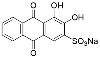
Alizarin Red S is a water-soluble sodium salt of Alizarin sulfonic acid with a chemical formula of C
14H
7NaO
7S. Alizarin Red S was discovered by Graebe and Libermann in 1871. In the field of histology alizarin Red S is used to stain calcium deposits in tissues, and in geology to stain and differentiate carbonate minerals.
Tissue clearing refers to a group of chemical techniques used to turn tissues transparent. This allows deep insight into these tissues, while preserving spatial resolution. Many tissue clearing methods exist, each with different strengths and weaknesses. Some are generally applicable, while others are designed for specific applications. Tissue clearing is usually combined with one or more labeling techniques and subsequently imaged, most often by optical sectioning microscopy techniques. Tissue clearing has been applied to many areas in biological research.