Dilute Russell's viper venom time

Last updated
Russell's viper, Daboia russelii Daboia head.jpg
Russell's viper, Daboia russelii

Dilute Russell's viper venom time (dRVVT) is a laboratory test often used for detection of lupus anticoagulant (LA). It is an assessment of the time for blood to clot in the presence of a diluted amount of venom from Russell's viper (Daboia russelii), a highly venomous snake native to the Indian subcontinent and named after the herpetologist Patrick Russell. [1]

Contents

History

Russell's viper venom (RVV) was known to clot blood many years ago. [2] It was widely used as a styptic to clot minor wounds when razor blades were more commonly used for shaving (e.g. “Stypven”, Burroughs-Wellcome Pharma). RVV came to be useful in laboratory tests for blood clotting factors V, X, prothrombin and phospholipid. [3]

It was first used in clotting tests for lupus anticoagulant (LA) in an individual case in 1975. [4] The “dilute Russells Viper Venom time (dRVVT)” test was then applied in 1985 to diagnose LA in a large number of patients and it became more widely used for this purpose. This multi-step method involved adding individual solutions of dilute phospholipid, RVV and calcium chloride to a test plasma and then measuring how long it took for the mixture to clot. [5]

In 1989, researchers at Westmead Hospital developed a simpler assay by combining the venom, phospholipid, and calcium into a single reagent. Its first use on LA patients was reported in 1990. [6] It was commercialized as “LA Screen” by Gradipore Ltd, Sydney (later Life Diagnostics) and distributed widely by American Diagnostica Inc (New York) as “dVVTest”.

The reagent was improved in 1992 by making it resistant to the widely used interfering anticoagulant heparin. Also a new LA resistant version with increased phospholipid was released at that time. This was introduced as “LA-Confirm” by Gradipore and “dVVConfirm” by American Diagnostica. Results with this high phospholipid reagent were not prolonged by most LA but remained similarly affected as in the "screen" test by all other variables in test plasmas (Gradipore product information). The combination of screening and confirmatory dRVVT reagents made identification of LA more simple. [7] Manufacture of these reagents has since passed on to the major diagnostic companies such as Diagnostica Stago, Precision Biologic, and IL/Werfen.

Mechanism

The dRVVT assay relies on the venom of the Russelli viper. The venom contains enzymes (RVV-X) that directly activates clotting factor X (bypassing intrinsic and extrinsic cascades). [8] In the presence of calcium and phospholipids, these factors convert prothrombin into thrombin, leading to fibrin clot formation. The assay uses low concentrations of venom and phospholipids, resulting in a standard clotting time of 35 to 37 seconds. The test has three phases: In screening phase a lower phospholipid quantity is used (35–37 seconds clotting time) to enhance sensitivity to lupus anticoagulant. [9] In confirmatory phase full phospholipid dose (30–35 seconds clotting time) helps validate results. In mixing Study, the Patient plasma is mixed with normal pooled plasma (NPP) in1:1 ratio to assess clotting factor deficiency. If clotting time prolongs during screening, lupus antibodies may be present. Mixing study differentiates between lupus antibodies and factor deficiency. Excess phospholipids in the confirmatory phase shorten clotting time if lupus antibodies are present. [10]

Interpretation

Through the three phases, following ratios are calculated which help interpret the diagnosis:

i) dRVVT Screen Ratio: Patient plasma dRVVT/NPP dRVVT

ii) dRVVT Confirm Ratio (Phospholipid Neutralization): Patient confirm dRVVT/NPP confirm dRVVT

iii) Normalized Phospholipid Neutralization Ratio: dRVVT screen ratio/dRVVT confirm ratio

iv) dRVVT Mix Ratio: (Patient plasma + NPP mix dRVVT)/(NPP dRVVT)

dRVVT Interpretation/Algorithm
dRVVT Screen RatioNormalized Phospholipid Neutralization ratiodRVVT Mix RatioInterpretation
<1.2Not TestedNot TestedNegative
≥1.2≥1.2Not testedProbable Lupus Anticoagulant
≥1.2<1.2<1.20Possible factor deficiency or anticoagulant therapy
≥1.2<1.2≥1.2Possible Factor inhibitor or anticoagulation therapy

Additional calculations are made using percentage correction of dRVVT and Normalizaed percentage correction as under:

i) Percentage correction = [Patient Screen dRVVT/Patient confirm dRVVT] x 100

ii) Normalized percentage correction = [(dRVVT Screen Ratio - dRVVT confirm ratio) /dRVVT screen ratio] x 100 [11]

Normalized percentage correction of >10% is taken as positive and suggests the presence of Lupus anticoagulant. [11]

Limitations

However dRVVT tests are strongly affected by the new direct oral anticoagulants (DOACs) and false positive LA results are obtained particularly with rivaroxaban. [12] It is now possible to specifically remove DOACs from test plasmas with activated carbon and enable the correct diagnosis of LA with the dRVVT system despite their initial presence. [13]

Use in diagnosis

The dRVVT is one component of a workup of a suspected antiphospholipid antibody, the other component being the serological testing for anticardiolipin antibodies and anti-β2 glycoprotein-I antibodies using ELISA technology. The Sapporo criteria require at least one of the above laboratory tests to be positive and the patient to have at least one clinical manifestation of antiphospholipid syndrome, such as vascular thrombosis or fetal mortality/morbidity, in order to diagnose the antiphospholipid syndrome. [14] Positive laboratory test results should be seen on two occasions at least 12 weeks apart in order for diagnosis. Antiphospholipid antibody syndrome is an important marker for recurrent thrombosis, and often warrants indefinite anticoagulant (blood thinner) therapy. Warfarin appears to be preferable to DOACs as the latter have recently been found less effective than expected. [15]

The criteria were defined in 1999, and revised in 2006. [16]

Related Research Articles

<span class="mw-page-title-main">Anticoagulant</span> Class of drugs

An anticoagulant, commonly known as a blood thinner, is a chemical substance that prevents or reduces the coagulation of blood, prolonging the clotting time. Some occur naturally in blood-eating animals, such as leeches and mosquitoes, which help keep the bite area unclotted long enough for the animal to obtain blood.

<span class="mw-page-title-main">Coagulation</span> Process of formation of blood clots

Coagulation, also known as clotting, is the process by which blood changes from a liquid to a gel, forming a blood clot. It results in hemostasis, the cessation of blood loss from a damaged vessel, followed by repair. The process of coagulation involves activation, adhesion and aggregation of platelets, as well as deposition and maturation of fibrin.

<span class="mw-page-title-main">Antiphospholipid syndrome</span> Medical condition

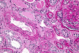
Antiphospholipid syndrome, or antiphospholipid antibody syndrome, is an autoimmune, hypercoagulable state caused by antiphospholipid antibodies. APS can lead to blood clots (thrombosis) in both arteries and veins, pregnancy-related complications, and other symptoms like low platelets, kidney disease, heart disease, and rash. Although the exact etiology of APS is still not clear, genetics is believed to play a key role in the development of the disease. Diagnosis is made based on symptoms and testing, but sometimes research criteria are used to aid in diagnosis. The research criteria for definite APS requires one clinical event and two positive blood test results spaced at least three months apart that detect lupus anticoagulant, anti-apolipoprotein antibodies, and/or anti-cardiolipin antibodies.

<span class="mw-page-title-main">Venereal Disease Research Laboratory test</span> Blood test for syphilis

The Venereal Disease Research Laboratory test (VDRL) is a blood test for syphilis and related non-venereal treponematoses that was developed by the eponymous US laboratory. The VDRL test is used to screen for syphilis, whereas other, more specific tests are used to diagnose the disease.

<span class="mw-page-title-main">Prothrombin time</span> Assay for evaluating the extrinsic pathway & common pathway of coagulation

The prothrombin time (PT) – along with its derived measures of prothrombin ratio (PR) and international normalized ratio (INR) – is an assay for evaluating the extrinsic pathway and common pathway of coagulation. This blood test is also called protime INR and PT/INR. They are used to determine the clotting tendency of blood, in such things as the measure of warfarin dosage, liver damage, and vitamin K status. PT measures the following coagulation factors: I (fibrinogen), II (prothrombin), V (proaccelerin), VII (proconvertin), and X.

<span class="mw-page-title-main">Heparin-induced thrombocytopenia</span> Low platelet count due to heparin, associated with a risk of thrombosis

Heparin-induced thrombocytopenia (HIT) is the development of thrombocytopenia, due to the administration of various forms of heparin, an anticoagulant. HIT predisposes to thrombosis. When thrombosis is identified the condition is called heparin-induced thrombocytopenia and thrombosis (HITT). HIT is caused by the formation of abnormal antibodies that activate platelets, which release microparticles that activate thrombin, leading to thrombosis. If someone receiving heparin develops new or worsening thrombosis, or if the platelet count falls, HIT can be confirmed with specific blood tests.

<span class="mw-page-title-main">Partial thromboplastin time</span> Test for coagulation of blood

The partial thromboplastin time (PTT), also known as the activated partial thromboplastin time, is a blood test that characterizes coagulation of the blood. A historical name for this measure is the kaolin-cephalin clotting time (KCCT), reflecting kaolin and cephalin as materials historically used in the test. Apart from detecting abnormalities in blood clotting, partial thromboplastin time is also used to monitor the treatment effect of heparin, a widely prescribed drug that reduces blood's tendency to clot.

Mixing studies are tests performed on blood plasma of patients or test subjects to distinguish factor deficiencies from factor inhibitors, such as lupus anticoagulant, or specific factor inhibitors, such as antibodies directed against factor VIII. Mixing studies are screening tests widely performed in coagulation laboratories. The basic purpose of these tests is to determine the cause of prolongation of Prothrombin Time (PT), Partial Thromboplastin Time, or sometimes of thrombin time (TT). Mixing studies take advantage of the fact that factor levels that are 50 percent of normal should give a normal Prothrombin time (PT) or Partial thromboplastin time (PTT) result. Factor deficient plasmas are used in mixing studies. Plasma with known factor deficiencies are commercially available but are very expensive, so they are often prepared in the laboratory and can then be used for mixing tests.

Lupus anticoagulant is an immunoglobulin that binds to phospholipids and proteins associated with the cell membrane. Its name is a partial misnomer, as it is actually a prothrombotic antibody in vivo. The name derives from their properties in vitro, as these antibodies increase coagulation times in laboratory tests such as the activated partial thromboplastin time (aPTT). Investigators speculate that the antibodies interfere with phospholipids used to induce in vitro coagulation. In vivo, the antibodies are thought to interact with platelet membrane phospholipids, increasing adhesion and aggregation of platelets, which accounts for the in vivo prothrombotic characteristics.

<span class="mw-page-title-main">Thrombophilia</span> Abnormality of blood coagulation

Thrombophilia is an abnormality of blood coagulation that increases the risk of thrombosis. Such abnormalities can be identified in 50% of people who have an episode of thrombosis that was not provoked by other causes. A significant proportion of the population has a detectable thrombophilic abnormality, but most of these develop thrombosis only in the presence of an additional risk factor.

<span class="mw-page-title-main">Russell's viper</span> Species of venomous snake

Russell's viper is a highly venomous snake in the family Viperidae native to the Indian subcontinent and one of the big four snakes in India. It was described in 1797 by George Shaw and Frederick Polydore Nodder, and named after Patrick Russell, who wrote about it in his 1796 work An account of Indian serpents, collected on the coast of Coromandel.

<span class="mw-page-title-main">Protein C deficiency</span> Medical condition

Protein C deficiency is a rare genetic trait that predisposes to thrombotic disease. It was first described in 1981. The disease belongs to a group of genetic disorders known as thrombophilias. Protein C deficiency is associated with an increased incidence of venous thromboembolism, whereas no association with arterial thrombotic disease has been found.

Catastrophic antiphospholipid syndrome (CAPS), also known as Asherson's syndrome, is a rare autoimmune disease in which widespread, intravascular clotting causes multi-organ failure. The syndrome is caused by antiphospholipid antibodies that target a group of proteins in the body that are associated with phospholipids. These antibodies activate endothelial cells, platelets, and immune cells, ultimately causing a large inflammatory immune response and widespread clotting. CAPS was first described by Ronald Asherson in 1992. The syndrome exhibits thrombotic microangiopathy, multiple organ thromboses, and in some cases tissue necrosis and is considered an extreme or catastrophic variant of the antiphospholipid syndrome.

<span class="mw-page-title-main">Thrombin time</span>

The thrombin time (TT), also known as the thrombin clotting time (TCT), is a blood test that measures the time it takes for a clot to form in the plasma of a blood sample containing anticoagulant, after an excess of thrombin has been added. It is used to diagnose blood coagulation disorders and to assess the effectiveness of fibrinolytic therapy. This test is repeated with pooled plasma from normal patients. The difference in time between the test and the 'normal' indicates an abnormality in the conversion of fibrinogen to fibrin, an insoluble protein.

<span class="mw-page-title-main">Ecarin clotting time</span>

Ecarin clotting time (ECT) is a laboratory test used to monitor anticoagulation during treatment with hirudin, an anticoagulant medication which was originally isolated from leech saliva. Ecarin, the primary reagent in this assay, is derived from the venom of the saw-scaled viper, Echis carinatus.

Ecarin is an enzyme that is derived from the venom of the Indian saw-scaled viper, Echis carinatus, It is the primary reagent in the Ecarin clotting time test.

<span class="mw-page-title-main">Anti-cardiolipin antibodies</span> Type of autoantibody

Anti-cardiolipin antibodies (ACA) are antibodies often directed against cardiolipin and found in several diseases, including syphilis, antiphospholipid syndrome, livedoid vasculitis, vertebrobasilar insufficiency, Behçet's syndrome, idiopathic spontaneous abortion, and systemic lupus erythematosus (SLE). They are a form of anti-mitochondrial antibody. In SLE, anti-DNA antibodies and anti-cardiolipin antibodies may be present individually or together; the two types of antibodies act independently. This is in contrast to rheumatoid arthritis with systemic sclerosis (scleroderma) because anti-cardiolipin antibodies are present in both conditions, and therefore may tie the two conditions together.

In autoimmune disease, anti-apolipoprotein H (AAHA) antibodies, also called anti-β2 glycoprotein I antibodies, comprise a subset of anti-cardiolipin antibodies and lupus anticoagulant. These antibodies are involved in sclerosis and are strongly associated with thrombotic forms of lupus. As a result, AAHA are strongly implicated in autoimmune deep vein thrombosis.

Blood clots are a relatively common occurrence in the general population and are seen in approximately 1-2% of the population by age 60. Typically, blood clots develop in the deep veins of the lower extremities, deep vein thrombosis (DVT) or as a blood clot in the lung, pulmonary embolism. A very small number of people who develop blood clots have a more serious and often life-threatening condition, known as thrombotic storm (TS). TS is characterized by the development of more than one blood clot in a short period of time. These clots often occur in multiple and sometimes unusual locations in the body and are often difficult to treat. TS may be associated with an existing condition or situation that predisposes a person to blood clots, such as injury, infection, or pregnancy. In many cases, a risk assessment will identify interventions that will prevent the formation of blood clots.

Kaolin clotting time (KCT) is a sensitive test to detect lupus anticoagulants. There is evidence that suggests it is the most sensitive test for detecting lupus anticoagulants. It can also detect factor VIII inhibitors but is sensitive to unfractionated heparin as well.

References

  1. Favaloro EJ (4 August 2019). "The Russell viper venom time (RVVT) test for investigation of lupus anticoagulant (LA)". American Journal of Hematology. 94 (11): 1290–1296. doi: 10.1002/ajh.25606 . PMID   31379004. S2CID   199438687.
  2. Macfarlane RG (July 1967). "Russell's viper venom, 1934-64". British Journal of Haematology. 13 (4): 437–51. doi:10.1111/j.1365-2141.1967.tb00754.x. PMID   6067638. S2CID   2208466.
  3. Marsh NA (July 1998). "Use of snake venom fractions in the coagulation laboratory". Blood Coagulation & Fibrinolysis. 9 (5): 395–404. doi:10.1097/00001721-199807000-00001. PMID   9712287.
  4. Exner T, Rickard KA, Kronenberg H (October 1975). "Studies on phospholipids in the action of a lupus coagulation inhibitor". Pathology. 7 (4): 319–28. doi:10.3109/00313027509081688. PMID   1223721. S2CID   24552164.
  5. Thiagarajan P, Pengo V, Shapiro SS (October 1986). "The use of the dilute Russell viper venom time for the diagnosis of lupus anticoagulants". Blood. 68 (4): 869–74. doi: 10.1182/blood.V68.4.869.869 . PMID   3092888.
  6. Exner T, Papadopoulos G, Koutts J (August 1990). "Use of a simplified dilute Russell's viper venom time (DRVVT) confirms heterogeneity among 'lupus anticoagulants'". Blood Coagulation & Fibrinolysis. 1 (3): 259–66. doi:10.1097/00001721-199008000-00002. PMID   2129412.
  7. Laboratory testing for the lupus anticoagulant : approved guideline. Clinical and Laboratory Standards Institute. 2014. ISBN   978-1-56238-959-8.
  8. Castro-Amorim J, Oliveira A, Mukherjee AK, Ramos MJ, Fernandes PA (2023-07-10). "Unraveling the Reaction Mechanism of Russell's Viper Venom Factor X Activator: A Paradigm for the Reactivity of Zinc Metalloproteinases?". Journal of Chemical Information and Modeling. 63 (13): 4056–4069. doi:10.1021/acs.jcim.2c01156. ISSN   1549-9596. PMC   10336966 . PMID   37092784.
  9. Hillarp A, Strandberg K, Gustafsson KM, Lindahl TL (August 2020). "Unveiling the complex effects of direct oral anticoagulants on dilute Russell's viper venom time assays". Journal of Thrombosis and Haemostasis. 18 (8): 1866–1873. doi:10.1111/jth.14829.
  10. Vandevelde A, Devreese KM (2022-04-13). "Laboratory Diagnosis of Antiphospholipid Syndrome: Insights and Hindrances". Journal of Clinical Medicine. 11 (8): 2164. doi: 10.3390/jcm11082164 . ISSN   2077-0383. PMC   9025581 . PMID   35456258.
  11. 1 2 Moore GW (March 2014). "Recent Guidelines and Recommendations for Laboratory Detection of Lupus Anticoagulants". Seminars in Thrombosis and Hemostasis. 40 (2): 163–171. doi:10.1055/s-0033-1364185. ISSN   0094-6176. PMID   24500573.
  12. Flieder T, Weiser M, Eller T, Dittrich M, von Bargen K, Alban S, Kuhn J, Knabbe C, Birschmann I (May 2018). "Interference of DOACs in different DRVVT assays for diagnosis of lupus anticoagulants". Thrombosis Research. 165: 101–106. doi:10.1016/j.thromres.2018.03.009. PMID   29627719.
  13. Favaloro EJ, Gilmore G, Arunachalam S, Mohammed S, Baker R (August 2019). "Neutralising rivaroxaban induced interference in laboratory testing for lupus anticoagulant (LA): A comparative study using DOAC Stop and andexanet alfa" (PDF). Thrombosis Research. 180: 10–19. doi:10.1016/j.thromres.2019.05.013. PMID   31158643. S2CID   174807712.
  14. Miyakis S, Lockshin MD, Atsumi T, Branch DW, Brey RL, Cervera R, Derksen RH, De Groot PG, et al. (2006). "International consensus statement on an update of the classification criteria for definite antiphospholipid syndrome (APS)". Journal of Thrombosis and Haemostasis. 4 (2): 295–306. doi:10.1111/j.1538-7836.2006.01753.x. hdl: 11379/21509 . PMID   16420554. S2CID   9752817.
  15. Kajy M, Mathew A, Ramappa P (9 October 2019). "Treatment Failures of Direct Oral Anticoagulants". American Journal of Therapeutics. 28 (1): e87–e95. doi:10.1097/MJT.0000000000001083. PMID   31599766. S2CID   204029056.
  16. Kaul M, Erkan D, Sammaritano L, Lockshin MD (July 2007). "Assessment of the 2006 revised antiphospholipid syndrome classification criteria". Ann. Rheum. Dis. 66 (7): 927–30. doi:10.1136/ard.2006.067314. PMC   2497429 . PMID   17337473.