Related Research Articles

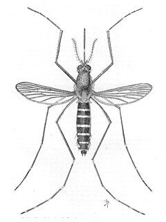
A mosquito is any member of a group of about 3,500 species of small insects belonging to the order Diptera (flies). Within Diptera, mosquitoes constitute the family Culicidae. The word "mosquito" is Spanish and Portuguese for "little fly". Mosquitoes have a slender segmented body, one pair of wings, one pair of halteres, three pairs of long hair-like legs, and elongated mouthparts.

Loa loa filariasis is a skin and eye disease caused by the nematode worm Loa loa. Humans contract this disease through the bite of a deer fly or mango fly, the vectors for Loa loa. The adult Loa loa filarial worm migrates throughout the subcutaneous tissues of humans, occasionally crossing into subconjunctival tissues of the eye where it can be easily observed. Loa loa does not normally affect one's vision but can be painful when moving about the eyeball or across the bridge of the nose. The disease can cause red itchy swellings below the skin called "Calabar swellings". The disease is treated with the drug diethylcarbamazine (DEC), and when appropriate, surgical methods may be employed to remove adult worms from the conjunctiva. Loiasis belongs to the so-called neglected diseases.

Filariasis is a parasitic disease caused by an infection with roundworms of the Filarioidea type. These are spread by blood-feeding insects such as black flies and mosquitoes. They belong to the group of diseases called helminthiases.

Wuchereria bancrofti is a human parasitic worm (Filariworm) that is the major cause of lymphatic filariasis. It is one of the three parasitic worms, together with Brugia malayi and B. timori, that infect the lymphatic system to cause lymphatic filariasis. These filarial worms are spread by a variety of mosquito vector species. W. bancrofti is the most prevalent of the three and affects over 120 million people, primarily in Central Africa and the Nile delta, South and Central America, the tropical regions of Asia including southern China, and the Pacific islands. If left untreated, the infection can develop into a chronic disease called lymphatic filariasis. In rare conditions, it also causes tropical eosinophilia, an asthmatic disease. No vaccine is commercially available, but high rates of cure have been achieved with various antifilarial regimens and lymphatic filariasis is the target of the WHO Global Program to Eliminate Lymphatic Filariasis with the aim to eradicate the disease as a public-health problem by 2020.

Anopheles is a genus of mosquito first described and named by J. W. Meigen in 1818. About 460 species are recognised; while over 100 can transmit human malaria, only 30–40 commonly transmit parasites of the genus Plasmodium, which cause malaria in humans in endemic areas. Anopheles gambiae is one of the best known, because of its predominant role in the transmission of the most dangerous malaria parasite species – Plasmodium falciparum.
Gnathostomiasis is the human infection caused by the nematode (roundworm) Gnathostoma spinigerum and/or Gnathostoma hispidum, which infects vertebrates.

Brugia malayi is a nematode (roundworm), one of the three causative agents of lymphatic filariasis in humans. Lymphatic filariasis, also known as elephantiasis, is a condition characterized by swelling of the lower limbs. The two other filarial causes of lymphatic filariasis are Wuchereria bancrofti and Brugia timori, which both differ from B. malayi morphologically, symptomatically, and in geographical extent.

Dirofilaria immitis, also known as heartworm or dog heartworm, is a parasitic roundworm that is a type of filarial worm, a small thread-like worm, that causes dirofilariasis. It is spread from host to host through the bites of mosquitoes. There are four genera of mosquitoes that transmit dirofilariasis, Aedes, Culex, Anopheles, and Mansonia. The definitive host is the dog, but it can also infect cats, wolves, coyotes, jackals, foxes, ferrets, red deers, pudús, bears, seals, sea lions and, under rare circumstances, humans.
Genus Acanthocheilonema refers to a taxonomic genus within the family Onchocercidae which comprises mainly tropical parasitic worms. Cobbold created the genus Acanthocheilonema with only one species, Acanthocheilonema dracunculoides, which was collected from aardwolf in the region of South Africa in nineteenth century. These parasites has a wide range of mammalian species, namely Carnivora, Macroscelidea, Rodentia, Pholidota, Edentata, and Marsupialia. Although many species among several genera of filarioids exhibit high degree of endemecity in studies done on mammalian species in Japan region. However, no concrete evidence has confirmed any endemic species in the genus Acanthocheilonema.
Sparganosis is a parasitic infection caused by the plerocercoid larvae of the genus Spirometra including S. mansoni, S. ranarum, S. mansonoides and S. erinacei. It was first described by Patrick Manson from China in 1882, and the first human case was reported by Charles Wardell Stiles from Florida in 1908. The infection is transmitted by ingestion of contaminated water, ingestion of a second intermediate host such as a frog or snake, or contact between a second intermediate host and an open wound or mucous membrane. Humans are the accidental hosts in the life cycle, while dogs, cats, and other mammals are definitive hosts. Copepods are the first intermediate hosts, and various amphibians and reptiles are second intermediate hosts.

Dirofilariasis is an infection by parasites of the genus Dirofilaria. It is transmitted through a mosquito bite; its main hosts include dogs and wild canids. These can give rise to granulomas in the pulmonary artery. Some common symptoms include cough, fever and pleural effusion. It may also appear on x-rays of the chest.
In population ecology, density-dependent processes occur when population growth rates are regulated by the density of a population. This article will focus on density-dependence in the context of macroparasite life cycles.
Mosquito-borne diseases or mosquito-borne illnesses are diseases caused by bacteria, viruses or parasites transmitted by mosquitoes. Nearly 700 million people get a mosquito-borne illness each year resulting in over one million deaths.

Dirofilaria is a genus of nematodes, or roundworms, in the family Onchocercidae. Some species cause dirofilariasis, a state of parasitic infection, in humans and other animals.
Dirofilaria repens is a filarial nematode that affects dogs and other carnivores such as cats, wolves, coyotes, foxes, and sea lions, as well as muskrats. It is transmitted by mosquitoes. Although humans may become infected as aberrant hosts, the worms fail to reach adulthood while infecting a human body.

The Filarioidea are a superfamily of highly specialised parasitic nematodes. Species within this superfamily are known as filarial worms or filariae. Infections with parasitic filarial worms cause disease conditions generically known as filariasis. Drugs against these worms are known as filaricides.

Anopheles albimanus is a species of mosquito in the order Diptera. It is found in coastal Central and South America, the Caribbean, and Mexico. It is a generalist species and capable of wide dispersion. A. albimanus is a common malaria vector.
Taenia serialis, also known as a canid tapeworm, is found within canines such as foxes and dogs. Adult T. serialis are parasites of carnivores, particularly dogs, with herbivorous lagomorph mammals such as rabbits and hares, serving as intermediate hosts. In definitive hosts, T. serialis is acquired by eating tissues from a variety of intermediate hosts. Accidental infection of humans though, can occur when eggs are ingested from food or water contaminated with dog feces and the human then becomes the T. serialis intermediate host.
Setaria is a genus of parasitic roundworms that infect domesticated mammals such as pigs, camels, cattle and horses. Some species also infect wild mammals such as deer and antelope. The genus consists of about 43 species. Members of the genus are uniquely parasites in the abdominal cavity of the body. They are mostly large-sized roundworms, possessing an elaborate head (cephalic) region that is characterised by spines, presence of four lips, and well-guarded mouth. Little is known about their pathogenic effects, but some are known to affect nervous system and eye. The larval infective forms are transmitted from one animal to another by the bite of mosquitoes and flies. In addition Setaria marshalli can be transmitted from the womb to new-born calf.
Aedes taeniorhynchus, or the black salt marsh mosquito, is a mosquito in the family Culicidae. It is a carrier for encephalitic viruses including Venezuelan equine encephalitis and can transmit Dirofilaria immitis. It resides in the Americas and is known to bite mammals, reptiles, and birds. Like other mosquitoes, Ae. taeniorhynchus adults survive on a combination diet of blood and sugar, with females generally requiring a blood meal before laying eggs.
References
- ↑ Asa C. Chandler. The Helminths of Raccoons in East Texas. The Journal of Parasitology. Vol. 28, No. 4 (Aug., 1942), pp. 255–268. Published by: The American Society of Parasitologists
- 1 2 Capinera, John L. (2008). Encyclopedia of Entomology Vol. 4. New York: Springer. p. 1228.
- ↑ Studies on the Development of Dirofilaria tenuis Chandler 1942. Warren R. Pistey, The Journal of Parasitology, Vol. 44, No. 6 (Dec., 1958), pp. 613–626
- ↑ Watts KJ, Courteny CH, Reddy GR (December 1999). "Development of a PCR- and probe-based test for the sensitive and specific detection of the dog heartworm, Dirofilaria immitis, in its mosquito intermediate host". Mol. Cell. Probes. 13 (6): 425–30. doi:10.1006/mcpr.1999.0270. PMID 10657147.
- 1 2 3 Pistey, Warren R. (December 1958). "Studies on the Development of Dirofilaria Tenuis Chandler 1942". The Journal of Parasitology. 44 (6): 613–626. doi:10.2307/3274546. JSTOR 3274546.
- 1 2 Beaver, Paul C.; Thomas C. Orihel (November 1965). "Morphology and Relationship of Dirofilaria Tenuis and Dirofilaria Conjuntivae". The American Journal of Tropical Medicine and Hygiene. 14 (6): 1030–1043. doi:10.4269/ajtmh.1965.14.1030. PMID 5840636.
- 1 2 3 4 "Biology-Life Cycle of D. Tenuis". Center for Disease Control and Prevention. Retrieved 23 Oct 2013.
- ↑ Wong, Ming M.; K. C. Lim (January 1976). "Experimental Dirofilariasis in Macaques: Susceptibility and Host Responses to Dirofilaria tenuis of Raccoons". The American Journal of Tropical Medicine and Hygiene. 25 (1): 94–98. doi:10.4269/ajtmh.1976.25.94.
- 1 2 Warthan, Mandy; Warthan, T.; Hearne, Ron; Polk, Aaron; Warthan, Molly (2007). "Human Dirofilariasis: Raccoon Heartworm Causing a Leg Nodule". Cutis. 80 (2): 125–128.