
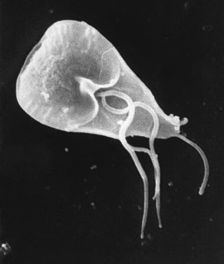
Isosporiasis, also known as cystoisosporiasis, is a human intestinal disease caused by the parasite Cystoisospora belli. It is found worldwide, especially in tropical and subtropical areas. Infection often occurs in immuno-compromised individuals, notably AIDS patients, and outbreaks have been reported in institutionalized groups in the United States. The first documented case was in 1915. It is usually spread indirectly, normally through contaminated food or water (CDC.gov).

Fasciolosis is a parasitic worm infection caused by the common liver fluke Fasciola hepatica as well as by Fasciola gigantica. The disease is a plant-borne trematode zoonosis, and is classified as a neglected tropical disease (NTD). It affects humans, but its main host is ruminants such as cattle and sheep. The disease progresses through four distinct phases; an initial incubation phase of between a few days up to three months with little or no symptoms; an invasive or acute phase which may manifest with: fever, malaise, abdominal pain, gastrointestinal symptoms, urticaria, anemia, jaundice, and respiratory symptoms. The disease later progresses to a latent phase with less symptoms and ultimately into a chronic or obstructive phase months to years later. In the chronic state the disease causes inflammation of the bile ducts, gall bladder and may cause gall stones as well as fibrosis. While chronic inflammation is connected to increased cancer rates, it is unclear whether fasciolosis is associated with increased cancer risk.

Coccidia (Coccidiasina) are a subclass of microscopic, spore-forming, single-celled obligate intracellular parasites belonging to the apicomplexan class Conoidasida. As obligate intracellular parasites, they must live and reproduce within an animal cell. Coccidian parasites infect the intestinal tracts of animals, and are the largest group of apicomplexan protozoa.
Coccidiosis is a parasitic disease of the intestinal tract of animals caused by coccidian protozoa. The disease spreads from one animal to another by contact with infected feces or ingestion of infected tissue. Diarrhea, which may become bloody in severe cases, is the primary symptom. Most animals infected with coccidia are asymptomatic, but young or immunocompromised animals may suffer severe symptoms and death.

Eimeria tenella is a species of Eimeria that causes hemorrhagic cecal coccidiosis in young poultry. It is found worldwide.

Eimeria is a genus of apicomplexan parasites that includes various species capable of causing the disease coccidiosis in animals such as cattle, poultry and smaller ruminants including sheep and goats. Eimeria species are considered to be monoxenous because the life cycle is completed within a single host, and stenoxenous because they tend to be host specific, although a number of exceptions have been identified. Species of this genus infect a wide variety of hosts. Thirty-one species are known to occur in bats (Chiroptera), two in turtles, and 130 named species infect fish. Two species infect seals. Five species infect llamas and alpacas: E. alpacae, E. ivitaensis, E. lamae, E. macusaniensis, and E. punonensis. A number of species infect rodents, including E. couesii, E. kinsellai, E. palustris, E. ojastii and E. oryzomysi. Others infect poultry, rabbits and cattle. For full species list, see below.

Neospora caninum is a coccidian parasite that was identified as a species in 1988. Prior to this, it was misclassified as Toxoplasma gondii due to structural similarities. The genome sequence of Neospora caninum has been determined by the Wellcome Trust Sanger Institute and the University of Liverpool. Neospora caninum is an important cause of spontaneous abortion in infected livestock.

Sarcocystis is a genus of protozoan parasites, with many species infecting mammals, reptiles and birds. Its name is dervived from Greek sarx = flesh and kystis = bladder.
Neospora is a single celled parasite of livestock and companion animals. It was not discovered until 1984 in Norway, where it was found in dogs. Neosporosis, the disease that affects cattle and companion animals, has a worldwide distribution. Neosporosis causes abortions in cattle and paralysis in companion animals. It is highly transmissible and some herds can have up to a 90% prevalence. Up to 33% of pregnancies can result in aborted fetuses on one dairy farm. In many countries this organism is the main cause of abortion in cattle. Neosporosis is now considered as a major cause of abortion in cattle worldwide. Many reliable diagnostic tests are commercially available. Neospora caninum does not appear to be infectious to humans. In dogs, Neospora caninum can cause neurological signs, especially in congenitally infected puppies, where it can form cysts in the central nervous system.
Eimeria stiedae is a species of Eimeria that causes hepatic coccidiosis in rabbits. It was observed for the first time by Antonie van Leeuwenhoek in 1674.
Protozoan infections are parasitic diseases caused by organisms formerly classified in the kingdom Protozoa. These organisms are now classified in the supergroups Excavata, Amoebozoa, Harosa, and Archaeplastida. They are usually contracted by either an insect vector or by contact with an infected substance or surface.
Neospora hughesi is an obligate protozoan apicomplexan parasite that causes myelitis and equine protozoal myeloencephalitis (EPM) in horses, and has only been documented in North America. EPM is a neurological disease from lesions in the spinal cord, brain stem, or brain from parasites such as N. hughesi or Sarcocystis neurona. Signs that a horse may have EPM include ataxia, muscle atrophy, difficulty swallowing, and head tilt. There are antiprotozoal drugs, such as the 28-day course of ponazuril, to treat the disease, as well as anti-inflammatories to alleviate neurologic symptoms
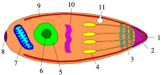
Apicomplexans, a group of intracellular parasites, have life cycle stages that allow them to survive the wide variety of environments they are exposed to during their complex life cycle. Each stage in the life cycle of an apicomplexan organism is typified by a cellular variety with a distinct morphology and biochemistry.

Cystoisospora belli, previously known as Isospora belli, is a parasite that causes an intestinal disease known as cystoisosporiasis. This protozoan parasite is opportunistic in immune suppressed human hosts. It primarily exists in the epithelial cells of the small intestine, and develops in the cell cytoplasm. The distribution of this coccidian parasite is cosmopolitan, but is mainly found in tropical and subtropical areas of the world such as the Caribbean, Central and S. America, India, Africa, and S.E. Asia. In the U.S., it is usually associated with HIV infection and institutional living.
Cystoisospora canis, previously known as Isospora canis, is a microscopic, coccidian parasite that causes an intestinal tract infection in dogs. The intestinal tract infection is coccidiosis caused by a protozoa called coccidia.
Ostertagia ostertagi, commonly known as the medium stomach worm or brown stomach worm, is a parasitic nematode of cattle. O. ostertagi can also be found to a lesser extent in sheep, goats, wild ruminants, and horses. It causes ostertagiosis, which is potentially fatal in cattle. It is found worldwide and is economically important to cattle industries, particularly those found in temperate climates.
Growell India is a multinational feed additive company headquartered in Pune, Maharashtra, India. Growell India manufactures and exports phytogenic feed additives for livestock. The company is a pioneer in phytogenic feed additives and has established a strong presence worldwide since its inception in 1995.

Sarcocystis neurona is primarily a neural parasite of horses and its management is of concern in veterinary medicine. The protozoan Sarcocystis neurona is a protozoan of single celled character and belongs to the family Sarcocystidae, in a group called coccidia. The protozoan, S. neurona, is a member of the genus Sarcocystis, and is most commonly associated with equine protozoal myeloencephalitis (EPM). The protozoan, S. neurona, can be easily cultivated and genetically manipulated, hence its common use as a model to study numerous aspects of cell biology.
Eimeria arlongi is a species of Eimeria that causes clinical coccidiosis in goats. It and Eimeria ninakohlyakimovae are two of the most pathogenic species for goats. It is particularly prevalent in goat kids in Iran. Issues with coccidiosis specifically due to Eimeria arloingi have also been reported in Egypt and Portugal. It is unclear whether this species is present in the Americas as most of the case reports of coccidiosis in these areas do not differentiate the species causing the disease. Infections with this species are commonly compounded by infections with other Eimeria species in "mixed infections." This species is closely related to Eimeria bovis and Eimeria zuernii which are both highly pathogenic in cattle' Infections with this species are characterized by lesions specifically in the jejunum, but also the ilium and cecum which results in diarrhea. Oocysts begin shedding between 16 and 18 days after the animal is infected which is when the parasite is spread. The shedding can last as long as 15 days. This parasite causes an immune response in its host that includes accumulation of fluid in body cavities, presence of large numbers of leukocytes in the small intestine, and necrosis of the tissue of the small intestine. Pale yellow plaques can be seen on the small intestine of severely affected kids at necropsy.
Eimeria bovis is a parasite belonging to the genus Eimeria and is found globally. The pathogen can cause a diarrheic disease in cattle referred to as either eimeriosis or coccidiosis. The infection predominantly cause disease in younger animals.