Elbow dysplasia

Last updated

Ununited anconeal process X-ray image of an isolates processus anconaeus in a 5 year old German shepherd dog.jpg
Ununited anconeal process

Elbow dysplasia is a condition involving multiple developmental abnormalities of the elbow-joint in the dog, specifically the growth of cartilage or the structures surrounding it. These abnormalities, known as 'primary lesions', give rise to osteoarthritic processes. [1] Elbow dysplasia is a common condition of certain breeds of dogs.

Contents

Most primary lesions are related to osteochondrosis, a disease of the joint cartilage, and osteochondritis dissecans (OCD), the separation of a flap of cartilage on the joint surface. Other common causes of elbow dysplasia include an ununited anconeal process (UAP) and fragmented or ununited medial coronoid process (FCP or FMCP). [1]

Osteochondritis dissecans is difficult to diagnose clinically as the animal may only exhibit an unusual gait. Consequently, OCD may be masked by, or misdiagnosed as, other skeletal and joint conditions such as hip dysplasia. [2] The problem develops in puppyhood, although often is subclinical, and there may be pain or stiffness, discomfort on extension, or other compensating characteristics. [2] Diagnosis generally depends on X-rays, arthroscopy, or MRI scans. [2] While cases of OCD of the stifle go undetected and heal spontaneously, others are exhibited in acute lameness. [2] Surgery is recommended once the animal has been deemed lame; otherwise, non-surgical control is usually used. [2]

The disease

Elbow dysplasia is a significant genetically determined problem in many breeds of dog, often manifesting from puppyhood and continuing for life. [1] In elbow dysplasia, the complex elbow joint suffers from a structural defect, often related to its cartilage. [1] This initial condition, known as a "primary lesion", [1] causes an abnormal level of wear and tear and gradual degradation of the joint, at times disabling or with chronic pain. Secondary processes such as inflammation and osteoarthritis can arise from this damage [1] which increase the problem and add further problems of their own.

Causes

Human femur head showing a flap of cartilage (osteochondritis dissecans) due to avascular necrosis. Similar cartilage damage in dogs causes wear in the joint leading to osteoarthritic processes and is a common cause of elbow dysplasia. Head of femur avascular necrosis.jpg
Human femur head showing a flap of cartilage (osteochondritis dissecans) due to avascular necrosis. Similar cartilage damage in dogs causes wear in the joint leading to osteoarthritic processes and is a common cause of elbow dysplasia.

The most common cause is osteochondrosis, which is a disease of the joint cartilage, and osteochondritis dissecans (OCD or OD), the separation of a flap of cartilage from the joint surface [1] as a result of avascular necrosis, which in turn arises from failed blood flow in the subchondral bone. [3]

In OCD, the normal change of cartilage to bone in the development of the joint fails or is delayed. The cartilage continues to grow and may split or become necrotic. The cause is uncertain, but possibly includes genetics, trauma, and nutrition (including excessive calcium and decreased vitamin C intake). [4] OCD lesions found in the elbow at the medial epicondyle of the humerus are caused by disturbed endochondral fusion of the epiphysis of the medial epicondyle with the distal end of the humerus, which may in turn be caused by avulsion of the epiphysis. [5] Specific conditions related to OCD include fragmentation of the medial coronoid process of the ulna (FMCP) and an ununited anconeal process of the ulna (UAP). All types of OCD of the elbow are most typically found in large breed dogs, with symptoms starting between the ages of 4 and 8 months. [4] Males are affected twice as often as females. The disease often affects both elbows (30 to 70 percent of the time), and symptoms include intermittent lameness, joint swelling, and external rotation and abduction of the paw. [6] Osteoarthritis will develop later in most cases.

UAP is caused by a separation from the ulna of the ossification center of the anconeal process. [7] FMCP is caused by a failure of the coronoid process to unite with the ulna.

Diagnosis and treatment

Diagnosis is through x-rays, arthroscopy or CT (computed tomography). In cases with significant lameness, surgery is the best option, especially with UAP. However, conservative treatment is often enough for cases of FMCP and OCD of the medial humeral epicondyle. The dogs are exercised regularly and given pain medication, and between the ages of 12 and 18 months the lameness will often improve or disappear. [4] Control of body weight is important in all cases of elbow dysplasia, and prevention of quick growth spurts in puppies may help to prevent the disease.

Surgery for FMCP consists of removal of cartilage and bone fragments and correction of any incongruity of the joint. Reattachment of UAP with a screw is usually attempted before the age of 24 weeks, and after that age the typical treatment is removal of the UAP. [8] Without surgery, UAP rapidly progresses to osteoarthritis, but with FMCP osteoarthritis typically occurs with or without surgery. Osteoarthritis is also a common sequela of OCD of the humerus despite medical or surgical treatment. [8] Elbow replacement surgery has been developed and can be an option for treatment. [9]

Fragmentation of the medial coronoid process Elbowdysplasia-dog.jpg
Fragmentation of the medial coronoid process

Non-surgical treatment

Conservative therapies include NSAIDs, pain medication, weight management and exercise restrictions. [10]

Total elbow replacement

The elbow is a complex joint, bears 60% of body load, and tolerates problems less well than the hips. [11] As a result, elbow replacement is more complex than hip replacement, rehabilitation can take significantly longer, and some degree of lameness will remain. [11] The surgery is classified as of 2010 as a "salvage" operation - that is, a last resort for an otherwise viable animal. [12] Success rates of around 80 - 85% (approximately 5 in 6) were being discussed by sources in 2005 [11] and again in 2008. [13]

There are also fewer options if the replacement fails; arthrodesis (surgical fusion of the joint) can result in a pain-free but lame gait. [11] However, arthrodesis is itself a complex surgery with a long recovery time, [11] and if arthrodesis is required, additional strain will be borne by other nearby joints, so other leg and shoulder conditions such as osteoarthritis may become more significant. [11]

Grading for breeding purposes

The Orthopedic Foundation for Animals in the United States will grade elbow X-rays of dogs intended for breeding.

Related Research Articles

<span class="mw-page-title-main">Knee</span> Region around the kneecap

In humans and other primates, the knee joins the thigh with the leg and consists of two joints: one between the femur and tibia, and one between the femur and patella. It is the largest joint in the human body. The knee is a modified hinge joint, which permits flexion and extension as well as slight internal and external rotation. The knee is vulnerable to injury and to the development of osteoarthritis.

<span class="mw-page-title-main">Ulna</span> Medial bone from forearm

The ulna is a long bone found in the forearm that stretches from the elbow to the wrist, and when in anatomical position, is found on the medial side of the forearm. That is, the ulna is on the same side of the forearm as the little finger. It runs parallel to the radius, the other long bone in the forearm. The ulna is usually slightly longer than the radius, but the radius is thicker. Therefore, the radius is considered to be the larger of the two.

<span class="mw-page-title-main">Humerus</span> Long bone of the upper arm

The humerus is a long bone in the arm that runs from the shoulder to the elbow. It connects the scapula and the two bones of the lower arm, the radius and ulna, and consists of three sections. The humeral upper extremity consists of a rounded head, a narrow neck, and two short processes. The body is cylindrical in its upper portion, and more prismatic below. The lower extremity consists of 2 epicondyles, 2 processes, and 3 fossae. As well as its true anatomical neck, the constriction below the greater and lesser tubercles of the humerus is referred to as its surgical neck due to its tendency to fracture, thus often becoming the focus of surgeons.

<span class="mw-page-title-main">Ulnar nerve</span> Nerve which runs near the ulna bone

In human anatomy, the ulnar nerve is a nerve that runs near the ulna bone. The ulnar collateral ligament of elbow joint is in relation with the ulnar nerve. The nerve is the largest in the human body unprotected by muscle or bone, so injury is common. This nerve is directly connected to the little finger, and the adjacent half of the ring finger, innervating the palmar aspect of these fingers, including both front and back of the tips, perhaps as far back as the fingernail beds.

<span class="mw-page-title-main">Epiphysis</span> End of a long bone that ossifies from a secondary center

An epiphysis is one of the rounded ends or tips of a long bone that ossify from a secondary center of ossification. Between the epiphysis and diaphysis lies the metaphysis, including the epiphyseal plate. At the joint, the epiphysis is covered with articular cartilage; below that covering is a zone similar to the epiphyseal plate, known as subchondral bone.

<span class="mw-page-title-main">Hip dysplasia (canine)</span> Joint abnormality in dogs

In dogs, hip dysplasia is an abnormal formation of the hip socket that, in its more severe form, can eventually cause lameness and arthritis of the joints. It is a genetic (polygenic) trait that is affected by environmental factors. It is common in many dog breeds, particularly the larger breeds, and is the most common single cause of arthritis of the hips.

<span class="mw-page-title-main">Hock (anatomy)</span>

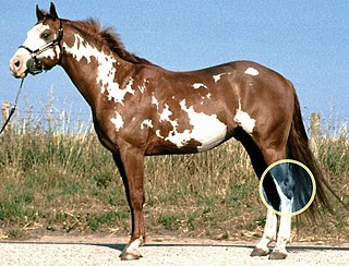
The hock, or gambrel, is the joint between the tarsal bones and tibia of a digitigrade or unguligrade quadrupedal mammal, such as a horse, cat, or dog. This joint may include articulations between tarsal bones and the fibula in some species, while in others the fibula has been greatly reduced and is only found as a vestigial remnant fused to the distal portion of the tibia. It is the anatomical homologue of the ankle of the human foot. While homologous joints occur in other tetrapods, the term is generally restricted to mammals, particularly long-legged domesticated species.

<span class="mw-page-title-main">Ulnar collateral ligament of elbow joint</span> Ligament on the elbow

The ulnar collateral ligament (UCL) or internal lateral ligament is a thick triangular ligament at the medial aspect of the elbow uniting the distal aspect of the humerus to the proximal aspect of the ulna.

Osteochondrosis is a family of orthopedic diseases of the joint that occur in children, adolescents and rapidly growing animals, particularly pigs, horses, dogs, and broiler chickens. They are characterized by interruption of the blood supply of a bone, in particular to the epiphysis, followed by localized bony necrosis, and later, regrowth of the bone. This disorder is defined as a focal disturbance of endochondral ossification and is regarded as having a multifactorial cause, so no one thing accounts for all aspects of this disease.

Osteochondritis is a painful type of osteochondrosis where the cartilage or bone in a joint is inflamed.

<span class="mw-page-title-main">Osteochondritis dissecans</span> Ischemic bone disease

Osteochondritis dissecans is a joint disorder primarily of the subchondral bone in which cracks form in the articular cartilage and the underlying subchondral bone. OCD usually causes pain during and after sports. In later stages of the disorder there will be swelling of the affected joint which catches and locks during movement. Physical examination in the early stages does only show pain as symptom, in later stages there could be an effusion, tenderness, and a crackling sound with joint movement.

<span class="mw-page-title-main">Trochlea of humerus</span> Articular surface of the elbow joint which articulates with the trochlear notch of the ulna

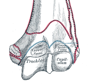
In the human arm, the humeral trochlea is the medial portion of the articular surface of the elbow joint which articulates with the trochlear notch on the ulna in the forearm.

<span class="mw-page-title-main">Anterior ligament of elbow</span>

The anterior ligament of the elbow is a broad and thin fibrous layer covering the anterior surface of the joint.

The coronoid process of the ulna is a triangular process projecting forward from the anterior proximal portion of the ulna.

<span class="mw-page-title-main">Elbow</span> Joint between the upper and lower parts of the arm

The elbow is the region between the upper arm and the forearm that surrounds the elbow joint. The elbow includes prominent landmarks such as the olecranon, the cubital fossa, and the lateral and the medial epicondyles of the humerus. The elbow joint is a hinge joint between the arm and the forearm; more specifically between the humerus in the upper arm and the radius and ulna in the forearm which allows the forearm and hand to be moved towards and away from the body. The term elbow is specifically used for humans and other primates, and in other vertebrates forelimb plus joint is used.

<span class="mw-page-title-main">Knee pain</span> Medical condition

Knee pain is pain in or around the knee.

Panner disease is an osteochondrosis of the capitellum of the elbow. Panner disease is primarily seen in boys between the ages of five and ten years old. Panner disease is often caused by excessive throwing due to valgus stress. The disease causes pain and stiffness in the affected elbow and may limit extension; the affected elbow is usually on the dominant arm the child uses. The disease may be associated with pitching and athletic activity. On radiographs, the capitellum may appear irregular with areas of radiolucency. Treatment is symptomatic, with a good prognosis. Treatment is minimal and includes restricting athletic activity to allow for the elbow to heal and for pain to be relieved. The disease is named after the Danish radiologist Hans Jessen Panner (1871–1930).

<span class="mw-page-title-main">Ulnar collateral ligament injury of the elbow</span> Medical condition

Ulnar collateral ligament injuries can occur during certain activities such as overhead baseball pitching. Acute or chronic disruption and/or attenuation of the ulnar collateral ligament often result in medial elbow pain, valgus instability, neurologic deficiency, and impaired throwing performance. There are both non-surgical and surgical treatment options.

<span class="mw-page-title-main">Index of trauma and orthopaedics articles</span>

Orthopedic surgery is the branch of surgery concerned with conditions involving the musculoskeletal system. Orthopedic surgeons use both surgical and nonsurgical means to treat musculoskeletal injuries, sports injuries, degenerative diseases, infections, bone tumours, and congenital limb deformities. Trauma surgery and traumatology is a sub-specialty dealing with the operative management of fractures, major trauma and the multiply-injured patient.

The treatment of equine lameness is a complex subject. Lameness in horses has a variety of causes, and treatment must be tailored to the type and degree of injury, as well as the financial capabilities of the owner. Treatment may be applied locally, systemically, or intralesionally, and the strategy for treatment may change as healing progresses. The end goal is to reduce the pain and inflammation associated with injury, to encourage the injured tissue to heal with normal structure and function, and to ultimately return the horse to the highest level of performance possible following recovery.

References

  1. 1 2 3 4 5 6 7 Quoted from: Matthew Pead; Sue Guthrie. "Elbow Dysplasia in dogs - a new scheme explained" (PDF). British Veterinary Association (BVA). Archived from the original (PDF) on 2 October 2011. Retrieved 16 July 2010.
  2. 1 2 3 4 5 Lenehan TM, Van Sickle DC (1985). "Chapter 84: Canine osteochondrosis". In Nunamaker DM, Newton CD (eds.). Textbook of small animal orthopaedics. Philadelphia: Lippincott. ISBN   0-397-52098-0.
  3. Shiel WC Jr. "Definition of Osteochondritis dissecans". MedicineNet, Inc. Retrieved 20 February 2009.
  4. 1 2 3 Ettinger, Stephen J.; Feldman, Edward C. (1995). Textbook of Veterinary Internal Medicine (4th ed.). W.B. Saunders Company. ISBN   0-7216-6795-3.
  5. "Elbow Dysplasia". The Merck Veterinary Manual. 2006. Retrieved 4 February 2007.
  6. Hazewinkel, H.A.W. (2003). "Elbow Dysplasia; Clinical Aspects and Screening Programs". Proceedings of the 28th World Congress of the World Small Animal Veterinary Association. Retrieved 24 August 2006.
  7. "Ununited Anoconeal Process". Fitzpatrick Referrals. 2014. Retrieved 14 January 2014.
  8. 1 2 Demko J, McLaughlin R (2005). "Developmental orthopedic disease". Vet Clin North Am Small Anim Pract. 35 (5): 1111–35, v. doi:10.1016/j.cvsm.2005.05.002. PMID   16129135.
  9. "Orthopaedics Fact Sheets" . Retrieved 29 July 2023.
  10. Innovations asgvets.com [ dead link ]
  11. 1 2 3 4 5 6 Brown; Gilbert; Broome (November 2005). "Total Elbow Replacement in Dogs" (PDF). Grove Orthopaedic Referrals. Archived from the original (PDF) on 9 March 2012. Retrieved 17 July 2010.
  12. "Elbow dysplasia 2 / OC and OCD". Fitzpatrick referrals. Retrieved 14 January 2014.
  13. "Total Elbow Replacement". University of Minnesota Veterinary Clinical Sciences dept. Retrieved 17 July 2010.