Fetal movement

Last updated
The human fetus moves throughout its entire development. Views of a Foetus in the Womb detail.jpg
The human fetus moves throughout its entire development.

Fetal movement refers to motion of a fetus caused by its own muscle activity. Locomotor activity begins during the late embryological stage and changes in nature throughout development. Muscles begin to move as soon as they are innervated. These first movements are not reflexive, but arise from self-generated nerve impulses originating in the spinal cord. As the nervous system matures, muscles can move in response to stimuli. [1]

Contents

Generally speaking, fetal motility can be classified as either elicited or spontaneous, and spontaneous movements may be triggered by either the spine or the brain. Whether a movement is supraspinally determined can be inferred by comparison to movements of an anencephalic fetus. [2]

This article primarily deals with voluntary and reflex movements. Ages are given as age from fertilization rather than as gestational age.

Some sources contend that there is no voluntary movement until after birth. [3] Other sources say that purposive movement begins months earlier. [4] 3D ultrasound has been used to create motion pictures of fetal movement, which are called "4D ultrasound". [5]

Movement during development

First trimester

Embryonic stage

An embryo of a gestational age of 9 weeks and 0 days. The head is directed to the right in the image. The heart is discerned in the center of the embryo. A hand is visible slightly above. Movements at gestational age of 9 weeks.gif
An embryo of a gestational age of 9 weeks and 0 days. The head is directed to the right in the image. The heart is discerned in the center of the embryo. A hand is visible slightly above.

Even before the fetal stage begins, a six-week-old human embryo can arch its back and neck. [6] By seven weeks, movement in the arms and legs can be detected by ultrasound. [6]

Fetal stage

Fetal movements at the end of first trimester (early fetal stage) detected by 3D ultrasound

The parts of the fetal brain that control movement will not fully form until late in the second trimester, and the first part of the third trimester. [7] Control of movement is limited at birth, and purposeful voluntary movements develop during the long period up until puberty. [8] According to an overview produced by the Royal College of Physicians of Edinburgh, purposive movement begins at about 18 weeks, gradually replacing reflex movements, and purposeful voluntary movements then develop further after birth. [4]

In these early movements, the limbs move together; they begin to move independently by the ninth week as the controlling neurons in the spinal cord develop. [9] At week 11, the fetus can open its mouth and suck its fingers; at week 12, it begins to swallow amniotic fluid. [10]

In addition to sideward bendings of the head, complex and generalized movements occur at the beginning of the fetal stage, with movements and startles that involve the whole body. [11] Movement of hands, hips and knees have been observed at nine weeks, [12] stretches and yawns at ten weeks, [13] and isolated limb movements beginning shortly thereafter. [14]

Second trimester

By about the twelfth week, the fetus is able to kick and curl its toes, and may grasp its feet or scratch itself with its fingernails. [10] It can also move in response to a touch on its skin. [15] Also starting about week 12, the thoracic diaphragm moves up and down as if the fetus were breathing, but this movement disappears about week 16 and does not resume until the third trimester. [16]

Movements such as kicking continue, and the mother usually feels movement for the first time, an event called quickening, during the fifth month. [17] Around this time, limb movements become more complex, with flexing of the joints and ribs. This activity assists with proper joint development. [15] Women who have already given birth have more relaxed uterine muscles that are consequently more sensitive to fetal motion, and for them fetal motion can sometimes be felt as early as 14 weeks. [18]

By about week 21, the fetus begins to develop a regular schedule of movement. [17] The startle reflex is present in half of all fetuses by week 24 and in all fetuses by week 28. [19] Movement is restricted around this time because the fetus has grown so large it has little space for kicking or changing body position. [20]

Third trimester

In later pregnancy, a complex motion called "stepping" develops. This movement consists of circular "bicycling" motion of legs, which helps the fetus move to a head-down position in preparation for birth. [15]

Variation in activity level

Fetuses aged 14 to 18 weeks show a pronounced circadian rhythm in their activity level, which can be detected both by fetal electrocardiogram and by measuring locomotor activity. Active and quiet periods for the fetus do not correspond to those of the mother; fetuses are most active from 9 a.m. to 2 p.m. and again from 7 p.m. to 4 a.m. [21] During the last four to six weeks before birth, most of the fetus's kicking and jabbing movements occur while it is sleeping lightly. [22]

Monitoring fetal movement

After quickening, a pregnant woman may choose to count the number and types of movements she feels her fetus make. This tally is informally known as a kick count. The American Pregnancy Association states that advantages of conducting kick counts range from giving a pregnant woman an opportunity to bond with her baby to reducing the risk of stillbirth; kick counts are especially recommended in high risk pregnancies. [23] However, instructing women to monitor fetal movements is potentially associated with increased maternal anxiety. [24] 70% of pregnancies with a single episode of reduced fetal movements are uncomplicated. [24] [25]

In the UK, the Royal College of Obstetricians and Gynaecologists say that if you think your baby has stopped moving or is moving less and you are over 28 weeks pregnant you should contact your midwife. Do not wait until the next day to contact the hospital or midwife. [26]

Notes

  1. Vaughan 1996, p. 208.
  2. De Vries, Johanna et al. "Fetal Motility in the First Half of Pregnancy", in Continuity of Neural Functions from Prenatal to Postnatal Life, pages 4 and 63 (1984 Cambridge University Press, edited by Heinz F. R. Prechtl).
  3. White, Lois. Foundations Of Maternal & Pediatric Nursing, pages 10 and 128 (2004): "By the end of the 12th week, skeletal muscles begin involuntary movements....The newborn may cry and have muscular activity when cold, but there is no voluntary control of muscular activity."
  4. 1 2 Becher, Julie-Claire. "Insights into Early Fetal Development". Archived from the original on 2013-06-01. (Royal College of Physicians of Edinburgh and Royal College of Physicians and Surgeons of Glasgow October 2004): "Purposive movement depends on brain maturation. This begins at about 18 weeks' gestation and progressively replaces reflex movements, which disappear by about 8 months after birth....Reflexes are very different from purposeful voluntary movements which develop during the first year of life."
  5. Prenatal Image Gallery Index, The Endowment for Human Development (providing numerous 4D ultrasounds that can be viewed online).
  6. 1 2 Vaughan, p. 207.
  7. The development of cerebral connections during the first 20–45 weeks’ gestation. Seminars in Fetal and Neonatal Medicine, Volume 11, Issue 6, Pages 415-422
  8. Stanley, Fiona et al. "Cerebral Palsies: Epidemiology and Causal Pathways", page 48 (2000 Cambridge University Press).
  9. Vaughan 1996, pp. 208-209.
  10. 1 2 Vaughan 1996, p. 74.
  11. Prechtl, Heinz. "Prenatal and Early Postnatal Development of Human Motor Behavior" in Handbook of brain and behaviour in human development, Kalverboer and Gramsbergen eds., pp. 415-418 (2001 Kluwer Academic Publishers): "The first movements to occur are sideward bendings of the head....At 9-10 weeks postmestrual age complex and generalized movements occur. These are the so-called general movements (Prechtl et al., 1979) and the startles. Both include the whole body, but the general movements are slower and have a complex sequence of involved body parts, while the startle is a quick, phasic movement of all limbs and trunk and neck."
  12. Valman HB, Pearson JF (January 1980). "What the fetus feels". Br Med J. 280 (6209): 233–4. doi:10.1136/bmj.280.6209.233. PMC   1600041 . PMID   7427089. Nine weeks after conception...fingers [bend] round an object in the palm of his hand. In response to a touch on the sole of his foot...hips and knees [bend] to move away from the touching object.
  13. Butterworth, George and Harris, Margaret. Principles of developmental psychology , page 48 (Psychology Press 1994): "stretch and yawn pattern at 10 weeks."
  14. Prechtl, Heinz. "Prenatal and Early Postnatal Development of Human Motor Behavior" in Handbook of brain and behaviour in human development, Kalverboer and Gramsbergen eds., pp. 415-418 (2001 Kluwer Academic Publishers): "At 9-10 weeks postmenstrual age complex and generalized movements occur... [I]solated movements of one arm or leg emerge 1 week later."
  15. 1 2 3 Vaughan 1996, p. 209.
  16. Vaughan 1996, pp. 106-107.
  17. 1 2 Vaughan 1996, p. 134.
  18. Van Der Ziel, Cornelia & Tourville, Jacqueline. Big, Beautiful & Pregnant: Expert Advice And Comforting Wisdom for the Expecting Plus-size Woman (Marlowe 2006). Retrieved 2007-02-15.
  19. Vaughan 1996, p. 160, p. 189.
  20. Vaughan 1996, p. 190.
  21. Kintraia et al. 2005.
  22. Vaughan 1996, p. 138.
  23. American Pregnancy Association, 2007.
  24. 1 2 RCOG release: Reduced fetal movements – New Green-top Guideline from Royal College of Obstetricians and Gynaecologists. Date published: 25/02/2011
  25. Mangesi, L; Hofmeyr, GJ; Smith, V; Smyth, RM (15 October 2015). "Fetal movement counting for assessment of fetal wellbeing". The Cochrane Database of Systematic Reviews. 2015 (10): CD004909. doi:10.1002/14651858.CD004909.pub3. hdl: 10019.1/104296 . PMC   9270931 . PMID   26467769.
  26. "Reduced Fetal Movements" (PDF). Retrieved 25 April 2016.

Related Research Articles

<span class="mw-page-title-main">Amniocentesis</span> Sampling of amniotic fluid done mainly to detect fetal chromosomal abnormalities

Amniocentesis is a medical procedure used primarily in the prenatal diagnosis of genetic conditions. It has other uses such as in the assessment of infection and fetal lung maturity. Prenatal diagnostic testing, which includes amniocentesis, is necessary to conclusively diagnose the majority of genetic disorders, with amniocentesis being the gold-standard procedure after 15 weeks' gestation.

<span class="mw-page-title-main">Stillbirth</span> Death of a fetus before or during delivery, resulting in delivery of a dead baby

Stillbirth is typically defined as fetal death at or after 20 or 28 weeks of pregnancy, depending on the source. It results in a baby born without signs of life. A stillbirth can often result in the feeling of guilt or grief in the mother. The term is in contrast to miscarriage, which is an early pregnancy loss, and Sudden Infant Death Syndrome, where the baby dies a short time after being born alive.

Prenatal psychology can be seen as a part of developmental psychology, although historically it was developed in the heterogenous field of psychoanalysis. Its scope is the description and explanation of experience and behaviour of the individual before birth and postnatal consequences as well. In so far as the actual birth process is involved one can consider this perinatal psychology. Pre- and perinatal aspects are often discussed together.

<span class="mw-page-title-main">Obstetric ultrasonography</span> Use of medical ultrasonography in pregnancy

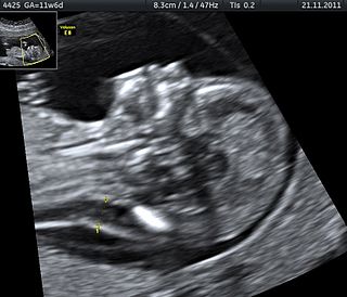
Obstetric ultrasonography, or prenatal ultrasound, is the use of medical ultrasonography in pregnancy, in which sound waves are used to create real-time visual images of the developing embryo or fetus in the uterus (womb). The procedure is a standard part of prenatal care in many countries, as it can provide a variety of information about the health of the mother, the timing and progress of the pregnancy, and the health and development of the embryo or fetus.

<span class="mw-page-title-main">Prenatal testing</span> Testing for diseases or conditions in a fetus

Nearly every pregnant individual undergoes some type of prenatal testing. There are varying levels of simplicity, invasiveness, risk, and information that correlate to each test. These tests can be as basic as taking the mothers weight (routine) to as complex and invasive as taking internal fetal blood samples for chromosomal analysis (genetic). Routine tests are conducted repeatedly throughout an individual’s pregnancy at prenatal checkups to monitor health and progression of the mother and the baby. Genetic testing is now a common practice offered to pregnant women by their healthcare professional at specific stages of their pregnancy to test for birth defects and chromosomal abnormalities, or the possibility of carrying these genetic traits. Testing capabilities and practices are becoming safer and less invasive as this area of research continues to evolve.

<span class="mw-page-title-main">Amniotic fluid</span> Fluid surrounding a fetus within the amnion

The amniotic fluid is the protective liquid contained by the amniotic sac of a gravid amniote. This fluid serves as a cushion for the growing fetus, but also serves to facilitate the exchange of nutrients, water, and biochemical products between mother and fetus.

In obstetrics, gestational age is a measure of the age of a pregnancy taken from the beginning of the woman's last menstrual period (LMP), or the corresponding age of the gestation as estimated by a more accurate method, if available. Such methods include adding 14 days to a known duration since fertilization, or by obstetric ultrasonography. The popularity of using this measure of pregnancy is largely due to convenience: menstruation is usually noticed, while there is generally no convenient way to discern when fertilization or implantation occurred.

<span class="mw-page-title-main">Hydronephrosis</span> Medical condition

Hydronephrosis describes hydrostatic dilation of the renal pelvis and calyces as a result of obstruction to urine flow downstream. Alternatively, hydroureter describes the dilation of the ureter, and hydronephroureter describes the dilation of the entire upper urinary tract.

<span class="mw-page-title-main">Pregnancy</span> Time of offspring development in mothers body

Pregnancy is the time during which one or more offspring develops (gestates) inside a woman's uterus (womb). A multiple pregnancy involves more than one offspring, such as with twins.

Prenatal development includes the development of the embryo and of the fetus during a viviparous animal's gestation. Prenatal development starts with fertilization, in the germinal stage of embryonic development, and continues in fetal development until birth.

<span class="mw-page-title-main">Fetal surgery</span> Growing branch of maternal-fetal medicine

Fetal surgery also known as antenatal surgery, prenatal surgery, is a growing branch of maternal-fetal medicine that covers any of a broad range of surgical techniques that are used to treat congenital abnormalities in fetuses who are still in the pregnant uterus. There are three main types: open fetal surgery, which involves completely opening the uterus to operate on the fetus; minimally invasive fetoscopic surgery, which uses small incisions and is guided by fetoscopy and sonography; and percutaneous fetal therapy, which involves placing a catheter under continuous ultrasound guidance.

<span class="mw-page-title-main">Ventriculomegaly</span> Increased size of the lateral ventricles

Ventriculomegaly is a brain condition that mainly occurs in the fetus when the lateral ventricles become dilated. The most common definition uses a width of the atrium of the lateral ventricle of greater than 10 mm. This occurs in around 1% of pregnancies. When this measurement is between 10 and 15 mm, the ventriculomegaly may be described as mild to moderate. When the measurement is greater than 15mm, the ventriculomegaly may be classified as more severe.

In pregnancy terms, quickening is the moment in pregnancy when the pregnant woman starts to feel the fetus's movement in the uterus.

<span class="mw-page-title-main">Velamentous cord insertion</span> Velamentous placenta

Velamentous cord insertion is a complication of pregnancy where the umbilical cord is inserted in the fetal membranes. It is a major cause of antepartum hemorrhage that leads to loss of fetal blood and associated with high perinatal mortality. In normal pregnancies, the umbilical cord inserts into the middle of the placental mass and is completely encased by the amniotic sac. The vessels are hence normally protected by Wharton's jelly, which prevents rupture during pregnancy and labor. In velamentous cord insertion, the vessels of the umbilical cord are improperly inserted in the chorioamniotic membrane, and hence the vessels traverse between the amnion and the chorion towards the placenta. Without Wharton's jelly protecting the vessels, the exposed vessels are susceptible to compression and rupture.

<span class="mw-page-title-main">Palmar grasp reflex</span> Primitive and involuntary reflex

The palmar grasp reflex is a primitive and involuntary reflex found in infants of humans and most primates. When an object, such as an adult finger, is placed in an infant's palm, the infant's fingers reflexively grasp the object. Placement of the object triggers a spinal reflex, resulting from stimulation of tendons in the palm, that gets transmitted through motor neurons in the median and ulnar sensory nerves. The reverse motion can be induced by stroking the back or side of the hand. A fetus exhibits the reflex in utero by 28 weeks into gestation, and persists until development of rudimentary fine motor skills between two and six months of age.

Fetal echocardiography, or Fetal echocardiogram, is the name of the test used to diagnose cardiac conditions in the fetal stage. Cardiac defects are amongst the most common birth defects. Their diagnosis is important in the fetal stage as it might help provide an opportunity to plan and manage the baby as and when the baby is born. Not all pregnancies need to undergo fetal echo.

A fetus or foetus is the unborn offspring that develops from an animal embryo. Following embryonic development the fetal stage of development takes place. In human prenatal development, fetal development begins from the ninth week after fertilization and continues until birth. Prenatal development is a continuum, with no clear defining feature distinguishing an embryo from a fetus. However, a fetus is characterized by the presence of all the major body organs, though they will not yet be fully developed and functional and some not yet situated in their final anatomical location.

The MOMS Trial was a clinical trial that studied treatment of a birth defect called myelomeningocele, which is the most severe form of spina bifida. The study looked at prenatal and postnatal surgery to repair this defect. The first major phase concluded that prenatal surgery had strong, long-term benefits and some risks.

<span class="mw-page-title-main">Circumvallate placenta</span> Medical condition

Circumvallate placenta is a rare condition affecting about 1-2% of pregnancies, in which the amnion and chorion fetal membranes essentially "double back" on the fetal side around the edges of the placenta. After delivery, a circumvallate placenta has a thick ring of membranes on its fetal surface. Circumvallate placenta is a placental morphological abnormality associated with increased fetal morbidity and mortality due to the restricted availability of nutrients and oxygen to the developing fetus.

Prenatal memory, also called fetal memory, is important for the development of memory in humans. Many factors can impair fetal memory and its functions, primarily maternal actions. There are multiple techniques available not only to demonstrate the existence of fetal memory but to measure it. Fetal memory is vulnerable to certain diseases so much so that exposure can permanently damage the development of the fetus and even terminate the pregnancy by aborting the fetus. Maternal nutrition and the avoidance of drugs, alcohol and other substances during all nine months of pregnancy is important to the development of the fetus and its memory systems. The use of certain substances can entail long-term permanent effects on the fetus that can carry on throughout their lifespan.

References