Fetal rhabdomyoma

Last updated

Rhabdomyoma is a benign mesenchymal tumor of skeletal muscle, separated into two major categories based on site: Cardiac and extracardiac. They are further separated by histology: fetal (myxoid and cellular), juvenile (intermediate), and adult types. Genital types are recognized, but are often part of either the fetal or juvenile types. The fetal type is thought to recapitulate immature skeletal muscle at about week six to ten of gestational development.

Contents

Signs and symptoms

Most fetal rhabdomyomas are tumors that develop in the head and neck or in the genital region. [1] There are a number of cases which have been seen in association with Gorlin syndrome. [2] [3] However, cardiac myxomas are known to be associated with tuberous sclerosis.

Pathophysiology

Gross pathology

The tumor may be seen within the subcutaneous tissues (below the skin), [4] mucosal surfaces or in soft tissue. Within the head and neck, the posterior ear region, skin of the face, and the tongue are the most commonly affected sites (about a 2:1 ratio of soft tissue to mucosa). [5] [6] The tumors are well defined, non-specific usually solitary masses, but when seen in the head and neck (or genital region), they may be polypoid. Tumors range in dimension from a few millimeters up to 12.5 cm, with a mean of about 3.0 cm. Although there are isolated case reports, multifocality is very rare.

Microscopic pathology

A hematoxylin and eosin stained image (high power) of the short spindle cells of a fetal rhabdomyoma. Fetal Rhabdomyoma Ear HE LDRT.tif
A hematoxylin and eosin stained image (high power) of the short spindle cells of a fetal rhabdomyoma.

Fetal rhabdomyoma are separated into two histologic types: Myxoid and cellular. [7] [8] However, irrespective of histologic type, these tumors almost never show necrosis or increased mitoses. However, a cambium layer, abnormal mitoses and nuclear pleomorphism is not seen.

Cellular fetal rhabdomyomas are composed of bland, primitive spindled cells. The spindle cells are haphazardly arranged primitive, elongated skeletal muscle cells. The cells are set within a well-developed fibromyxoid stroma. A different pattern (intermediate type) is predominantly composed of cells with better differentiation towards skeletal muscle. There are often large ganglion cell-like rhabdomyoblasts showing prominent nucleoli within nuclei that show vesicular chromatin distribution. Another population includes strap-like rhabdomyoblasts with darkly staining pink cytoplasm. Nearly all tumors show short to more sweeping fascicles of spindled rhabdomyoblasts. The tumor cells may infiltrate into adjacent skeletal muscle or fat. It is not uncommon to see peri-neural association, although not perineural infiltration. [9]

Diagnosis

These tumors may be detected prenatally by ultrasound and MRI. [10] Additionally, preoperative fine needle aspiration can be used to diagnose the tumor. [11]

Ancillary testing

A phosphotungstic acid hematoxylin stain may be used to highlight cross striations in the cytoplasm of the tumor cells. PAS with diastase will highlight the presence of glycogen in the tumor cells' cytoplasm. Immunohistochemistry will yield a positive reaction with a variety of myoid markers, including desmin, myoglobin, myogenin, MYOD1 and muscle specific actin. They may also be positive with vimentin, smooth muscle actin, and Leu-7. However, the tumor cells are almost always negative with glial fibrillary acidic protein, S100 protein, cytokeratin, epithelial membrane antigen, CD68, FLI1, CD99 and CD56. Although not used as frequently now, electron microscopy will show thick and thin myofilaments, Z-bands: these are features of sarcometric differentiation. [12]

Differential diagnosis

The differential diagnosis histologically includes rhabdomyosarcoma, granular cell tumor, alveolar soft part sarcoma, hibernoma, oncocytoma, and crystal storing histiocytosis, among others. [13]

Management

Surgical excision is the treatment of choice. [14] Recurrences are reported, but this is usually due to incomplete removal initially. [15] [16] There is no role for chemotherapy or radiation therapy.

Epidemiology

They present over a wide age range (birth to about 65 years), but within the head and neck region, about 50% of cases develop in patients younger than 15 years of age. Within the head and neck, males are affected about 2–3 times more often than females. [17]

Related Research Articles

<span class="mw-page-title-main">Rhabdomyoma</span> Medical condition

A rhabdomyoma is a benign tumor of striated muscle. Rhabdomyomas may be either cardiac or extracardiac. Extracardiac forms of rhabdomyoma are sub-classified into three distinct types: adult type, fetal type, and genital type.

<span class="mw-page-title-main">Hemangioendothelioma</span> Medical condition

Hemangioendotheliomas are a family of vascular neoplasms of intermediate malignancy.

<span class="mw-page-title-main">Canalicular adenoma</span> Benign salivary gland tumor

Canalicular adenoma is a benign, epithelial salivary gland neoplasm arranged in interconnecting cords of columnar cells. This is a very rare benign neoplasm, that makes up about 1% of all salivary gland tumors, or about 4% of all benign salivary gland tumors.

<span class="mw-page-title-main">Schwannoma</span> Medical condition

A schwannoma is a usually benign nerve sheath tumor composed of Schwann cells, which normally produce the insulating myelin sheath covering peripheral nerves.

<span class="mw-page-title-main">Granular cell tumor</span> Medical condition

Granular cell tumor is a tumor that can develop on any skin or mucosal surface, but occurs on the tongue 40% of the time.

Alveolar rhabdomyosarcoma (ARMS) is a subtype of the rhabdomyosarcoma soft tissue cancer family whose lineage is from mesenchymal cells and are related to skeletal muscle cells. ARMS tumors resemble the alveolar tissue in the lungs. Tumor location varies from patient to patient, but is commonly found in the head and neck region, male and female urogenital tracts, the torso, and extremities. Two fusion proteins can be associated with ARMS, but are not necessary, PAX3-FKHR. and PAX7-FKHR. In children and adolescents ARMS accounts for about 1 percent of all malignancies, has an incidence rate of 1 per million, and most cases occur sporadically with no genetic predisposition. PAX3-FOXO1 is now known to drive cancer-promoting gene expression programs through creation of distant genetic elements called super enhancers.

An ossifying fibromyxoid tumor is a type of myxoma. It presents in the extremities more frequently than the trunk. It is derived from mesenchyme. Appearance in the head and neck is rare, but has been reported. Its malignancy has been characterized as "intermediate".

<span class="mw-page-title-main">Gliosarcoma</span> Medical condition

Gliosarcoma is a rare type of glioma, a cancer of the brain that comes from glial, or supportive, brain cells, as opposed to the neural brain cells. Gliosarcoma is a malignant cancer, and is defined as a glioblastoma consisting of gliomatous and sarcomatous components. Primary gliosarcoma (PGS) is classified as a grade IV tumor and a subtype of glioblastoma multiforme in the 2007 World Health Organization classification system (GBM). Because of a lack of specific and clear diagnostic criteria, the word "gliosarcoma" was frequently used to refer to glial tumours with mesenchymal properties, such as the ability to make collagen and reticulin.

<span class="mw-page-title-main">Hibernoma</span> Medical condition

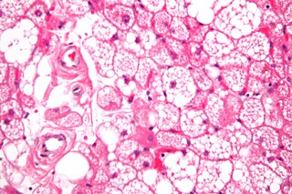
A hibernoma is a benign neoplasm of vestigial brown fat. The term was originally used by the French anatomist Louis Gery in 1914.

<span class="mw-page-title-main">Neurothekeoma</span>

A Neurothekeoma (NT) is a type of rare benign cutaneous tumor that usually develops on the head and neck. They often occur in the second and early third decades of life and tend to afflict women more frequently than men. First described by Gallager and Helwig, who proposed the term in order to reflect the presumed origin of the lesion from nerve sheath. Microscopically, the lesions described closely resembled the tumor, "nerve sheath myxoma (NSM)", an entity first described by Harkin and Reed. The latter had, through the years, been variously described as "bizarre cutaneous neurofibroma", "myxoma of nerve sheath", and "pacinian neurofibroma".

<span class="mw-page-title-main">Fetal adenocarcinoma</span> Rare subtype of pulmonary adenocarcinoma

Fetal adenocarcinoma (FA) of the lung is a rare subtype of pulmonary adenocarcinoma that exhibits tissue architecture and cell characteristics that resemble fetal lung tissue upon microscopic examination. It is currently considered a variant of solid adenocarcinoma with mucin production.

<span class="mw-page-title-main">Giant-cell carcinoma of the lung</span> Medical condition

Giant-cell carcinoma of the lung (GCCL) is a rare histological form of large-cell lung carcinoma, a subtype of undifferentiated lung cancer, traditionally classified within the non-small-cell lung carcinomas (NSCLC).

<span class="mw-page-title-main">Myoepithelioma of the head and neck</span> Medical condition

Myoepithelioma of the head and neck, also myoepithelioma, is a salivary gland tumour of the head and neck that is usually benign. When malignant, which is exceedingly rare, they are known as malignant myoepithelioma or Myoepithelial carcinoma, and they account for 1% of the salivary tumors with poor prognosis.

A rhabdomyoblast is a cell type which is found in some rhabdomyosarcomas. When found histologically, a rhabdomyoblast aids the diagnosis of embryonal, alveolar, spindle cell/sclerosing, and pleomorphic rhabdomyosarcomas; however, in a tumor, expression of the rhabdomyoblast phenotype is not the only factor in diagnosing a rhabdomyosarcoma. Mesenchymal malignancies can exhibit this phenotype as well. Immunohistochemistry techniques allow for the sensitive detection of desmin, vimentin, muscle specific actin, and MyoD1. Similarly the rhabdomyoblast phenotype can be detected morphologically. Rhabdomyoblasts are early stage mesenchymal cells, having the potential to differentiate into a wide range of skeletal cells. Each stage of differentiation exhibits unique and distinguishable histological characteristics. In its initial from, stellate cells with amphiphilic cytoplasm and ovular central nuclei are observed. Commonly referred to as rhabdoid features, the maturing rhabdomyoblast will likely exhibit low levels of eosinophilic cytoplasm in proximal distances to the nucleus. As maturation and differentiation progress, the cell's cytoplasmic levels of white blood cells increase; additionally, elongated shapes, commonly depicted as “tadpole”, “strap” and "spider cells", are observed. In the concluding phase of differentiation, the white blood cell rich cytoplasm appears bright and exhibits cross-striation. The highly regulated organization of actin and myosin microfilaments in contractile proteins results in this appearance.

A sialoblastoma is a low-grade salivary gland neoplasm that recapitulates primitive salivary gland anlage. It has previously been referred to as congenital basal cell adenoma, embryoma, or basaloid adenocarcinoma. It is an extremely rare tumor, with less than 100 cases reported worldwide.

A ceruminous adenoma is a benign glandular neoplasm which arises from the ceruminous glands located within the external auditory canal. These glands are found within the outer one third to one half of the external auditory canal, more common along the posterior surface; therefore, the tumor develops within a very specific location.

Metanephric dysplastic hematoma of the sacral region (MDHSR) has been described by Cozzutto and Lazzaroni-Fossati in 1980, by Posalaki et al. in 1981 and by Cozzutto et al. in 1982. Three additional cases were seen by Finegold.

<span class="mw-page-title-main">Sclerosing polycystic adenosis</span> Type of salivary gland tumor

Sclerosing polycystic adenosis is a rare salivary gland tumor first described in 1996 by Dr. Brion Smith. The major salivary glands, specifically the parotid gland and the submandibular gland, are affected most commonly. Patients usually come to clinical attention with a mass or swelling in their salivary glands in the 5th decade of life, with females affected much more commonly than males. Nearly all of the cases reported so far have a benign behavior, although there is a single case that has had an associated malignant transformation.

Ectomesenchymal chondromyxoid tumor (ECT) is a benign intraoral tumor with presumed origin from undifferentiated (ecto)mesenchymal cells. There are some who think it is a myoepithelial tumor type.

Renal anaplastic sarcoma is a rare tumour of the kidney. By 2017 about 25 cases have been reported. This tumour occurs in children and young adults and is more common in females than males.

References

  1. Lapner PC, Chou S, Jimenez C. Perianal fetal rhabdomyoma: case report. Pediatr Surg Int 1997;12(7):544-7.
  2. Watson J, Depasquale K, Ghaderi M, Zwillenberg S. Nevoid basal cell carcinoma syndrome and fetal rhabdomyoma: a case study. Ear Nose Throat J 2004;83(10):716-8.
  3. Hardisson D, Jimenez-Heffernan JA, Nistal M, Picazo ML, Tovar JA, Contreras F. Neural variant of fetal rhabdomyoma and naevoid basal cell carcinoma syndrome. Histopathology 1996;29(3):247-52.
  4. Walsh SN, Hurt MA. Cutaneous fetal rhabdomyoma: a case report and historical review of the literature. Am J Surg Pathol 2008:Mar;32(3):485-91.
  5. Kapadia SB, Meis JM, Frisman DM, Ellis GL, Heffner DK. Fetal rhabdomyoma of the head and neck: a clinicopathologic and immunophenotypic study of 24 cases. Hum Pathol 1993;24(7):754-65.
  6. Willis J, Abdul-Karim FW, di Sant'Agnese PA. Extracardiac rhabdomyomas. Semin Diagn Pathol 1994;11(1):15-25.
  7. Willis J, Abdul-Karim FW, di Sant'Agnese PA. Extracardiac rhabdomyomas. Semin Diagn Pathol 1994;11(1):15-25.
  8. Kapadia SB, Meis JM, Frisman DM, Ellis GL, Heffner DK. Fetal rhabdomyoma of the head and neck: a clinicopathologic and immunophenotypic study of 24 cases. Hum Pathol 1993;24(7):754-65.
  9. Kapadia SB, Meis JM, Frisman DM, Ellis GL, Heffner DK. Fetal rhabdomyoma of the head and neck: a clinicopathologic and immunophenotypic study of 24 cases. Hum Pathol 1993;24(7):754-65.
  10. O'Callaghan MG, House M, Ebay S, Bhadelia R. Rhabdomyoma of the head and neck demonstrated by prenatal magnetic resonance imaging. J Comput Assist Tomogr 2005;29(1):130-2
  11. al Rikabi AC, al Kharfy T, al Sohaibani MO, al Samarrai AI. Fetal rhabdomyoma. A case report with the diagnosis suggested by intraoperative cytology. Acta Cytol 1996;40(4):786-8.
  12. Kapadia SB, Meis JM, Frisman DM, Ellis GL, Heffner DK. Fetal rhabdomyoma of the head and neck: a clinicopathologic and immunophenotypic study of 24 cases. Hum Pathol 1993;24(7):754-65.
  13. Kapadia SB, Meis JM, Frisman DM, Ellis GL, Heffner DK. Fetal rhabdomyoma of the head and neck: a clinicopathologic and immunophenotypic study of 24 cases. Hum Pathol 1993;24(7):754-65.
  14. Kapadia SB, Meis JM, Frisman DM, Ellis GL, Heffner DK. Fetal rhabdomyoma of the head and neck: a clinicopathologic and immunophenotypic study of 24 cases. Hum Pathol 1993;24(7):754-65.
  15. Valdez TA, Desai U, Volk MS. Recurrent fetal rhabdomyoma of the head and neck. Int J Pediatr Otorhinolaryngol 2006:70(6);1115-8.
  16. Smith NM, Thornton CM. Fetal rhabdomyoma: two instances of recurrence. Pediatr Pathol Lab Med 1996;16(4):673-80.
  17. Kapadia SB, Meis JM, Frisman DM, Ellis GL, Heffner DK. Fetal rhabdomyoma of the head and neck: a clinicopathologic and immunophenotypic study of 24 cases. Hum Pathol 1993;24(7):754-65.