Fluid replacement

Last updated
Fluid replacement
Cholera rehydration nurses.jpg
A person with cholera drinking oral rehydration solution (ORS)
Other namesFluid resuscitation

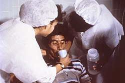
Fluid replacement or fluid resuscitation is the medical practice of replenishing bodily fluid lost through sweating, bleeding, fluid shifts or other pathologic processes. Fluids can be replaced with oral rehydration therapy (drinking), intravenous therapy, rectally such as with a Murphy drip, or by hypodermoclysis , the direct injection of fluid into the subcutaneous tissue. Fluids administered by the oral and hypodermic routes are absorbed more slowly than those given intravenously.

Contents

By mouth

Oral rehydration therapy (ORT) is a simple treatment for dehydration associated with diarrhea, particularly gastroenteritis/gastroenteropathy, such as that caused by cholera or rotavirus. ORT consists of a solution of salts and sugars which is taken by mouth. For most mild to moderate dehydration in children, the preferable treatment in an emergency department is ORT over intravenous replacement of fluid. [1]

It is used around the world, but is most important in the developing world, where it saves millions of children a year from death due to diarrhea—the second leading cause of death in children under five. [2]

Intravenous

Daily requirements
Water30 ml/kg/24 h
Na+ ~ 1 mmol/kg/24 h
K+ ~ 1 mmol/kg/24 h
Glucose 5 (3 to 8) g/hour

Similar precaution should be taken in administration of resuscitation fluid as to drug prescription. Fluid replacement should be considered as part of the complex physiological in the human body. Therefore, fluid requirements should be adjusted from time to time in those who are severely ill. [3]

In severe dehydration, intravenous fluid replacement is preferred, and may be lifesaving. It is especially useful where there is depletion of fluid both in the intracellular space and the vascular spaces.[ citation needed ]

Fluid replacement is also indicated in fluid depletion due to hemorrhage, extensive burns and excessive sweating (as from a prolonged fever), and prolonged diarrhea (cholera).[ citation needed ]

During surgical procedures, fluid requirement increases by increased evaporation, fluid shifts, or excessive urine production, among other possible causes. Even a small surgery may cause a loss of approximately 4 ml/kg/hour, and a large surgery approximately 8 ml/kg/hour, in addition to the basal fluid requirement.[ citation needed ]

The table to the right shows daily requirements for some major fluid components. If these cannot be given enterally, they may need to be given entirely intravenously. If continued long-term (more than approx. 2 days), a more complete regimen of total parenteral nutrition may be required.[ citation needed ]

Types

Resuscitation fluid can be broadly classified into: albumin solution, semisynthetic colloids, and crystalloids. [3]

The types of intravenous fluids used in fluid replacement are generally within the class of volume expanders. Physiologic saline solution, or 0.9% sodium chloride solution, is often used because it is isotonic, and therefore will not cause potentially dangerous fluid shifts. Also, if it is anticipated that blood will be given, normal saline is used because it is the only fluid compatible with blood administration.[ citation needed ]

Blood transfusion is the only approved fluid replacement capable of carrying oxygen; some oxygen-carrying blood substitutes are under development.

Lactated Ringer's solution is another isotonic crystalloid solution and it is designed to match most closely blood plasma. If given intravenously, isotonic crystalloid fluids will be distributed to the intravascular and interstitial spaces.[ citation needed ]

Plasmalyte is another isotonic crystalloid.[ citation needed ]

Blood products, non-blood products and combinations are used in fluid replacement, including colloid and crystalloid solutions. Colloids are increasingly used but they are more expensive than crystalloids. A systematic review found no evidence that resuscitation with colloids, instead of crystalloids, reduces the risk of death in patients with trauma or burns, or following surgery. [4]

Maintenance

Maintenance fluids are used in those who are currently normally hydrated but unable to drink enough to maintain this hydration. In children isotonic fluids are generally recommended for maintaining hydration. [5] [6] Potassium chloride and dextrose should be included. [6] The amount of maintenance IV fluid required in 24 hours is based on the weight of the patient using the Holliday-Segar formula. [7] [8] For weights ranging from 0 to 10 kg, the caloric expenditure is 100 cal/kg/day; from 10 to 20 kg the caloric expenditure is 1000 cal plus 50 cal/kg for each kilogram of body weight more than 10; over 20 kg the caloric expenditure is 1500 cal plus 20 cal/kg for each kilogram more than 20. More complex calculations (e.g., those using body surface area) are rarely required. [9]

Procedure

It is important to achieve a fluid status that is good enough to avoid low urine production. Low urine output has various limits, and varies for children, infants, and adults (see low urine production). The Parkland formula is not perfect and fluid therapy will need to be titrated to hemodynamic values and urine output.[ citation needed ]

The speed of fluid replacement may differ between procedures. For example, the planning of fluid replacement for burn patients is based on the Parkland formula (4mL Lactated Ringers X weight in kg X % total body surface area burned = Amount of fluid ( in ml) to give over 24 hours). The Parkland formula gives the minimum amount to be given in 24 hours. Half of the volume is given over the first eight hours after the time of the burn (not from time of admission to hospital) and the other half over the next 16 hours. In dehydration, 2/3 of the deficit may be given in 4 hours, and the rest during approximately 20 hours.[ citation needed ]

Clinical uses

Septic shock

Fluid replacement in patients with septic shock can be divided into four stages as shown below:

Acute kidney injury

Sepsis accounts for 50% of acute kidney injury patients in (intensive care unit) (ICU). [11] Intravenous crystalloid is recommended as the first line therapy to prevent or to treat acute kidney injury (AKI) when compared to colloids as colloids increases the risk of AKI. [13] 4% human albumin may be used in cirrhotic patients with spontaneous bacterial peritonitis as it can reduce the rate of kidney failure and improve survival. [11] However, fluid overload can exacerbate acute kidney injury. The use of diuretics does not prevent or treat AKI even with the help of renal replacement therapy. The 2012 KDIGO (Kidney Disease: Improving Global Outcomes) guidelines stated that diuretics should not be used to treat AKI, except for the management of volume overload. [13] In acute respiratory distress syndrome (ARDS), conservative fluid management is associated with better oxygenation and lung function with less prevalence of dialysis in the first 60 days of hospitalization when compared with liberal fluid management. [11]

Surgery (perioperative fluid therapy)

Managing fluids during major surgical procedures is an important aspect of surgical care. [14] The goal of fluid therapy is to maintain fluid and electrolyte levels and restore levels that may be depleted. [14] Intravenous fluid therapy is used when a person cannot control their own fluid intake and it can also reduce nausea and vomiting. [14] Goal-directed fluid therapy is a perioperative strategy in which the person is administered fluids continuously and the amount of fluids given are based on the person's physiological and haemodynamic (blood flow) measurements. [14] A second approach to fluid management during surgical procedures is called perioperative restrictive fluid therapy, also known as near-zero or zero-balance perioperative fluid approach; this approach recommends lower amounts of fluids during surgery, replacing fluids when the person is low (basal fluid requirements) or loses fluid due to a surgical procedure or bleed. [14] The effectiveness of goal-directed fluid therapy compared to restrictive fluid therapy is not clear as evidence comparing both approaches have very low certainty. [14]

Fluid overload

Fluid overload is defined as an increase in body weight of over 10%. [11] Aggressive fluid resuscitation can lead to fluid overload which can lead to damage of multiple organs: cerebral oedema, which leads to delirium; pulmonary oedema and pleural effusion, which lead to respiratory distress; myocardial oedema and pericardial effusion, which lead to impaired contractility of the heart; gastrointestinal oedema, which leads to malabsorption; hepatic congestion, which leads to cholestasis and acute kidney injury; and tissue oedema, which leads to poor wound healing. All these effects can cause disability and death, and increase in hospitalisation costs. [10]

Fluid overload causes cardiac dilation, which leads to increased ventricular wall stress, mitral insufficiency and leads to cardiac dysfunction. Pulmonary hypertension can lead to tricuspid insufficiency. Excess administration of fluid causes accumulation of extracellular fluid, leading to pulmonary oedema and lack of oxygen delivery to tissues. The use of mechanical ventilation in such case can cause barotrauma, infection, and oxygen toxicity, leading to acute respiratory distress syndrome. [11] Fluid overload also stretches the arterial endothelium, which causes damage to the glycocalyx, leading to capillary leakage and worsens the acute kidney injury. [15]

Other treatments

Proctoclysis, an enema, is the administration of fluid into the rectum as a hydration therapy. It is sometimes used for very ill persons with cancer. [16] The Murphy drip is a device by means of which this treatment may be performed.

See also

Related Research Articles

<span class="mw-page-title-main">Oncotic pressure</span> Measure of pressure exerted by large dissolved molecules in biological fluids

Oncotic pressure, or colloid osmotic-pressure, is a type of osmotic pressure induced by the plasma proteins, notably albumin, in a blood vessel's plasma that causes a pull on fluid back into the capillary. Participating colloids displace water molecules, thus creating a relative water molecule deficit with water molecules moving back into the circulatory system within the lower venous pressure end of capillaries.

<span class="mw-page-title-main">Shock (circulatory)</span> Medical condition of insufficient blood flow

Shock is the state of insufficient blood flow to the tissues of the body as a result of problems with the circulatory system. Initial symptoms of shock may include weakness, fast heart rate, fast breathing, sweating, anxiety, and increased thirst. This may be followed by confusion, unconsciousness, or cardiac arrest, as complications worsen.

<span class="mw-page-title-main">Dehydration</span> Deficit of total body water

In physiology, dehydration is a lack of total body water, with an accompanying disruption of metabolic processes. It occurs when free water loss exceeds free water intake, usually due to exercise, disease, or high environmental temperature. Mild dehydration can also be caused by immersion diuresis, which may increase risk of decompression sickness in divers.

<span class="mw-page-title-main">Sepsis</span> Life-threatening organ dysfunction triggered by infection

Sepsis is a potentially life-threatening condition that arises when the body's response to infection causes injury to its own tissues and organs.

<span class="mw-page-title-main">Intravenous therapy</span> Medication administered into a vein

Intravenous therapy is a medical technique that administers fluids, medications and nutrients directly into a person's vein. The intravenous route of administration is commonly used for rehydration or to provide nutrients for those who cannot, or will not—due to reduced mental states or otherwise—consume food or water by mouth. It may also be used to administer medications or other medical therapy such as blood products or electrolytes to correct electrolyte imbalances. Attempts at providing intravenous therapy have been recorded as early as the 1400s, but the practice did not become widespread until the 1900s after the development of techniques for safe, effective use.

<span class="mw-page-title-main">Septic shock</span> Dangerously low blood pressure due to damage from an organ infection

Septic shock is a potentially fatal medical condition that occurs when sepsis, which is organ injury or damage in response to infection, leads to dangerously low blood pressure and abnormalities in cellular metabolism. The Third International Consensus Definitions for Sepsis and Septic Shock (Sepsis-3) defines septic shock as a subset of sepsis in which particularly profound circulatory, cellular, and metabolic abnormalities are associated with a greater risk of mortality than with sepsis alone. Patients with septic shock can be clinically identified by requiring a vasopressor to maintain a mean arterial pressure of 65 mm Hg or greater and having serum lactate level greater than 2 mmol/L (>18 mg/dL) in the absence of hypovolemia. This combination is associated with hospital mortality rates greater than 40%.

<span class="mw-page-title-main">Hypovolemia</span> Low blood volume

Hypovolemia, also known as volume depletion or volume contraction, is a state of abnormally low extracellular fluid in the body. This may be due to either a loss of both salt and water or a decrease in blood volume. Hypovolemia refers to the loss of extracellular fluid and should not be confused with dehydration.

<span class="mw-page-title-main">Acute kidney injury</span> Medical condition

Acute kidney injury (AKI), previously called acute renal failure (ARF), is a sudden decrease in kidney function that develops within 7 days, as shown by an increase in serum creatinine or a decrease in urine output, or both.

<span class="mw-page-title-main">Hypovolemic shock</span> Medical condition

Hypovolemic shock is a form of shock caused by severe hypovolemia. It could be the result of severe dehydration through a variety of mechanisms or blood loss. Hypovolemic shock is a medical emergency; if left untreated, the insufficient blood flow can cause damage to organs, leading to multiple organ failure.

Hyperchloremia is an electrolyte disturbance in which there is an elevated level of chloride ions in the blood. The normal serum range for chloride is 96 to 106 mEq/L, therefore chloride levels at or above 110 mEq/L usually indicate kidney dysfunction as it is a regulator of chloride concentration. As of now there are no specific symptoms of hyperchloremia; however, it can be influenced by multiple abnormalities that cause a loss of electrolyte-free fluid, loss of hypotonic fluid, or increased administration of sodium chloride. These abnormalities are caused by diarrhea, vomiting, increased sodium chloride intake, renal dysfunction, diuretic use, and diabetes. Hyperchloremia should not be mistaken for hyperchloremic metabolic acidosis as hyperchloremic metabolic acidosis is characterized by two major changes: a decrease in blood pH and bicarbonate levels, as well as an increase in blood chloride levels. Instead those with hyperchloremic metabolic acidosis are usually predisposed to hyperchloremia.

<span class="mw-page-title-main">Crush syndrome</span> Medical condition

Crush syndrome is a medical condition characterized by major shock and kidney failure after a crushing injury to skeletal muscle. Crush injury is compression of the arms, legs, or other parts of the body that causes muscle swelling and/or neurological disturbances in the affected areas of the body, while crush syndrome is localized crush injury with systemic manifestations. Cases occur commonly in catastrophes such as earthquakes, to individuals that have been trapped under fallen or moving masonry.

End organ damage is severe impairment of major body organs due to high blood pressure or states of low blood pressure or low blood volume. This can present as a heart attack or heart failure, pulmonary edema, neurologic deficits including a stroke, or acute kidney failure.

Capillary leak syndrome, or vascular leak syndrome, is characterized by the escape of blood plasma through capillary walls, from the blood circulatory system to surrounding tissues, muscle compartments, organs or body cavities. It is a phenomenon most commonly witnessed in sepsis, and less frequently in autoimmune diseases, differentiation syndrome, engraftment syndrome, hemophagocytic lymphohistiocytosis, the ovarian hyperstimulation syndrome, viral hemorrhagic fevers, and snakebite and ricin poisoning. Pharmaceuticals, including the chemotherapy medications gemcitabine and denileukin diftitox, as well as certain interleukins and monoclonal antibodies, can also cause capillary leaks. These conditions and factors are sources of secondary capillary leak syndrome.

<span class="mw-page-title-main">Hydroxyethyl starch</span> Pharmaceutical drug

Hydroxyethyl starch (HES/HAES), sold under the brand name Voluven among others, is a nonionic starch derivative, used as a volume expander in intravenous therapy. The use of HES on critically ill patients is associated with an increased risk of death and kidney problems.

Early goal-directed therapy was introduced by Emanuel P. Rivers in The New England Journal of Medicine in 2001 and is a technique used in critical care medicine involving intensive monitoring and aggressive management of perioperative hemodynamics in patients with a high risk of morbidity and mortality. In cardiac surgery, goal-directed therapy has proved effective when commenced after surgery. The combination of GDT and Point-of-Care Testing has demonstrated a marked decrease in mortality for patients undergoing congenital heart surgery. Furthermore, a reduction in morbidity and mortality has been associated with GDT techniques when used in conjunction with an electronic medical record.

A volume expander is a type of intravenous therapy that has the function of providing volume for the circulatory system. It may be used for fluid replacement or during surgery to prevent nausea and vomiting after surgery.

<span class="mw-page-title-main">Transfusion-associated circulatory overload</span> Medical condition

In transfusion medicine, transfusion-associated circulatory overload is a transfusion reaction resulting in signs or symptoms of excess fluid in the circulatory system (hypervolemia) within 12 hours after transfusion. The symptoms of TACO can include shortness of breath (dyspnea), low blood oxygen levels (hypoxemia), leg swelling, high blood pressure (hypertension), and a high heart rate (tachycardia).

<span class="mw-page-title-main">Acute decompensated heart failure</span> Medical condition

Acute decompensated heart failure (ADHF) is a sudden worsening of the signs and symptoms of heart failure, which typically includes difficulty breathing (dyspnea), leg or feet swelling, and fatigue. ADHF is a common and potentially serious cause of acute respiratory distress. The condition is caused by severe congestion of multiple organs by fluid that is inadequately circulated by the failing heart. An attack of decompensation can be caused by underlying medical illness, such as myocardial infarction, an abnormal heart rhythm, infection, or thyroid disease.

An antihypotensive agent, also known as a vasopressor agent or simply vasopressor, or pressor, is any substance, whether endogenous or a medication, that tends to raise low blood pressure. Some antihypotensive drugs act as vasoconstrictors to increase total peripheral resistance; some drugs sensitize adrenoreceptors to catecholamines; and others increase cardiac output.

Permissive hypotension or hypotensive resuscitation is the use of restrictive fluid therapy, specifically in the trauma patient, that increases systemic blood pressure without reaching normotension. The goal blood pressure for these patients is a mean arterial pressure of 40-50 mmHg or systolic blood pressure of less than or equal to 80. This goes along with certain clinical criteria. Following traumatic injury, some patients experience hypotension that is usually due to blood loss (hemorrhage) but can be due to other causes as well. In the past, physicians were very aggressive with fluid resuscitation to try to bring the blood pressure to normal values. Recent studies have found that there is some benefit to allowing specific patients to experience some degree of hypotension in certain settings. This concept does not exclude therapy by means of i.v. fluid, inotropes or vasopressors, the only restriction is to avoid completely normalizing blood pressure in a context where blood loss may be enhanced. When a person starts to bleed the body starts a natural coagulation process that eventually stops the bleed. Issues with fluid resuscitation without control of bleeding are thought to be secondary to dislodgement of the thrombus that is helping to control further bleeding. Thrombus dislodgement was found to occur at a systolic pressure greater than 80mm Hg. In addition, fluid resuscitation will dilute coagulation factors that help form and stabilize a clot, hence making it harder for the body to use its natural mechanisms to stop the bleeding. These factors are aggravated by hypothermia.

References

  1. "Ten Things Physicians and Patients Should Question", Choosing Wisely , American College of Emergency Physicians, October 27, 2014 [October 14, 2013], retrieved April 6, 2015, which cites:
  2. The State of the World's Children 2008: Child Survival (PDF). UNICEF. December 2007. p. 8. ISBN   9789280641912. Archived from the original (PDF) on December 8, 2017. Retrieved February 16, 2009.
  3. 1 2 Myburgh, John A.; Mythen, Michael G. (2013-09-26). "Resuscitation Fluids". New England Journal of Medicine. 369 (13): 1243–1251. doi: 10.1056/NEJMra1208627 . ISSN   0028-4793. PMID   24066745.
  4. Lewis, Sharon R.; Pritchard, Michael W.; Evans, David Jw; Butler, Andrew R.; Alderson, Phil; Smith, Andrew F.; Roberts, Ian (3 August 2018). "Colloids versus crystalloids for fluid resuscitation in critically ill people". The Cochrane Database of Systematic Reviews. 8 (8): CD000567. doi:10.1002/14651858.CD000567.pub7. ISSN   1469-493X. PMC   6513027 . PMID   30073665.
  5. McNab, S; Ware, RS; Neville, KA; Choong, K; et al. (2014). "Isotonic versus hypotonic solutions for maintenance intravenous fluid administration in children" (PDF). Cochrane Database of Systematic Reviews (12): CD009457. doi:10.1002/14651858.CD009457.pub2. hdl: 10072/172363 . PMID   25519949.
  6. 1 2 Feld, LG; Neuspiel, DR; Foster, BA; Leu, MG; Garber, MD; Austin, K; Basu, RK; Conway EE, Jr; Fehr, JJ; Hawkins, C; Kaplan, RL; Rowe, EV; Waseem, M; Moritz, ML; SUBCOMMITTEE ON FLUID AND ELECTROLYTE, THERAPY. (December 2018). "Clinical Practice Guideline: Maintenance Intravenous Fluids in Children". Pediatrics. 142 (6): e20183083. doi: 10.1542/peds.2018-3083 . PMID   30478247.
  7. Holliday, M. A.; Segar, W. E. (1957). "The maintenance need for water in parenteral fluid therapy". Pediatrics. 19 (5): 823–832. doi:10.1542/peds.19.5.823. ISSN   0031-4005. PMID   13431307. S2CID   245056959.
  8. "Table: Holliday-Segar Formula for Maintenance Fluid Requirements by Weight". Merck Manuals Professional Edition. Retrieved 2021-01-12.
  9. "Dehydration in Children - Pediatrics". Merck Manuals Professional Edition. Retrieved 2021-01-12.
  10. 1 2 3 4 5 6 C Ogbu, Ogbonna; J Murphy, David; S Martin, Greg (August 2015). "How to Avoid Fluid Overload". Current Opinion in Critical Care. 21 (4): 315–321. doi:10.1097/MCC.0000000000000211. PMC   4691845 . PMID   26103147.
  11. 1 2 3 4 5 6 W Schrier, Robert (2010). "Fluid Administration in Critically Ill Patients with Acute Kidney Injury". Clinical Journal of the American Society of Nephrology. 5 (4): 733–739. doi: 10.2215/CJN.00060110 . PMID   20167687.
  12. Vincent, J; Weil, M (2006). "Fluid challenge revisited". Critical Care Medicine . 34 (5): 1333–7. doi:10.1097/01.CCM.0000214677.76535.A5. PMID   16557164. S2CID   41215136.
  13. 1 2 Claire, Annie; Fredette, Nadeau; Bouchard, Josée (January 2013). "Fluid Management and Use of Diuretics in Acute Kidney Injury". Advances in Chronic Kidney Disease. 20 (1): 45–55. doi:10.1053/j.ackd.2012.09.005. PMID   23265596.
  14. 1 2 3 4 5 6 Wrzosek, Anna; Jakowicka-Wordliczek, Joanna; Zajaczkowska, Renata; Serednicki, Wojciech T.; Jankowski, Milosz; Bala, Malgorzata M.; Swierz, Mateusz J.; Polak, Maciej; Wordliczek, Jerzy (2019-12-12). "Perioperative restrictive versus goal-directed fluid therapy for adults undergoing major non-cardiac surgery". The Cochrane Database of Systematic Reviews. 12 (12): CD012767. doi:10.1002/14651858.CD012767.pub2. ISSN   1469-493X. PMC   6953415 . PMID   31829446.
  15. Ostermann, Marlies; Oudemans-van Straaten, Heleen M; G. Forni, Lui (27 December 2015). "Fluid overload and acute kidney injury: cause or consequence?". Critical Care. 19: 443. doi: 10.1186/s13054-015-1163-7 . PMC   4699346 . PMID   26707872.
  16. Bruera, E; Pruvost, M; Schoeller, T; Montejo, G; et al. (April 1998). "Proctoclysis for hydration of terminally ill cancer patients". Journal of Pain and Symptom Management . 15 (4): 216–9. doi: 10.1016/s0885-3924(97)00367-9 . PMID   9601155.