An epileptic seizure, informally known as a seizure, is a period of symptoms due to abnormally excessive or synchronous neuronal activity in the brain. Outward effects vary from uncontrolled shaking movements involving much of the body with loss of consciousness, to shaking movements involving only part of the body with variable levels of consciousness, to a subtle momentary loss of awareness. Most of the time these episodes last less than two minutes and it takes some time to return to normal. Loss of bladder control may occur.

Lissencephaly is a set of rare brain disorders whereby the whole or parts of the surface of the brain appear smooth. It is caused by defective neuronal migration during the 12th to 24th weeks of gestation resulting in a lack of development of brain folds (gyri) and grooves (sulci). It is a form of cephalic disorder. Terms such as agyria and pachygyria are used to describe the appearance of the surface of the brain.

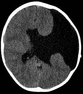
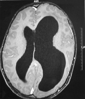
Schizencephaly is a rare birth defect characterized by abnormal clefts lined with grey matter that form the ependyma of the cerebral ventricles to the pia mater. These clefts can occur bilaterally or unilaterally. Common clinical features of this malformation include epilepsy, motor deficits, and psychomotor retardation.

Tuberous sclerosis complex (TSC) is a rare multisystem autosomal dominant genetic disease that causes non-cancerous tumours to grow in the brain and on other vital organs such as the kidneys, heart, liver, eyes, lungs and skin. A combination of symptoms may include seizures, intellectual disability, developmental delay, behavioral problems, skin abnormalities, lung disease, and kidney disease.

Myoclonus is a brief, involuntary, irregular twitching of a muscle or a group of muscles, different from clonus, which is rhythmic or regular. Myoclonus describes a medical sign and, generally, is not a diagnosis of a disease. These myoclonic twitches, jerks, or seizures are usually caused by sudden muscle contractions or brief lapses of contraction. The most common circumstance under which they occur is while falling asleep. Myoclonic jerks occur in healthy people and are experienced occasionally by everyone. However, when they appear with more persistence and become more widespread they can be a sign of various neurological disorders. Hiccups are a kind of myoclonic jerk specifically affecting the diaphragm. When a spasm is caused by another person it is known as a provoked spasm. Shuddering attacks in babies fall in this category.

Polymicrogyria (PMG) is a condition that affects the development of the human brain by multiple small gyri (microgyri) creating excessive folding of the brain leading to an abnormally thick cortex. This abnormality can affect either one region of the brain or multiple regions.
Epilepsia partialis continua is a rare type of brain disorder in which a patient experiences recurrent motor epileptic seizures that are focal, and recur every few seconds or minutes for extended periods. It is sometimes called Kozhevnikov's epilepsia named after Russian psychiatrist Aleksei Yakovlevich Kozhevnikov who first described this type of epilepsy.
Bilateral frontoparietal polymicrogyria is a genetic disorder with autosomal recessive inheritance that causes a cortical malformation. Our brain has folds in the cortex to increase surface area called gyri and patients with polymicrogyri have an increase number of folds and smaller folds than usual. Polymicrogyria is defined as a cerebral malformation of cortical development in which the normal gyral pattern of the surface of the brain is replaced by an excessive number of small, fused gyri separated by shallow sulci and abnormal cortical lamination. From ongoing research, mutation in GPR56, a member of the adhesion G protein-coupled receptor (GPCR) family, results in BFPP. These mutations are located in different regions of the protein without any evidence of a relationship between the position of the mutation and phenotypic severity. It is also found that GPR56 plays a role in cortical pattering.

In neuroanatomy, a gyrus is a ridge on the cerebral cortex. It is generally surrounded by one or more sulci. Gyri and sulci create the folded appearance of the brain in humans and other mammals.

Temporal lobe epilepsy (TLE) is a chronic disorder of the nervous system which is characterized by recurrent, unprovoked focal seizures that originate in the temporal lobe of the brain and last about one or two minutes. TLE is the most common form of epilepsy with focal seizures. A focal seizure in the temporal lobe may spread to other areas in the brain when it may become a focal to bilateral seizure.
Pachygyria is a congenital malformation of the cerebral hemisphere. It results in unusually thick convolutions of the cerebral cortex. Typically, children have developmental delay and seizures, the onset and severity depending on the severity of the cortical malformation. Infantile spasms are common in affected children, as is intractable epilepsy.
Frontal lobe epilepsy (FLE) is a neurological disorder which is a subtype of the larger group of epilepsy and then focal epilepsy is characterized by brief, recurring seizures that arise in the frontal lobes of the brain, often while the patient is sleeping. It is the second most common type of focal epilepsy after temporal lobe epilepsy (TLE), and is related to the temporal form by the fact that both forms are characterized by the occurrence of partial (focal) seizures. Partial seizures occurring in the frontal lobes can occur in one of two different forms: either “focal aware”, the old term was simple partial seizures “focal unaware” the old term was complex partial seizures. The symptoms and clinical manifestations of frontal lobe epilepsy can differ depending on which specific area of the frontal lobe is affected.

Gray matter heterotopias are neurological disorders caused by clumps of gray matter located in the wrong part of the brain. A grey matter heterotopia is characterized as a type of focal cortical dysplasia. The neurons in heterotopia appear to be normal, except for their mislocation; nuclear studies have shown glucose metabolism equal to that of normally positioned gray matter. The condition causes a variety of symptoms, but usually includes some degree of epilepsy or recurring seizures, and often affects the brain's ability to function on higher levels. Symptoms range from nonexistent to profound; the condition is occasionally discovered as an incidentaloma when brain imaging performed for an unrelated problem and has no apparent ill effect on the patient. At the other extreme, heterotopia can result in severe seizure disorder, loss of motor skills, and mental retardation. Fatalities are practically unknown, other than the death of unborn male fetuses with a specific genetic defect.
Juvenile myoclonic epilepsy (JME), also known as Janz syndrome, is a common form of genetic generalized epilepsy, representing 5-10% of all epilepsy cases. This disorder typically first presents between the ages of 12 and 18 with myoclonic seizure manifesting as sudden brief involuntary single or multiple episodes of muscle(s) contractions caused by an abnormal excessive or synchronous neuronal activity in the brain. These events typically occur after awakening from sleep, during the evening or upon sleep deprivation. JME is also characterized by generalized tonic-clonic seizures and a minority also have absence seizures. The genetics of JME are complex and rapidly evolving as over 20 chromosomal loci and multiple genes have been identified thus far. Given the genetic and clinical heterogeneity of JME some authors have suggested that it should be thought of as a spectrum disorder.

Hemimegalencephaly (HME), or unilateral megalencephaly, is a rare congenital disorder affecting all or a part of a cerebral hemisphere. It causes severe seizures, which are often frequent and hard to control. A minority might have seizure control with medicines, but most will need removal or disconnection of the affected hemisphere as the best chance. Uncontrolled, they often cause progressive intellectual disability and brain damage and stop development.

Dysembryoplastic neuroepithelial tumour is a type of brain tumor. Most commonly found in the temporal lobe, DNTs have been classified as benign tumours. These are glioneuronal tumours comprising both glial and neuron cells and often have ties to focal cortical dysplasia.
Ohtahara syndrome (OS), also known as early infantile epileptic encephalopathy (EIEE) is a progressive epileptic encephalopathy. The syndrome is outwardly characterized by tonic spasms and partial seizures within the first few months of life, and receives its more elaborate name from the pattern of burst activity on an electroencephalogram (EEG). It is an extremely debilitating progressive neurological disorder, involving intractable seizures and severe intellectual disabilities. No single cause has been identified, although in many cases structural brain damage is present.

Macrocephaly-capillary malformation (M-CM) is a multiple malformation syndrome causing abnormal body and head overgrowth and cutaneous, vascular, neurologic, and limb abnormalities. Though not every patient has all features, commonly found signs include macrocephaly, congenital macrosomia, extensive cutaneous capillary malformation, body asymmetry, polydactyly or syndactyly of the hands and feet, lax joints, doughy skin, variable developmental delay and other neurologic problems such as seizures and low muscle tone.

Neuronal migration disorder (NMD) refers to a heterogenous group of disorders that, it is supposed, share the same etiopathological mechanism: a variable degree of disruption in the migration of neuroblasts during neurogenesis. The neuronal migration disorders are termed cerebral dysgenesis disorders, brain malformations caused by primary alterations during neurogenesis; on the other hand, brain malformations are highly diverse and refer to any insult to the brain during its formation and maturation due to intrinsic or extrinsic causes that ultimately will alter the normal brain anatomy. However, there is some controversy in the terminology because virtually any malformation will involve neuroblast migration, either primarily or secondarily.

Microlissencephaly (MLIS) is a rare congenital brain disorder that combines severe microcephaly with lissencephaly. Microlissencephaly is a heterogeneous disorder, i.e. it has many different causes and a variable clinical course. Microlissencephaly is a malformation of cortical development (MCD) that occurs due to failure of neuronal migration between the third and fifth month of gestation as well as stem cell population abnormalities. Numerous genes have been found to be associated with microlissencephaly, however, the pathophysiology is still not completely understood.


