Related Research Articles

In aphasia, a person is unable to comprehend or unable to formulate language because of damage to specific brain regions. The major causes are stroke and head trauma; prevalence is hard to determine but aphasia due to stroke is estimated to be 0.1–0.4% in the Global North. Aphasia can also be the result of brain tumors, brain infections, or neurodegenerative diseases.
A hallucination is a perception in the absence of an external stimulus that has the qualities of a real perception. Hallucinations are vivid, substantial, and are perceived to be located in external objective space. Hallucination is a combination of two conscious states of brain wakefulness and REM sleep. They are distinguishable from several related phenomena, such as dreaming, which does not involve wakefulness; pseudohallucination, which does not mimic real perception, and is accurately perceived as unreal; illusion, which involves distorted or misinterpreted real perception; and mental imagery, which does not mimic real perception, and is under voluntary control. Hallucinations also differ from "delusional perceptions", in which a correctly sensed and interpreted stimulus is given some additional significance. Many hallucinations happen also during sleep paralysis.

Apraxia is a motor disorder caused by damage to the brain, which causes difficulty with motor planning to perform tasks or movements. The nature of the damage determines the disorder's severity, and the absence of sensory loss or paralysis helps to explain the level of difficulty. Children may be born with apraxia; its cause is unknown, and symptoms are usually noticed in the early stages of development. Apraxia occurring later in life, known as acquired apraxia, is typically caused by traumatic brain injury, stroke, dementia, Alzheimer's disease, brain tumor, or other neurodegenerative disorders. The multiple types of apraxia are categorized by the specific ability and/or body part affected.

Neurotrauma, brain damage or brain injury (BI) is the destruction or degeneration of brain cells. Brain injuries occur due to a wide range of internal and external factors. In general, brain damage refers to significant, undiscriminating trauma-induced damage.

The parietal lobe is one of the four major lobes of the cerebral cortex in the brain of mammals. The parietal lobe is positioned above the temporal lobe and behind the frontal lobe and central sulcus.
The temporal lobe is one of the four major lobes of the cerebral cortex in the brain of mammals. The temporal lobe is located beneath the lateral fissure on both cerebral hemispheres of the mammalian brain.
Anosognosia is a condition in which a person with a disability is cognitively unaware of having it due to an underlying physical or psychological condition. Anosognosia can result from physiological damage to brain structures, typically to the parietal lobe or a diffuse lesion on the fronto-temporal-parietal area in the right hemisphere, and is thus a neuropsychiatric disorder. A deficit of self-awareness, it was first named by the neurologist Joseph Babinski in 1914. Phenomenologically, anosognosia has similarities to denial, which is a psychological defense mechanism; attempts have been made at a unified explanation. Anosognosia is sometimes accompanied by asomatognosia, a form of neglect in which patients deny ownership of body parts such as their limbs. The term is from Ancient Greek ἀ- a-, 'without', νόσος nosos, 'disease' and γνῶσις gnōsis, 'knowledge'. It is also considered a disorder that makes the treatment of the patient more difficult, since it may affect negatively the therapeutic relationship.
Cerebral atrophy is a common feature of many of the diseases that affect the brain. Atrophy of any tissue means a decrement in the size of the cell, which can be due to progressive loss of cytoplasmic proteins. In brain tissue, atrophy describes a loss of neurons and the connections between them. Brain atrophy can be classified into two main categories: generalized and focal atrophy. Generalized atrophy occurs across the entire brain whereas focal atrophy affects cells in a specific location. If the cerebral hemispheres are affected, conscious thought and voluntary processes may be impaired.
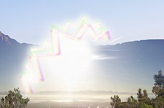
An aura is a perceptual disturbance experienced by some with epilepsy or migraine. An epileptic aura is a seizure.
Monoplegia is paralysis of a single limb, usually an arm. Common symptoms associated with monoplegic patients are weakness, numbness, and pain in the affected limb. Monoplegia is a type of paralysis that falls under hemiplegia. While hemiplegia is paralysis of half of the body, monoplegia is localized to a single limb or to a specific region of the body. Monoplegia of the upper limb is sometimes referred to as brachial monoplegia, and that of the lower limb is called crural monoplegia. Monoplegia in the lower extremities is not as common of an occurrence as in the upper extremities. Monoparesis is a similar, but less severe, condition because one limb is very weak, not paralyzed. For more information, see paresis.
Focal seizures are seizures which affect initially only one hemisphere of the brain. The brain is divided into two hemispheres, each consisting of four lobes – the frontal, temporal, parietal and occipital lobes. A focal seizure is generated in and affects just one part of the brain – a whole hemisphere or part of a lobe. Symptoms will vary according to where the seizure occurs. When seizures occur in the frontal lobe the patient may experience a wave-like sensation in the head. When seizures occur in the temporal lobe, a feeling of déjà vu may be experienced. When seizures are localized to the parietal lobe, a numbness or tingling may occur. With seizures occurring in the occipital lobe, visual disturbances or hallucinations have been reported.
This glossary covers terms found in the psychiatric literature; the word origins are primarily Greek, but there are also Latin, French, German, and English terms. Many of these terms refer to expressions dating from the early days of psychiatry in Europe.
In the field of neurology, seizure types are categories of seizures defined by seizure behavior, symptoms, and diagnostic tests. The International League Against Epilepsy (ILAE) 2017 classification of seizures is the internationally recognized standard for identifying seizure types. The ILAE 2017 classification of seizures is a revision of the prior ILAE 1981 classification of seizures. Distinguishing between seizure types is important since different types of seizures may have different causes, outcomes, and treatments.

Foix–Chavany–Marie Syndrome (FCMS), also known as bilateral opercular syndrome, is a neuropathological disorder characterized by paralysis of the facial, tongue, pharynx, and masticatory muscles of the mouth that aid in chewing. The disorder is primarily caused by thrombotic and embolic strokes, which cause a deficiency of oxygen in the brain. As a result, bilateral lesions may form in the junctions between the frontal lobe and temporal lobe, the parietal lobe and cortical lobe, or the subcortical region of the brain. FCMS may also arise from defects existing at birth that may be inherited or nonhereditary. Symptoms of FCMS can be present in a person of any age and it is diagnosed using automatic-voluntary dissociation assessment, psycholinguistic testing, neuropsychological testing, and brain scanning. Treatment for FCMS depends on the onset, as well as on the severity of symptoms, and it involves a multidisciplinary approach.
Todd's paresis is focal weakness in a part or all of the body after a seizure. This weakness typically affects the limbs and is localized to either the left or right side of the body. It usually subsides completely within 48 hours. Todd's paresis may also affect speech, eye position (gaze), or vision.
Transient epileptic amnesia (TEA) is a rare but probably underdiagnosed neurological condition which manifests as relatively brief and generally recurring episodes of amnesia caused by underlying temporal lobe epilepsy. Though descriptions of the condition are based on fewer than 100 cases published in the medical literature, and the largest single study to date included 50 people with TEA, TEA offers considerable theoretical significance as competing theories of human memory attempt to reconcile its implications.

A neurological disorder is any disorder of the nervous system. Structural, biochemical or electrical abnormalities in the brain, spinal cord or other nerves can result in a range of symptoms. Examples of symptoms include paralysis, muscle weakness, poor coordination, loss of sensation, seizures, confusion, pain and altered levels of consciousness. There are many recognized neurological disorders, some relatively common, but many rare. They may be assessed by neurological examination, and studied and treated within the specialities of neurology and clinical neuropsychology.
The neuroanatomy of memory encompasses a wide variety of anatomical structures in the brain.
Amnesia is a deficit in memory caused by brain damage or disease, but it can also be caused temporarily by the use of various sedatives and hypnotic drugs. The memory can be either wholly or partially lost due to the extent of damage that was caused. There are two main types of amnesia: retrograde amnesia and anterograde amnesia. Retrograde amnesia is the inability to retrieve information that was acquired before a particular date, usually the date of an accident or operation. In some cases the memory loss can extend back decades, while in others the person may lose only a few months of memory. Anterograde amnesia is the inability to transfer new information from the short-term store into the long-term store. People with anterograde amnesia cannot remember things for long periods of time. These two types are not mutually exclusive; both can occur simultaneously.
A somatosensory disorder is an impairment of the somatosensory system.
References
- ↑ Thiruppathy, S. P.; Muthukumar, N. (2004). "Mild head injury: Revisited". Acta Neurochirurgica. 146 (10): 1075–82, discussion 1082-3. doi:10.1007/s00701-004-0335-z. PMID 15744844. S2CID 13150034.
- ↑ Thal, G. D.; Szabo, M. D.; Lopez-Bresnahan, M.; Crosby, G. (1996). "Exacerbation or unmasking of focal neurologic deficits by sedatives". Anesthesiology. 85 (1): 21–5, discussion 29A-30A. doi: 10.1097/00000542-199607000-00004 . PMID 8694368. S2CID 8984607.
- 1 2 3 4 Fountoulakis, KN; Panagiotidis, P; Kimiskidis, V; Nimatoudis, I; Gonda, X (February 2019). "Neurological soft signs in familial and sporadic schizophrenia". Psychiatry Research. 272: 222–229. doi:10.1016/j.psychres.2018.12.105. PMID 30590276. S2CID 56476015.
- ↑ Ferri, Fred F. (2019). Ferri's clinical advisor 2019 : 5 books in 1. pp. 1225–1226. ISBN 9780323530422.
Essentials of Kumar and Clark's Clinical Medicine, 5th Edition. Saunders Elsevier, UK. 2012. page 725