A microscope is a laboratory instrument used to examine objects that are too small to be seen by the naked eye. Microscopy is the science of investigating small objects and structures using a microscope. Microscopic means being invisible to the eye unless aided by a microscope.

A scanning electron microscope (SEM) is a type of electron microscope that produces images of a sample by scanning the surface with a focused beam of electrons. The electrons interact with atoms in the sample, producing various signals that contain information about the surface topography and composition of the sample. The electron beam is scanned in a raster scan pattern, and the position of the beam is combined with the intensity of the detected signal to produce an image. In the most common SEM mode, secondary electrons emitted by atoms excited by the electron beam are detected using a secondary electron detector. The number of secondary electrons that can be detected, and thus the signal intensity, depends, among other things, on specimen topography. Some SEMs can achieve resolutions better than 1 nanometer.
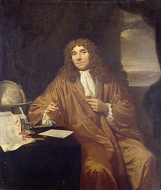
Antonie Philips van Leeuwenhoek was a Dutch microbiologist and microscopist in the Golden Age of Dutch science and technology. A largely self-taught man in science, he is commonly known as "the Father of Microbiology", and one of the first microscopists and microbiologists. Van Leeuwenhoek is best known for his pioneering work in microscopy and for his contributions toward the establishment of microbiology as a scientific discipline.

In biology, cell theory is a scientific theory first formulated in the mid-nineteenth century, that living organisms are made up of cells, that they are the basic structural/organizational unit of all organisms, and that all cells come from pre-existing cells. Cells are the basic unit of structure in all living organisms and also the basic unit of reproduction.

The optical microscope, also referred to as a light microscope, is a type of microscope that commonly uses visible light and a system of lenses to generate magnified images of small objects. Optical microscopes are the oldest design of microscope and were possibly invented in their present compound form in the 17th century. Basic optical microscopes can be very simple, although many complex designs aim to improve resolution and sample contrast.
The microscopic scale is the scale of objects and events smaller than those that can easily be seen by the naked eye, requiring a lens or microscope to see them clearly. In physics, the microscopic scale is sometimes regarded as the scale between the macroscopic scale and the quantum scale. Microscopic units and measurements are used to classify and describe very small objects. One common microscopic length scale unit is the micrometre, which is one millionth of a metre.

A magnifying glass is a convex lens that is used to produce a magnified image of an object. The lens is usually mounted in a frame with a handle. Beyond its primary function of magnification, this simple yet ingenious tool serves a variety of purposes. It can be employed to focus sunlight, harnessing the Sun's rays to create a concentrated hot spot at the lens's focus, which is often used for starting fires.

An eyepiece, or ocular lens, is a type of lens that is attached to a variety of optical devices such as telescopes and microscopes. It is named because it is usually the lens that is closest to the eye when someone looks through an optical device to observe an object or sample. The objective lens or mirror collects light from an object or sample and brings it to focus creating an image of the object. The eyepiece is placed near the focal point of the objective to magnify this image to the eyes. The amount of magnification depends on the focal length of the eyepiece.

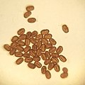
Macro photography is extreme close-up photography, usually of very small subjects and living organisms like insects, in which the size of the subject in the photograph is greater than life-size . By the original definition, a macro photograph is one in which the size of the subject on the negative or image sensor is life-size or greater. In some senses, however, it refers to a finished photograph of a subject that is greater than life-size.

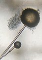
A micrograph is an image, captured photographically or digitally, taken through a microscope or similar device to show a magnified image of an object. This is opposed to a macrograph or photomacrograph, an image which is also taken on a microscope but is only slightly magnified, usually less than 10 times. Micrography is the practice or art of using microscopes to make photographs. A photographic micrograph is a photomicrograph, and one taken with an electron microscope is an electron micrograph.

An otoscope or auriscope is a medical device used by healthcare professionals to examine the ear canal and eardrum. This may be done as part of routine physical examinations, or for evaluating specific ear complaints, such as earaches, sense of fullness in the ear, or hearing loss.
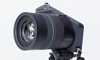
A light field camera, also known as a plenoptic camera, is a camera that captures information about the light field emanating from a scene; that is, the intensity of light in a scene, and also the precise direction that the light rays are traveling in space. This contrasts with conventional cameras, which record only light intensity at various wavelengths.

Dark-field microscopy describes microscopy methods, in both light and electron microscopy, which exclude the unscattered beam from the image. Consequently, the field around the specimen is generally dark.

A loupe is a simple, small magnification device used to see small details more closely. They generally have higher magnification than a magnifying glass, and are designed to be held or worn close to the eye. A loupe does not have an attached handle, and its focusing lens(es) are contained in an opaque cylinder or cone. On some loupes this cylinder folds into an enclosing housing that protects the lenses when not in use.

Paper models, also called card models or papercraft, are models constructed mainly from sheets of heavy paper, paperboard, card stock, or foam.

A digital microscope is a variation of a traditional optical microscope that uses optics and a digital camera to output an image to a monitor, sometimes by means of software running on a computer. A digital microscope often has its own in-built LED light source, and differs from an optical microscope in that there is no provision to observe the sample directly through an eyepiece. Since the image is focused on the digital circuit, the entire system is designed for the monitor image. The optics for the human eye are omitted.

The stereo, stereoscopic or dissecting microscope is an optical microscope variant designed for low magnification observation of a sample, typically using light reflected from the surface of an object rather than transmitted through it. The instrument uses two separate optical paths with two objectives and eyepieces to provide slightly different viewing angles to the left and right eyes. This arrangement produces a three-dimensional visualization of the sample being examined. Stereomicroscopy overlaps macrophotography for recording and examining solid samples with complex surface topography, where a three-dimensional view is needed for analyzing the detail.

A USB microscope is a low-powered digital microscope which connects to a computer's USB port. Microscopes essentially the same as USB models are also available with other interfaces either in addition to or instead of USB, such as via WiFi. They are widely available at low cost for use at home or in commerce. Their cost varies in the range of tens to thousands of dollars. In essence, a USB microscope is a webcam with a high-powered macro lens, and generally uses reflected rather than transmitted light, using built-in LED light sources surrounding the lens. The camera is usually sensitive enough not to need additional illumination beyond normal ambient lighting. The camera attaches directly to the USB port of a computer without the need for an eyepiece, and the images are shown directly on the computer's display.
Frugal innovation or frugal engineering is the process of reducing the complexity and cost of a good and its production. Usually this refers to removing nonessential features from a durable good, such as a car or telephone, in order to sell it in developing countries. Designing products for such countries may also call for an increase in durability and, when selling the products, reliance on unconventional distribution channels. When trying to sell to so-called "overlooked consumers", firms hope volume will offset razor-thin profit margins. Globalization and rising incomes in developing countries may also drive frugal innovation. Such services and products need not be of inferior quality but must be provided cheaply. While frugal innovation has been associated with good-enough performance, in some sectors such as in healthcare, frugal innovation must offer maximum performance without compromising on quality.

Manu Prakash is an Indian scientist who is a professor of bioengineering at Stanford University. Manu was born in Meerut, India. He is best known for his contributions to the Foldscope and Paperfuge. Prakash received the MacArthur Fellowship in September 2016. He and his team at Stanford University have developed a synchronous computer that operates using the physics of moving water droplets. His work focuses on frugal innovation that makes medicine, computing and microscopy accessible to more people across the world.