Frataxin

Last updated
FXN
Protein FXN PDB 1ekg.png
Available structures
PDB Ortholog search: PDBe RCSB
Identifiers
Aliases FXN , CyaY, FA, FARR, FRDA, X25, frataxin
External IDs OMIM: 606829 MGI: 1096879 HomoloGene: 47908 GeneCards: FXN
Orthologs
SpeciesHumanMouse
Entrez
Ensembl
UniProt
RefSeq (mRNA)

NM_181425
NM_000144
NM_001161706

NM_008044

RefSeq (protein)

NP_000135
NP_852090

NP_032070

Location (UCSC) Chr 9: 69.04 – 69.08 Mb Chr 19: 24.24 – 24.26 Mb
PubMed search [3] [4]
Wikidata
View/Edit Human View/Edit Mouse

Frataxin is a protein that in humans is encoded by the FXN gene. [5] [6]

It is located in the mitochondrion and Frataxin mRNA is mostly expressed in tissues with a high metabolic rate. The function of frataxin is not clear but it is involved in assembly of iron-sulfur clusters. It has been proposed to act as either an iron chaperone or an iron storage protein. Reduced expression of frataxin is the cause of Friedreich's ataxia.

Structure

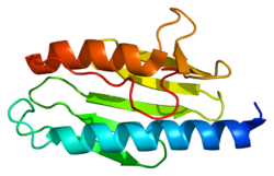
X-ray crystallography has shown that human frataxin consists of a β-sheet that supports a pair of parallel α-helices, forming a compact αβ sandwich. [7] Frataxin homologues in other species are similar, sharing the same core structure. However, the frataxin tail sequences, extending from the end of one helix, diverge in sequence and differ in length. Human frataxin has a longer tail sequence than frataxin found in bacteria or yeast. It is hypothesized that the purpose of the tail is to stabilize the protein. [7]

Like most mitochondrial proteins, frataxin is synthesized in cytoplasmic ribosomes as large precursor molecules with mitochondrial targeting sequences. Upon entry into mitochondria, the molecules are broken down by a proteolytic reaction to yield mature frataxin. [8]

Function

Frataxin is localized to the mitochondrion. The function of frataxin is not entirely clear, but it seems to be involved in assembly of iron-sulfur clusters. It has been proposed to act as either an iron chaperone or an iron storage protein. [9]

Frataxin mRNA is predominantly expressed in tissues with a high metabolic rate (including liver, kidney, brown fat and heart). Mouse and yeast frataxin homologues contain a potential N-terminal mitochondrial targeting sequence, and human frataxin has been observed to co-localise with a mitochondrial protein. Furthermore, disruption of the yeast gene has been shown to result in mitochondrial dysfunction. Friedreich's ataxia is thus believed to be a mitochondrial disease caused by a mutation in the nuclear genome (specifically, expansion of an intronic GAA triplet repeat in the FXN gene, which encodes the protein frataxin.). [5] [10] [11]

Clinical significance

Reduced expression of frataxin is the cause of Friedreich's ataxia (FRDA), a neurodegenerative disease. The reduction in frataxin gene expression may be attributable from either the silencing of transcription of the frataxin gene because of epigenetic modifications in the chromosomal entity [12] or from the inability of splicing the expanded GAA repeats in the first intron of the pre-mRNA as seen in bacteria [13] and Human cells [14] or both. The expansion of intronic trinucleotide repeat GAA results in Friedreich's ataxia. [15] This expanded repeat causes R-loop formation, and using a repeat-targeted oligonucleotide to disrupt the R-loop can reactivate frataxin expression. [16]

96% of FRDA patients have a GAA trinucleotide repeat expansion in intron 1 of both alleles of their FXN gene. [17] Overall, this leads to a decrease in frataxin mRNA synthesis and a decrease (but not absence) in frataxin protein in people with FRDA. (A subset of FRDA patients have GAA expansion in one chromosome and a point mutation in the FXN exon in the other chromosome.) In the typical case, the length of the allele with the shorter GAA expansion inversely correlates with frataxin levels. FRDA patients’ peripheral tissues typically have less than 10% of the frataxin levels exhibited by unaffected people. [17] Lower levels of frataxin result in earlier disease onset and faster progression.

FRDA is characterized by ataxia, sensory loss, and cardiomyopathy. The reason frataxin deficiency causes these symptoms is not entirely clear. On a cellular level, it is linked to iron accumulation in the mitochondria and increased oxidant sensitivity. For reasons that are not well understood, this primarily affects the tissue of the dorsal root ganglia, cerebellum, and heart muscle. [8]

Animal studies

In mice, complete inactivation of the FXN homolog (Frda) is lethal in the early embryonic stage. [18] Although nearly all organisms express a frataxin homologue, the GAA repeat in intron 1 only exists in humans and other primates, so the mutation that causes FDRA can't occur naturally in other animals. Scientists have developed several options to model this disease in mice. One approach is to silence frataxin expression in just one specific tissue type of interest: the heart (mice modified this way are called MCK), all neurons (NSE), or just the spinal cord and cerebellum (PRP). [19] Another approach involves inserting a GAA expansion into the first intron of the mouse FXN gene, which should inhibit frataxin production, just like in humans. Mice that are homozygous for this modified gene are called KIKI (knock-in knock-in), and the compound heterozygotes formed by crossing KIKI mice with frataxin knockout mice are called KIKO (knock-in knock-out). However, even KIKO mice still express 25-36% of the normal frataxin level, and show very mild symptoms. The final approach involves creating transgenic mice with a GAA-expanded version of the human frataxin gene. These mice are called YG22R (one GAA sequence of 190 repeats) and YG22R (two GAA sequences of 90 and 190 repeats). These mice show symptoms similar to human patients. [19]

An overexpression of frataxin in Drosophila has shown an increase in antioxidant capability, resistance to oxidative stress insults and longevity, [20] supporting the theory that the role of frataxin is to protect the mitochondria from oxidative stress and the ensuing cellular damage.

Fibroblasts from a mouse model of FRDA and FRDA patient fibroblasts show increased levels of DNA double-strand breaks. [21] A lentivirus gene delivery system was used to deliver the frataxin gene to the FRDA mouse model and human patient cells, and this resulted in long-term restored expression of frataxin mRNA and frataxin protein. This restored expression of the frataxin gene was accompanied by a substantial reduction in the number of DNA double-strand breaks. [21] The impaired frataxin in FRDA cells appears to cause reduced capacity for repair of DNA damage and this may contribute to neurodegeneration. [21]

Interactions

Frataxin has been shown to biologically interact with the enzyme PMPCB. [22]

Related Research Articles

Repeated sequences are short or long patterns of nucleic acids that occur in multiple copies throughout the genome. In many organisms, a significant fraction of the genomic DNA is repetitive, with over two-thirds of the sequence consisting of repetitive elements in humans. Some of these repeated sequences are necessary for maintaining important genome structures such as telomeres or centromeres.

<span class="mw-page-title-main">Friedreich's ataxia</span> Rare autosomal-recessive human disease

Friedreich's ataxia is an autosomal-recessive genetic disease that causes difficulty walking, a loss of coordination in the arms and legs, and impaired speech that worsens over time. Symptoms generally start between 5 and 20 years of age. Many develop hypertrophic cardiomyopathy and require a mobility aid such as a cane, walker, or wheelchair in their teens. As the disease progresses, some affected people lose their sight and hearing. Other complications may include scoliosis and diabetes mellitus.

A trinucleotide repeat expansion, also known as a triplet repeat expansion, is the DNA mutation responsible for causing any type of disorder categorized as a trinucleotide repeat disorder. These are labelled in dynamical genetics as dynamic mutations. Triplet expansion is caused by slippage during DNA replication, also known as "copy choice" DNA replication. Due to the repetitive nature of the DNA sequence in these regions, 'loop out' structures may form during DNA replication while maintaining complementary base pairing between the parent strand and daughter strand being synthesized. If the loop out structure is formed from the sequence on the daughter strand this will result in an increase in the number of repeats. However, if the loop out structure is formed on the parent strand, a decrease in the number of repeats occurs. It appears that expansion of these repeats is more common than reduction. Generally, the larger the expansion the more likely they are to cause disease or increase the severity of disease. Other proposed mechanisms for expansion and reduction involve the interaction of RNA and DNA molecules.

<span class="mw-page-title-main">Ataxin 1</span> Protein-coding gene in the species Homo sapiens

Ataxin-1 is a DNA-binding protein which in humans is encoded by the ATXN1 gene.

Ataxin is a type of nuclear protein. The class is called ataxin because mutated forms of these proteins and their corresponding genes were found to cause progressive ataxia.

<span class="mw-page-title-main">Dihydrolipoamide dehydrogenase</span> Protein-coding gene in the species Homo sapiens

Dihydrolipoamide dehydrogenase (DLD), also known as dihydrolipoyl dehydrogenase, mitochondrial, is an enzyme that in humans is encoded by the DLD gene. DLD is a flavoprotein enzyme that oxidizes dihydrolipoamide to lipoamide.

<span class="mw-page-title-main">Carnitine palmitoyltransferase II</span> Mammalian protein found in Homo sapiens

Carnitine O-palmitoyltransferase 2, mitochondrial is an enzyme that in humans is encoded by the CPT2 gene.

<span class="mw-page-title-main">POLG</span> Protein-coding gene in the species Homo sapiens

DNA polymerase subunit gamma is an enzyme that in humans is encoded by the POLG gene. Mitochondrial DNA polymerase is heterotrimeric, consisting of a homodimer of accessory subunits plus a catalytic subunit. The protein encoded by this gene is the catalytic subunit of mitochondrial DNA polymerase. Defects in this gene are a cause of progressive external ophthalmoplegia with mitochondrial DNA deletions 1 (PEOA1), sensory ataxic neuropathy dysarthria and ophthalmoparesis (SANDO), Alpers-Huttenlocher syndrome (AHS), and mitochondrial neurogastrointestinal encephalopathy syndrome (MNGIE).

<span class="mw-page-title-main">Ataxin 3</span> Protein-coding gene in the species Homo sapiens

Ataxin-3 is a protein that in humans is encoded by the ATXN3 gene.

<span class="mw-page-title-main">Twinkle (protein)</span> Mammalian protein found in Homo sapiens

Twinkle protein also known as twinkle mtDNA helicase is a mitochondrial protein that in humans is encoded by the TWNK gene located in the long arm of chromosome 10 (10q24.31).

<span class="mw-page-title-main">SCO1</span> Protein-coding gene in the species Homo sapiens

Protein SCO1 homolog, mitochondrial, also known as SCO1, cytochrome c oxidase assembly protein, is a protein that in humans is encoded by the SCO1 gene. SCO1 localizes predominantly to blood vessels, whereas SCO2 is barely detectable, as well as to tissues with high levels of oxidative phosphorylation. The expression of SCO2 is also much higher than that of SCO1 in muscle tissue, while SCO1 is expressed at higher levels in liver tissue than SCO2. Mutations in both SCO1 and SCO2 are associated with distinct clinical phenotypes as well as tissue-specific cytochrome c oxidase deficiency.

<span class="mw-page-title-main">PITRM1</span> Protein-coding gene in humans

Pitrilysin metallopeptidase 1 also known as presequence protease, mitochondrial (PreP) and metalloprotease 1 (MTP-1) is an enzyme that in humans is encoded by the PITRM1 gene. It is also sometimes called metalloprotease 1 (MP1).PreP facilitates proteostasis by utilizing an ~13300-A(3) catalytic chamber to degrade toxic peptides, including mitochondrial presequences and β-amyloid. Deficiency of PreP is found associated with Alzheimer’s disease. Reduced levels of PreP via RNAi mediated knockdown have been shown to lead to defective maturation of the protein Frataxin.

<span class="mw-page-title-main">COX10</span> Mammalian protein found in Homo sapiens

Protoheme IX farnesyltransferase, mitochondrial is an enzyme that in humans is encoded by the COX10 gene. Cytochrome c oxidase (COX), the terminal component of the mitochondrial respiratory chain, catalyzes the electron transfer from reduced cytochrome c to oxygen. This component is a heteromeric complex consisting of 3 catalytic subunits encoded by mitochondrial genes and multiple structural subunits encoded by nuclear genes. The mitochondrially-encoded subunits function in electron transfer, and the nuclear-encoded subunits may function in the regulation and assembly of the complex. This nuclear gene, COX10, encodes heme A: farnesyltransferase, which is not a structural subunit but required for the expression of functional COX and functions in the maturation of the heme A prosthetic group of COX. A gene mutation, which results in the substitution of a lysine for an asparagine (N204K), is identified to be responsible for cytochrome c oxidase deficiency. In addition, this gene is disrupted in patients with CMT1A duplication and with HNPP deletion.

<span class="mw-page-title-main">PMPCB</span> Protein-coding gene in humans

Mitochondrial-processing peptidase subunit beta is an enzyme that in humans is encoded by the PMPCB gene. This gene is a member of the peptidase M16 family and encodes a protein with a zinc-binding motif. This protein is located in the mitochondrial matrix and catalyzes the cleavage of the leader peptides of precursor proteins newly imported into the mitochondria, though it only functions as part of a heterodimeric complex.

<span class="mw-page-title-main">PMPCA</span> Protein-coding gene in humans

Mitochondrial-processing peptidase subunit alpha is an enzyme that in humans is encoded by the PMPCA gene. This gene PMPCA encoded a protein that is a member of the peptidase M16 family. This protein is located in the mitochondrial matrix and catalyzes the cleavage of the leader peptides of precursor proteins newly imported into the mitochondria, though it only functions as part of a heterodimeric complex.

<span class="mw-page-title-main">DNAJC19</span> Protein-coding gene in the species Homo sapiens

Mitochondrial import inner membrane translocase subunit TIM14 is an enzyme that in humans is encoded by the DNAJC19 gene on chromosome 3. TIM14 belongs to the DnaJ family, which has been involved in Hsp40/Hsp70 chaperone systems. As a mitochondrial chaperone, TIM14 functions as part of the TIM23 complex import motor to facilitate the import of nuclear-encoded proteins into the mitochondria. TIM14 also complexes with prohibitin complexes to regulate mitochondrial morphogenesis, and has been implicated in dilated cardiomyopathy with ataxia.

Mitochondrial processing peptidase is an enzyme complex found in mitochondria which cleaves signal sequences from mitochondrial proteins. In humans this complex is composed of two subunits encoded by the genes PMPCA, and PMPCB. The enzyme is also known as. This enzyme catalyses the following chemical reaction

Mitohondrial optic neuropathies are a heterogenous group of disorders that present with visual disturbances resultant from mitochondrial dysfunction within the anatomy of the Retinal Ganglion Cells (RGC), optic nerve, optic chiasm, and optic tract. These disturbances are multifactorial, their aetiology consisting of metabolic and/or structural damage as a consequence of genetic mutations, environmental stressors, or both. The three most common neuro-ophthalmic abnormalities seen in mitochondrial disorders are bilateral optic neuropathy, ophthalmoplegia with ptosis, and pigmentary retinopathy.

<span class="mw-page-title-main">ACO2</span> Protein-coding gene in the species Homo sapiens

Aconitase 2, mitochondrial is a protein that in humans is encoded by the ACO2 gene.

<span class="mw-page-title-main">Jean-Louis Mandel</span>

Jean-Louis Mandel, born in Strasbourg on February 12, 1946, is a French medical doctor and geneticist, and heads a research team at the Institute of Genetics and Molecular and Cellular Biology (IGBMC). He has been in charge of the genetic diagnosis laboratory at the University Hospitals of Strasbourg since 1992, as well as a professor at the Collège de France since 2003.

References

  1. 1 2 3 GRCh38: Ensembl release 89: ENSG00000165060 - Ensembl, May 2017
  2. 1 2 3 GRCm38: Ensembl release 89: ENSMUSG00000059363 - Ensembl, May 2017
  3. "Human PubMed Reference:". National Center for Biotechnology Information, U.S. National Library of Medicine.
  4. "Mouse PubMed Reference:". National Center for Biotechnology Information, U.S. National Library of Medicine.
  5. 1 2 Campuzano V, Montermini L, Moltò MD, Pianese L, Cossée M, Cavalcanti F, Monros E, Rodius F, Duclos F, Monticelli A, Zara F, Cañizares J, Koutnikova H, Bidichandani SI, Gellera C, Brice A, Trouillas P, De Michele G, Filla A, De Frutos R, Palau F, Patel PI, Di Donato S, Mandel JL, Cocozza S, Koenig M, Pandolfo M (Mar 1996). "Friedreich's ataxia: autosomal recessive disease caused by an intronic GAA triplet repeat expansion". Science. 271 (5254): 1423–7. Bibcode:1996Sci...271.1423C. doi:10.1126/science.271.5254.1423. PMID   8596916. S2CID   20303793.
  6. Carvajal JJ, Pook MA, dos Santos M, Doudney K, Hillermann R, Minogue S, Williamson R, Hsuan JJ, Chamberlain S (Oct 1996). "The Friedreich's ataxia gene encodes a novel phosphatidylinositol-4- phosphate 5-kinase". Nature Genetics. 14 (2): 157–62. doi:10.1038/ng1096-157. PMID   8841185. S2CID   6324358.
  7. 1 2 Dhe-Paganon S, Shigeta R, Chi YI, Ristow M, Shoelson SE (Oct 2000). "Crystal structure of human frataxin". The Journal of Biological Chemistry. 275 (40): 30753–6. doi: 10.1074/jbc.C000407200 . PMID   10900192.
  8. 1 2 Stemmler TL, Lesuisse E, Pain, Dancis (August 2010). "Frataxin and Mitochondrial FeS Cluster Biogenesis". Journal of Biological Chemistry. 285 (35): 26737–26743. doi: 10.1074/jbc.R110.118679 . PMC   2930671 . PMID   20522547.
  9. Adinolfi S, Iannuzzi C, Prischi F, Pastore C, Iametti S, Martin SR, Bonomi F, Pastore A (Apr 2009). "Bacterial frataxin CyaY is the gatekeeper of iron-sulfur cluster formation catalyzed by IscS". Nature Structural & Molecular Biology. 16 (4): 390–6. doi:10.1038/nsmb.1579. PMID   19305405. S2CID   205522816.
  10. Dürr A, Cossee M, Agid Y, Campuzano V, Mignard C, Penet C, Mandel JL, Brice A, Koenig M (Oct 1996). "Clinical and genetic abnormalities in patients with Friedreich's ataxia". The New England Journal of Medicine. 335 (16): 1169–75. doi: 10.1056/NEJM199610173351601 . PMID   8815938.
  11. Koutnikova H, Campuzano V, Foury F, Dollé P, Cazzalini O, Koenig M (Aug 1997). "Studies of human, mouse and yeast homologues indicate a mitochondrial function for frataxin". Nature Genetics. 16 (4): 345–51. doi:10.1038/ng0897-345. PMID   9241270. S2CID   5883249.
  12. Kim E, Napierala M, Dent SY (Oct 2011). "Hyperexpansion of GAA repeats affects post-initiation steps of FXN transcription in Friedreich's ataxia". Nucleic Acids Research. 39 (19): 8366–77. doi:10.1093/nar/gkr542. PMC   3201871 . PMID   21745819.
  13. Pan X, Ding Y, Shi L (Nov 2009). "The roles of SbcCD and RNaseE in the transcription of GAA x TTC repeats in Escherichia coli". DNA Repair. 8 (11): 1321–7. doi:10.1016/j.dnarep.2009.08.001. PMID   19733517.
  14. Baralle M, Pastor T, Bussani E, Pagani F (Jul 2008). "Influence of Friedreich ataxia GAA noncoding repeat expansions on pre-mRNA processing". American Journal of Human Genetics. 83 (1): 77–88. doi:10.1016/j.ajhg.2008.06.018. PMC   2443835 . PMID   18597733.
  15. "Entrez Gene: FXN frataxin".
  16. Li L, Matsui M, Corey DR (2016-01-01). "Activating frataxin expression by repeat-targeted nucleic acids". Nature Communications. 7: 10606. Bibcode:2016NatCo...710606L. doi:10.1038/ncomms10606. PMC   4742999 . PMID   26842135.
  17. 1 2 Clark E, Johnson J, Dong YN, Mercado-Ayon, Warren N, Zhai M, McMillan E, Salovin A, Lin H, Lynch DR (November 2018). "Role of frataxin protein deficiency and metabolic dysfunction in Friedreich ataxia, an autosomal recessive mitochondrial disease". Neuronal Signaling. 2 (4): NS20180060. doi: 10.1042/NS20180060 . PMC   7373238 . PMID   32714592.
  18. Cossée M, Puccio H, Gansmuller A, Koutnikova H, Dierich A, LeMeur M, Fischbeck K, Dollé P, Kœnig M (May 2000). "Inactivation of the Friedreich ataxia mouse gene leads to early embryonic lethality without iron accumulation". Human Molecular Genetics. 9 (8): 1219–1226. doi: 10.1093/hmg/9.8.1219 . PMID   10767347. Archived from the original on 2 June 2018. Retrieved 5 April 2019.
  19. 1 2 Perdomini M, Hick A, Puccio H (17 July 2013). "Animal and cellular models of Friedreich ataxia". Journal of Neurochemistry. 126: 65–79. doi: 10.1111/jnc.12219 . PMID   23859342. S2CID   1427817.
  20. Runko AP, Griswold AJ, Min KT (March 2008). "Overexpression of frataxin in the mitochondria increases resistance to oxidative stress and extends lifespan in Drosophila". FEBS Letters. 582 (5): 715–9. doi: 10.1016/j.febslet.2008.01.046 . PMID   18258192. S2CID   207603250.
  21. 1 2 3 Khonsari H, Schneider M, Al-Mahdawi S, Chianea YG, Themis M, Parris C, Pook MA, Themis M (December 2016). "Lentivirus-meditated frataxin gene delivery reverses genome instability in Friedreich ataxia patient and mouse model fibroblasts". Gene Ther. 23 (12): 846–856. doi:10.1038/gt.2016.61. PMC   5143368 . PMID   27518705.
  22. Koutnikova H, Campuzano V, Koenig M (Sep 1998). "Maturation of wild-type and mutated frataxin by the mitochondrial processing peptidase". Human Molecular Genetics. 7 (9): 1485–9. doi: 10.1093/hmg/7.9.1485 . PMID   9700204.

Further reading