Fundic gland polyposis

Last updated
Fundic gland polyposis
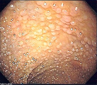
Endoscopic view of fundic gland polyps.jpg
Endoscopic image of fundic gland polyps taken on retroflexion of gastroscope.
Specialty Gastroenterology

Fundic gland polyposis is a medical syndrome where the fundus and the body of the stomach develop many fundic gland polyps. The condition has been described both in patients with familial adenomatous polyposis (FAP) and attenuated variants (AFAP), and in patients in whom it occurs sporadically. [1]

Contents

Presentation

Most patients with fundic gland polyps (FGPs) do not have any symptoms, and the diagnosis is made on gastroscopy done for other reasons. Retrospective analysis of patients with sporadic FGPs shows that a high percentage do have symptoms, but that this is more likely to be related to the underlying disease responsible for the polyposis. [2] These symptoms include:

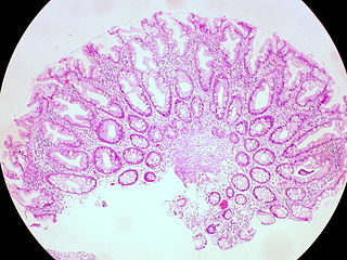
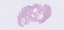
The polyps on endoscopy are usually tiny, numerous and sessile, [3] and usually scattered throughout the fundus of the stomach, where parietal cells are more numerous. They have the same colour as the gastric mucosa, and never have a stalk. [4] When the polyps are biopsied, the pathology typically shows shortened gastric pits, and both superficial and deep cystic lesions in the fundic glands, lined by all three types of cells of acid-producing mucosa: mucous, parietal and chief cells. As sometimes parietal cell hyperplasia may develop deep dilations of gland, [5] one should be really strict in the diagnosis of FGPs (i.e. the presence of deep and superficial dilations). Infrequently, the two lesions may coexist. [3] Foci of dysplasia can sometimes be seen. [4]

Disease associations

FGPs can be found in association with the following genetic conditions: [4]

Sporadic FGPs [1] have been associated with:

Pathophysiology

H&E stain of fundic gland polyp showing shortening of the gastric pits with cystic dilatation Fundic gland polyp (1).jpg
H&E stain of fundic gland polyp showing shortening of the gastric pits with cystic dilatation

The development of polyps depends on the underlying disorder. [17] In sporadic cases of FGPs, more than 90% of patients have activating mutations in the β-catenin gene, so that they may be considered "neoplastic" polyps. [18]

In familial adenomatous polyposis, the abnormality is a mutation in the APC gene, resulting in its inactivity. Attenuated FAP can occur from other mutations in the APC gene, and causes a phenotype wherein colonic polyps may be few in number. [4]

Both the β-catenin gene and the APC gene are involved in the same cell growth signalling pathway, but the APC gene is known to have a significantly higher association with the development of colorectal tumors. [19]

Diagnosis

The most important consideration in evaluating patients with FGPs is distinguishing between sporadic form (patients without any other gastrointestinal condition, usually in middle age with female prevalence) and syndromic form. This is to ascertain the risk of development of gastric cancer and to ascertain the risk of concomitant colon cancer. [20] [21]

Screening

There is a risk of development of cancer with fundic gland polyposis, [22] but it varies based on the underlying cause of the polyposis. [4] The risk is highest with congenital polyposis syndromes, and is lowest in acquired causes. [4] [23] As a result, it is recommended that patients with multiple fundic polyps have a colonoscopy to evaluate the colon. [4] If there are polyps seen on colonoscopy, genetic testing and testing of family members is recommended. [4] In the gastric adenocarcinoma associated with proximal polyposis of the stomach (GAPPS), there is a high risk of early development of proximal gastric adenocarcinoma. [9] [10]

It is still unclear which patients would benefit with surveillance gastroscopy, but most physicians recommend endoscopy every one to three years to survey polyps for dysplasia or cancer. [4] In the event of high grade dysplasia, polypectomy, which is done through the endoscopy, or partial gastrectomy may be recommended. One study showed the benefit of NSAID therapy in regression of gastric polyps, but the efficacy of this strategy (given the side effects of NSAIDs) is still dubious. [24]

Treatment

Related Research Articles

<span class="mw-page-title-main">Polyp (medicine)</span> Abnormal growth of tissue projecting from a mucous membrane

In anatomy, a polyp is an abnormal growth of tissue projecting from a mucous membrane. If it is attached to the surface by a narrow elongated stalk, it is said to be pedunculated; if it is attached without a stalk, it is said to be sessile. Polyps are commonly found in the colon, stomach, nose, ear, sinus(es), urinary bladder, and uterus. They may also occur elsewhere in the body where there are mucous membranes, including the cervix, vocal folds, and small intestine. Some polyps are tumors (neoplasms) and others are non-neoplastic, for example hyperplastic or dysplastic, which are benign. The neoplastic ones are usually benign, although some can be pre-malignant, or concurrent with a malignancy.

<span class="mw-page-title-main">Adenoma</span> Type of benign tumor

An adenoma is a benign tumor of epithelial tissue with glandular origin, glandular characteristics, or both. Adenomas can grow from many glandular organs, including the adrenal glands, pituitary gland, thyroid, prostate, and others. Some adenomas grow from epithelial tissue in nonglandular areas but express glandular tissue structure. Although adenomas are benign, they should be treated as pre-cancerous. Over time adenomas may transform to become malignant, at which point they are called adenocarcinomas. Most adenomas do not transform. However, even though benign, they have the potential to cause serious health complications by compressing other structures and by producing large amounts of hormones in an unregulated, non-feedback-dependent manner. Some adenomas are too small to be seen macroscopically but can still cause clinical symptoms.

<span class="mw-page-title-main">Gardner's syndrome</span> Medical condition

Gardner's syndrome is a subtype of familial adenomatous polyposis (FAP). Gardner syndrome is an autosomal dominant form of polyposis characterized by the presence of multiple polyps in the colon together with tumors outside the colon. The extracolonic tumors may include osteomas of the skull, thyroid cancer, epidermoid cysts, fibromas, as well as the occurrence of desmoid tumors in approximately 15% of affected individuals.
<span class="mw-page-title-main">Familial adenomatous polyposis</span> Medical condition

Familial adenomatous polyposis (FAP) is an autosomal dominant inherited condition in which numerous adenomatous polyps form mainly in the epithelium of the large intestine. While these polyps start out benign, malignant transformation into colon cancer occurs when they are left untreated. Three variants are known to exist, FAP and attenuated FAP are caused by APC gene defects on chromosome 5 while autosomal recessive FAP is caused by defects in the MUTYH gene on chromosome 1. Of the three, FAP itself is the most severe and most common; although for all three, the resulting colonic polyps and cancers are initially confined to the colon wall. Detection and removal before metastasis outside the colon can greatly reduce and in many cases eliminate the spread of cancer.

<span class="mw-page-title-main">Benign tumor</span> Mass of cells which cannot spread throughout the body

A benign tumor is a mass of cells (tumor) that does not invade neighboring tissue or metastasize. Compared to malignant (cancerous) tumors, benign tumors generally have a slower growth rate. Benign tumors have relatively well differentiated cells. They are often surrounded by an outer surface or stay contained within the epithelium. Common examples of benign tumors include moles and uterine fibroids.

<span class="mw-page-title-main">Hepatoblastoma</span> Liver cancer occurring in infants and children

Hepatoblastoma is a malignant liver cancer occurring in infants and children and composed of tissue resembling fetal liver cells, mature liver cells, or bile duct cells. They usually present with an abdominal mass. The disease is most commonly diagnosed during a child's first three years of life. Alpha-fetoprotein (AFP) levels are commonly elevated, but when AFP is not elevated at diagnosis the prognosis is poor.
<span class="mw-page-title-main">Adenomatous polyposis coli</span> Protein-coding gene in the species Homo sapiens

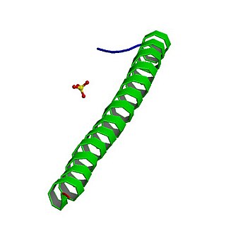
Adenomatous polyposis coli (APC) also known as deleted in polyposis 2.5 (DP2.5) is a protein that in humans is encoded by the APC gene. The APC protein is a negative regulator that controls beta-catenin concentrations and interacts with E-cadherin, which are involved in cell adhesion. Mutations in the APC gene may result in colorectal cancer and desmoid tumors.

<span class="mw-page-title-main">MUTYH</span> Protein-coding gene in the species Homo sapiens

MUTYH is a human gene that encodes a DNA glycosylase, MUTYH glycosylase. It is involved in oxidative DNA damage repair and is part of the base excision repair pathway. The enzyme excises adenine bases from the DNA backbone at sites where adenine is inappropriately paired with guanine, cytosine, or 8-oxo-7,8-dihydroguanine, a common form of oxidative DNA damage.

<span class="mw-page-title-main">Aggressive fibromatosis</span> Medical condition

Aggressive fibromatosis or desmoid tumor is a rare condition. Desmoid tumors are a type of fibromatosis and related to sarcoma, though without the ability to spread throughout the body (metastasize). The tumors arise from cells called fibroblasts, which are found throughout the body and provide structural support, protection to the vital organs, and play a critical role in wound healing. These tumors tend to occur in women in their thirties, but can occur in anyone at any age. They can be either relatively slow-growing or malignant. However, aggressive fibromatosis is locally aggressive and can cause life-threatening problems or even death when the tumors compress vital organs such as intestines, kidneys, lungs, blood vessels, or nerves. The condition is rarely fatal. Most cases are sporadic, but some are associated with familial adenomatous polyposis (FAP). Approximately 10% of individuals with Gardner's syndrome, a type of FAP with extracolonic features, have desmoid tumors.
<span class="mw-page-title-main">Cronkhite–Canada syndrome</span> Medical condition

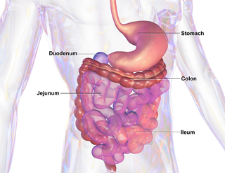
Cronkhite–Canada syndrome is a rare syndrome characterized by multiple polyps of the digestive tract. It is sporadic, and it is currently considered acquired and idiopathic.

<span class="mw-page-title-main">Juvenile polyposis syndrome</span> Medical condition

Juvenile polyposis syndrome is an autosomal dominant genetic condition characterized by the appearance of multiple juvenile polyps in the gastrointestinal tract. Polyps are abnormal growths arising from a mucous membrane. These usually begin appearing before age 20, but the term juvenile refers to the type of polyp, not to the age of the affected person. While the majority of the polyps found in juvenile polyposis syndrome are non-neoplastic, hamartomatous, self-limiting and benign, there is an increased risk of adenocarcinoma.

<span class="mw-page-title-main">Colorectal polyp</span> Growth found in bowel wall

A colorectal polyp is a polyp occurring on the lining of the colon or rectum. Untreated colorectal polyps can develop into colorectal cancer.

<span class="mw-page-title-main">Sessile serrated lesion</span> Medical condition

A sessile serrated lesion (SSL) is a premalignant flat lesion of the colon, predominantly seen in the cecum and ascending colon.

<span class="mw-page-title-main">Apc, wnt signaling pathway regulator</span> Mammalian protein found in Homo sapiens

APC, WNT signaling pathway regulator is a protein that in humans is encoded by the APC gene.

MUTYH-associated polyposis is an autosomal recessive polyposis syndrome. The disorder is caused by mutations in both alleles of the DNA repair gene, MUTYH. The MUTYH gene encodes a base excision repair protein, which corrects oxidative damage to DNA. Affected individuals have an increased risk of colorectal cancer, precancerous colon polyps (adenomas) and an increased risk of several additional cancers. About 1–2 percent of the population possess a mutated copy of the MUTYH gene, and less than 1 percent of people have the MUTYH associated polyposis syndrome. The presence of 10 or more colon adenomas should prompt consideration of MUTYH-associated polyposis, familial adenomatous polyposis and similar syndromes.
<span class="mw-page-title-main">Serrated polyposis syndrome</span> Medical condition

Serrated polyposis syndrome (SPS), previously known as hyperplastic polyposis syndrome, is a disorder characterized by the appearance of serrated polyps in the colon. While serrated polyposis syndrome does not cause symptoms, the condition is associated with a higher risk of colorectal cancer (CRC). The lifelong risk of CRC is between 25 and 40%. SPS is the most common polyposis syndrome affecting the colon, but is under recognized due to a lack of systemic long term monitoring. Diagnosis requires colonoscopy, and is defined by the presence of either of two criteria: ≥5 serrated lesions/polyps proximal to the rectum, or >20 serrated lesions/polyps of any size distributed throughout the colon with 5 proximal to the rectum.

Polymerase proofreading-associated polyposis (PPAP) is an autosomal dominant hereditary cancer syndrome, which is characterized by numerous polyps in the colon and an increased risk of colorectal cancer. It is caused by germline mutations in DNA polymerase ε (POLE) and δ (POLD1). Affected individuals develop numerous polyps called colorectal adenomas. Compared with other polyposis syndromes, Polymerase proofreading-associated polyposis is rare. Genetic testing can help exclude similar syndromes, such as Familial adenomatous polyposis and MUTYH-associated polyposis. Endometrial cancer, duodenal polyps and duodenal cancer may also occur.

Juvenile polyps are a type of polyp found in the colon. While juvenile polyps are typically found in children, they may be found in people of any age. Juvenile polyps are a type of hamartomatous polyps, which consist of a disorganized mass of tissue. They occur in about two percent of children. Juvenile polyps often do not cause symptoms (asymptomatic); when present, symptoms usually include gastrointestinal bleeding and prolapse through the rectum. Removal of the polyp (polypectomy) is warranted when symptoms are present, for treatment and definite histopathological diagnosis. In the absence of symptoms, removal is not necessary. Recurrence of polyps following removal is relatively common. Juvenile polyps are usually sporadic, occurring in isolation, although they may occur as a part of juvenile polyposis syndrome. Sporadic juvenile polyps may occur in any part of the colon, but are usually found in the distal colon. In contrast to other types of colon polyps, juvenile polyps are not premalignant and are not usually associated with a higher risk of cancer; however, individuals with juvenile polyposis syndrome are at increased risk of gastric and colorectal cancer., Unlike juvenile polyposis syndrome, solitary juvenile polyps do not require follow up with surveillance colonoscopy.

<span class="mw-page-title-main">Fundic gland polyp</span> Medical condition

A fundic gland polyp is a type of polyp, found in the fundus of the stomach. Fundic gland polyps are found in 0.8 to 1.9% of patients who undergo esophagogastroduodenoscopy, and are more common in middle-aged women.

Gardner fibroma (GF) is a benign fibroblastic tumor. GF tumors typically develop in the dermis and adjacent subcutaneous tissue lying just below the dermis. These tumors typically occur on the back, abdomen, and other superficial sites but in rare cases have been diagnoses in internal sites such as the retroperitoneum and around the large blood vessels in the upper thoracic cavity. The World Health Organization, 2020, classified Gardner fibroma as a benign tumor in the category of fibroblastic and myofibroblastic tumors.

References

  1. 1 2 Declich, P; Tavani, E; Ferrara, A; Caruso, S; Bellone, S (2005). "Sporadic fundic gland polyps: clinico-pathologic features and associated diseases". Polish Journal of Pathology. 56 (3): 131–7. PMID   16334981.
  2. Church, JM; McGannon, E; Hull-Boiner, S; Sivak, MV; Van Stolk, R; Jagelman, DG; Fazio, VW; Oakley, JR; et al. (1992). "Gastroduodenal polyps in patients with familial adenomatous polyposis". Diseases of the Colon and Rectum. 35 (12): 1170–3. doi:10.1007/BF02251971. PMID   1335405. S2CID   36152245.
  3. 1 2 Declich, P; Ambrosiani, L; Grassini, R; Tavani, E; Bellone, S; Bortoli, A; Gozzini, C; Prada, A (2000). "Fundic gland polyps: a still elusive entity on the eve of the year 2000". Pol J Pathol. 51 (1): 3–8. PMID   10833897.
  4. 1 2 3 4 5 6 7 8 9 Burt, RW (2003). "Gastric fundic gland polyps". Gastroenterology. 125 (5): 1462–9. doi:10.1016/j.gastro.2003.07.017. PMID   14598262.
  5. Declich, P; Ambrosiani, L; Bellone, S; Tavani, E; Grassini, R; Prada, A; Bortoli, A; Gozzini, C; Omazzi, B (2000). "Parietal cell hyperplasia with deep cystic dilations: a lesion closely mimicking fundic gland polyps". The American Journal of Gastroenterology. 95 (2): 566–8. doi:10.1016/S0002-9270(99)00873-4. PMID   10685784.
  6. "Familial Adenomatous Polyposis". The Lecturio Medical Concept Library. Retrieved 22 July 2021.
  7. Declich, P; Bellone, S; Ambrosiani, L; Bortoli, A; Gozzini, C; Tavani, E; Grassini, R; Prada, A (2000). "Fundic gland polyps: do they arise as a by-product of hypergastrinemia in patients with Zollinger-Ellison syndrome?". Human Pathology. 31 (7): 889–90. doi:10.1053/hupa.2000.8908. PMID   10923933.
  8. Aprile, MR; Azzoni, C; Gibril, F; Jensen, RT; Bordi, C (2000). "Intramucosal cysts in the gastric body of patients with Zollinger-Ellison syndrome". Human Pathology. 31 (2): 140–8. doi:10.1016/S0046-8177(00)80213-0. PMID   10685627.
  9. 1 2 Worthley DL, Phillips KD, Wayte N, Schrader KA, Healey S, Kaurah P, Shulkes A, Grimpen F, Clouston A, Moore D, Cullen D, Ormonde D, Mounkley D, Wen X, Lindor N, Carneiro F, Huntsman DG, Chenevix-Trench G, Suthers GK (May 2012). "Gastric adenocarcinoma and proximal polyposis of the stomach (GAPPS): a new autosomal dominant syndrome". Gut. 61 (5): 774–9. doi:10.1136/gutjnl-2011-300348. PMID   21813476. S2CID   206955098.
  10. 1 2 Li J, Woods SL, Healey S, Beesley J (5 May 2016). "Point Mutations in Exon 1B of APC Reveal Gastric Adenocarcinoma and Proximal Polyposis of the Stomach as a Familial Adenomatous Polyposis Variant". Am J Hum Genet. 98 (5): 830–42. doi:10.1016/j.ajhg.2016.03.001. PMC   4863475 . PMID   27087319.
  11. Freeman, HJ (2008). "Proton pump inhibitors and an emerging epidemic of gastric fundic gland polyposis". World Journal of Gastroenterology. 14 (9): 1318–20. doi: 10.3748/wjg.14.1318 . PMC   2693675 . PMID   18322941.
  12. Declich, P; Omazzi, B; Tavani, E; Bellone, S; Gozzini, C; Bortoli, A; Prada, A (2006). "Fundic gland polyps and PPI: the Mozart effect of gastrointestinal pathology?". Polish Journal of Pathology. 57 (4): 181–2. PMID   17285759.
  13. Vieth, M; Stolte, M (2001). "Fundic gland polyps are not induced by proton pump inhibitor therapy". American Journal of Clinical Pathology. 116 (5): 716–20. doi: 10.1309/XFWR-LXA7-7TK1-N3Q8 . PMID   11710689.
  14. Declich, P; Tavani, E; Ferrara, A; Prada, A; Caruso, S; Bellone, S; Bortoli, A; Porcellati, M; Gozzini, C (2001). "Sporadic fundic gland polyps, GERD and omeprazole usage: is there a little piece of information missed?". Polish Journal of Pathology. 52 (1–2): 63–4. PMID   11505682.
  15. Watanabe, N; Seno, H; Nakajima, T; Yazumi, S; Miyamoto, S; Matsumoto, S; Itoh, T; Kawanami, C; et al. (2002). "Regression of fundic gland polyps following acquisition of Helicobacter pylori". Gut. 51 (5): 742–5. doi:10.1136/gut.51.5.742. PMC   1773430 . PMID   12377817.
  16. Declich P, Tavani E, Bellone S, Porcellati M, Pastori L, Omazzi B, Gozzini C, Bortoli A, Prada A (November 2004). "Sporadic fundic gland polyps: what happened before?". Gut. 53 (11): 1721. PMC   1774292 . PMID   15479700.
  17. "Gastric Cancer: Pathology and Genetics". ScienceDirect. Retrieved 22 July 2021.
  18. Abraham, SC; Nobukawa, B; Giardiello, FM; Hamilton, SR; Wu, TT (2001). "Sporadic Fundic Gland Polyps : Common Gastric Polyps Arising Through Activating Mutations in the β-Catenin Gene". The American Journal of Pathology. 158 (3): 1005–10. doi:10.1016/S0002-9440(10)64047-3. PMC   1850357 . PMID   11238048.
  19. Chung, DC (2000). "The genetic basis of colorectal cancer: insights into critical pathways of tumorigenesis". Gastroenterology. 119 (3): 854–65. doi:10.1053/gast.2000.16507. PMID   10982779.
  20. "Gastric Cancer". The Lecturio Medical Concept Library. Retrieved 22 July 2021.
  21. "Colon Cancer Treatment (PDQ®)". NCI. May 12, 2014. Archived from the original on July 5, 2014. Retrieved June 29, 2014.
  22. Sebastian, S; Qasim, A; McLoughlin, R; O'Morain, CA; O'Connor, HJ (2004). "Fundic gland polyps: not so trivial entity and worth evaluation". Gastroenterology. 126 (5): 1497–8. doi: 10.1053/j.gastro.2004.03.044 . PMID   15131828.
  23. Jalving, M; Koornstra, JJ; Götz, JM; Van Der Waaij, LA; De Jong, S; Zwart, N; Karrenbeld, A; Kleibeuker, JH (2003). "High-grade dysplasia in sporadic fundic gland polyps: a case report and review of the literature" (PDF). European Journal of Gastroenterology & Hepatology. 15 (11): 1229–33. doi:10.1097/00042737-200311000-00013. PMID   14560158.
  24. Esaki, M; Matsumoto, T; Mizuno, M; Kobori, Y; Yoshimura, R; Yao, T; Iida, M (2002). "Effect of sulindac treatment for attenuated familial adenomatous polyposis with a new germline APC mutation at codon 161: report of a case". Diseases of the Colon and Rectum. 45 (10): 1397–402, discussion 1402–6. doi:10.1007/s10350-004-6432-5. PMID   12394442. S2CID   24467292.