Ghanaian bat henipavirus

Last updated
Ghanaian bat henipavirus
Virus classification OOjs UI icon edit-ltr.svg
(unranked): Virus
Realm: Riboviria
Kingdom: Orthornavirae
Phylum: Negarnaviricota
Class: Monjiviricetes
Order: Mononegavirales
Family: Paramyxoviridae
Genus: Henipavirus
Species:
Ghanaian bat henipavirus

Ghanaian bat henipavirus (also known Kumasi virus (KV) belongs to the genus Henipavirus in the family Paramyxoviridae . Human infections are caused by zoonotic events where the virus crosses over from another animal species. Therefore, humans are not the innate host for this virus family but instead become infected by peripheral viral reservoirs such as bats and other carriers of the virus. When these virus are spread to humans through zoonotic events they have been found to be one of the most deadly viruses with the capability to infect humans, with mortality rates between 50 and 100%. Therefore, these viruses have been classified as a biosafety level four (BSL-4) virus with regards to its pathogenesis when it infects humans. [1]

Contents

Compared to other henipaviruses, KV exhibits reduced surface expression of the attachment glycoprotein (KV-G). It is likely that KV-G expression is delayed in the endoplasmic reticulum and is not exported as readily to the cell surface due to defects in higher-order oligomerization. This may lead to reduced pathogenicity. [2]

Emergence

Emergence of Henipavirus was seen in 1994 when an outbreak in Australia caused an infectious outbreak in horses leading to severe respiratory disease. During this outbreak two people were infected and one died. [3] Henipavirus emergence is still relatively recent leading to a need for a greater range of genomic studies. Upon emergence in Australia it was found that Australian mainland flying foxes (bats) were the primary reservoir for the virus through analysis of their uterine fluid and urine. [4] However, it was discovered through subsequent break outs of Henipavirus that humans do not seem to contract the disease directly from flying foxes. In all human cases (4 of which have been fatal) the primary vector of transmission was infected horses. Therefore, it seems that horses contract the disease from flying foxes while humans contract the virus through close proximity to infected horses. This has also been further supported by a decrease in human contraction of Henipavirus after the development of an equestrian vaccine for the virus. [5] Epidemiological data show the presence of these viruses in Asia, Africa, and the South Pacific. In several studies it has been shown that bats, livestock, and humans carry neutralizing antibodies for Henipavirus in the Ghanaian region showing the potential for the existence of the virus within this population. [6] [7]

it was first detected in a zoological garden in Kumasi, Ghana in February 2008. Guano samples from a colony of an estimated 400,000 bats of the species Eidolon helvum were collected and screened for viral RNA. While 3 RNA genomes were obtained: BatPV/Eid.hel/GH10/2008; BatPV; Eid.hel/GH45/2008; and BatPV/Eid.hel/GH48/2008, only isolate BatPV/Eid.hel/GH10/2008 contained enough RNA to reliably quantified. BatPV/Eid.hel/GH10/2008 showed the highest sequence parsimony to established Nipah and Hendra henipavirus genomes. Infectious particles could not be isolated in cell culture; no cytopathic effects were observed and no viral RNA could be obtained. [8]

KV would be the first known henipavirus detected outside of the Austroasiatic geographic province that other known henipaviruses are known to circulate. [8] Serological evidence has previously suggested that henipaviruses likely have a much wider geographic range beyond areas of endemic Nipah and Hendra infection, [9] namely that undetected henipavirus infections may be common in South America and continental Africa. [10]

Genome

Henipavirus contain an enveloped single-strand negative-sense RNA genome. Therefore, they belong to the same genomic group of viruses such as measles and mumps which are more commonly found to cause pathogenesis and infection in humans; Measles, mumps, and other related viral pathogens also belong to the viral family Paramyxoviridae. There are two pathogenic members of the Henipavirus genome, Nipah Virus (NiV) and Hendra Virus (HeV). Ghanaian Bat Henipavirus (GhV) is phylogenetically related to both NiV and HeV although it is most closely associated with NiV. Both NiV and HeV consists of an 18.2kb genome encoding for six structural proteins; nucleoprotein (N), phosphoprotein (P), matrix protein (M), fusion protein (F), attachment glycoprotein (G), and the large protein or RNA polymerase protein (L). Furthermore, three nonstructural proteins are encoded by the P gene; Two RNA editing proteins (W and V) and on alternative open reading frame protein (C). [11] These genomes are conserved in most phylogenetic members of the Henipavirus genus. While members of the Henipavirus genus are similar in structure and protein makeup they do have subtle genomic differentiation in their nucleic acid sequences. While subtle differences exist both HeV and NiV are replicable in a variety of host species such as its natural reservoir bats, several forms of livestock, and of course humans. [12] However, the zoonotic potential for GhV is unknown as there have been no cases of transmission in Africa from the bat reservoir to any other organism.

GhV entry

Similar to all Henipavirus's GhV expresses two surface glycoproteins; The fusion protein (F) and the attachment glycoprotein (G). Both of these proteins mediate the entry process of GhV. Like other Henipavirus's the GhV glycoprotein G binds to the cellular surface receptor ephrin-B2. Ephrin-B2 is a transmembrane cellular surface receptor tyrosine kinase responsible for bi-directional signaling during tumorigenesis and other developmental events. Furthermore, this family of receptors has been shown to be highly conserved amongst humans and other animals explaining the broad Henipavirus transmission amongst several different species. [13] Like most enveloped viruses GhV follows the process of viral entry into the cell through two specific processes; Receptor binding and membrane fusion.

Attachment glycoprotein G is a tetrameric transmembrane domain protein that has a short cytoplasmic N-terminal tail and a large C-terminal globular head. This structure links the transmembrane and extracellular regions of this protein allowing it to bind to the Ephrin-B2 receptor on the cells surface. Furthermore, this creates a distinction between GhV and other Henipavirus's compared to other members of the Paramyxoviridae family as they do not exhibit hemagglutinate or neuraminidase activity and therefore do not bind sialic acid, only cell surface protein receptors. [14]

The Fusion protein F is synthesized as an inactive precursor F0 form before being cleaved by cellular proteases into the active form upon attachment by the G protein to the ephrin receptor. All domains of the fusion protein are conserved by NiV, HeV, and GhV as the protein contains a fusion cleavage sequence, fusion peptide, and the transmembrane anchor domain. [11]

The least understood element of the entry of GhV into the cell is the association between the attachment protein G and the fusion protein F. Therefore, two models have been established for the entry of this virus: The "clamp" model and the "provocateur" model. In the "clamp" model the F protein is activated by the removal of an inhibiter upon clamping of the attachment protein to the cell surface receptor. The "provocateur" model highlights a positive conformational change of the F protein upon the attachment of protein G to the cells surface receptor. [14] In either case the F protein is somehow activated by the attachment of the G protein to the cells surface receptor causing a release of the fusion peptide allowing the virus to be brought into the cell through endocytosis.

While this process is highly efficient in HeV and NiV leading to a broad tropism and effective infection of the virus within the host GhV exhibits a less than effective entry process. While HeV and NiV have strong fusion activity with a variety of host cells, GhV is restricted to a limited host range and only replicates in certain bat cells. This has been shown to be a result of an insufficient expression of GhV attachment protein G on the virus's surface. [13] With less attachment protein present the efficiency of cell-cell viral-mediated fusion would be less than optimal for viral transmission. Furthermore, lower expression of the G protein would limit its interaction with fusion protein F limiting the virus's capabilities of fusing with the host cell membrane reducing its capability for cellular entry via receptor-mediated endocytosis. In addition, a slight change in the cytoplasmic domain showed a limiting in the G proteins capabilities of activating the fusion protein F upon attachment to host cellular membrane receptor ephrin-B2. [15] Therefore, GhV is and has been limited to its innate reservoir of bat species limiting its zoonotic potential to infect other animals and humans.

Figure 1. Structure of Henipavirus shows the attachment protein (G) and the Fusion protein (F) located on the viral capsid. Furthermore, replication proteins N, L, and P are shown associated with the viral RNA. Structure d'un Henipavirus.svg
Figure 1. Structure of Henipavirus shows the attachment protein (G) and the Fusion protein (F) located on the viral capsid. Furthermore, replication proteins N, L, and P are shown associated with the viral RNA.

GhV replication

Upon virus entry into the cell, RNA replication can occur. As a negative-strand RNA virus, GhV brings its own RNA-dependent RNA-polymerase with it into the cell. Three proteins are essential for GhV replication: The nucleocapsid protein (N), the phosphoprotein (P), and the RNA-dependent RNA polymerase (RdRp) (L). It has been shown that GhV and other Henipavirus's RdRp's have a two-fold function, RNA transcription to mRNA and RNA replication of the genome. [16] The transcriptase complex is composed of L and P proteins and is responsible for the conversion of the negative-sense RNA genome into mRNA to be translated into viral proteins. As a replicase, the protein complex is responsible for the creation of genomic and anti-genomic RNA with its subsequent encapsulation in N proteins. The level of N proteins is a limiting component in the change from transcriptase to replicase; With more N protein there is greater replicase activity. [17] Furthermore, replication can occur in a variety of cell lines and types as the ephrin-B2 receptor is highly conserved amongst cell types. Therefore, replication is limited to cells expressing the ephrin-B2 receptor. This has been found in respiratory endothelial cells for HeV and NiV where pathogenesis is the greatest. However, this has not been discovered for GhV.

Henipavirus symptoms and pathogenesis

Infection with a Henipavirus, such as the more pathogenic HeV and NiV, can lead to vasculitis, necrosis, thrombosis, as well as brain parenchyma lesion associated with the formation of giant multi-nucleated cells. [18] Furthermore, with effects to vascular tissues Henipavirus causes severe respiratory pathogenesis. Infection of vascular endothelial cells leads to cellular disfunction and apoptosis causing large amounts of vascular inflammation. [19] While pathogenesis is severe in HeV and NiV, GhV has not been shown to cause these symptoms in other animals due to less of an ability to replicate in other hosts outside of the natural bat reservoir.

Related Research Articles

<i>Paramyxoviridae</i> Family of viruses

Paramyxoviridae is a family of negative-strand RNA viruses in the order Mononegavirales. Vertebrates serve as natural hosts. Diseases associated with this family include measles, mumps, and respiratory tract infections. The family has four subfamilies, 17 genera, three of which are unassigned to a subfamily, and 78 species.

<span class="mw-page-title-main">Coronavirus</span> Subfamily of viruses in the family Coronaviridae

Coronaviruses are a group of related RNA viruses that cause diseases in mammals and birds. In humans and birds, they cause respiratory tract infections that can range from mild to lethal. Mild illnesses in humans include some cases of the common cold, while more lethal varieties can cause SARS, MERS and COVID-19. In cows and pigs they cause diarrhea, while in mice they cause hepatitis and encephalomyelitis.

<span class="mw-page-title-main">SARS-related coronavirus</span> Species of coronavirus causing SARS and COVID-19

Severe-acute-respiratory-syndrome–related coronavirus is a species of virus consisting of many known strains. Two strains of the virus have caused outbreaks of severe respiratory diseases in humans: severe acute respiratory syndrome coronavirus 1, which caused the 2002–2004 outbreak of severe acute respiratory syndrome (SARS), and severe acute respiratory syndrome coronavirus 2 (SARS-CoV-2), which is causing the ongoing pandemic of COVID-19. There are hundreds of other strains of SARSr-CoV, which are only known to infect non-human mammal species: bats are a major reservoir of many strains of SARSr-CoV; several strains have been identified in Himalayan palm civets, which were likely ancestors of SARS-CoV-1.

<i>Henipavirus</i> Genus of RNA viruses

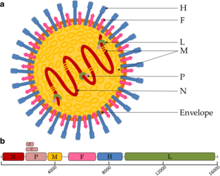
Henipavirus is a genus of negative-strand RNA viruses in the family Paramyxoviridae, order Mononegavirales containing six established species, and numerous others still under study. Henipaviruses are naturally harboured by several species of small mammals, notably pteropid fruit bats, microbats of several species, and shrews. Henipaviruses are characterised by long genomes and a wide host range. Their recent emergence as zoonotic pathogens capable of causing illness and death in domestic animals and humans is a cause of concern.

<i>Measles morbillivirus</i> Species of virus

Measles morbillivirus(MeV), also called measles virus (MV), is a single-stranded, negative-sense, enveloped, non-segmented RNA virus of the genus Morbillivirus within the family Paramyxoviridae. It is the cause of measles. Humans are the natural hosts of the virus; no animal reservoirs are known to exist.

<span class="mw-page-title-main">Rabies virus</span> Species of virus

Rabies virus, scientific name Rabies lyssavirus, is a neurotropic virus that causes rabies in animals, including humans. Rabies transmission can occur through the saliva of animals and less commonly through contact with human saliva. Rabies lyssavirus, like many rhabdoviruses, has an extremely wide host range. In the wild it has been found infecting many mammalian species, while in the laboratory it has been found that birds can be infected, as well as cell cultures from mammals, birds, reptiles and insects. Rabies is reported in more than 150 countries and on all continents except Antarctica. The main burden of disease is reported in Asia and Africa, but some cases have been reported also in Europe in the past 10 years, especially in returning travellers.

<span class="mw-page-title-main">Kaposi's sarcoma-associated herpesvirus</span> Species of virus

Kaposi's sarcoma-associated herpesvirus (KSHV) is the ninth known human herpesvirus; its formal name according to the International Committee on Taxonomy of Viruses (ICTV) is Human gammaherpesvirus 8, or HHV-8 in short. Like other herpesviruses, its informal names are used interchangeably with its formal ICTV name. This virus causes Kaposi's sarcoma, a cancer commonly occurring in AIDS patients, as well as primary effusion lymphoma, HHV-8-associated multicentric Castleman's disease and KSHV inflammatory cytokine syndrome. It is one of seven currently known human cancer viruses, or oncoviruses. Even after many years since the discovery of KSHV/HHV8, there is no known cure for KSHV associated tumorigenesis.

<i>Nipah virus</i> Species of virus

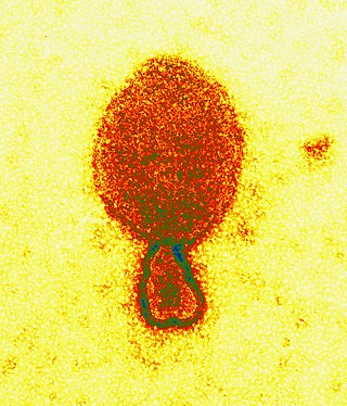
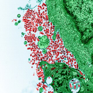
Nipah virus is a bat-borne, zoonotic virus that causes Nipah virus infection in humans and other animals, a disease with a very high mortality rate (40-75%). Numerous disease outbreaks caused by Nipah virus have occurred in South East Africa and Southeast Asia. Nipah virus belongs to the genus Henipavirus along with the Hendra virus, which has also caused disease outbreaks.

<i>Pestivirus</i> Genus of viruses

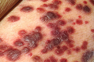
Pestivirus is a genus of viruses, in the family Flaviviridae. Viruses in the genus Pestivirus infect mammals, including members of the family Bovidae and the family Suidae. There are 11 species in this genus. Diseases associated with this genus include: hemorrhagic syndromes, abortion, and fatal mucosal disease.

<span class="mw-page-title-main">Viral envelope</span> Outermost layer of many types of the infectious agent

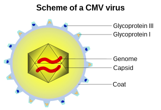
A viral envelope is the outermost layer of many types of viruses. It protects the genetic material in their life cycle when traveling between host cells. Not all viruses have envelopes. A viral envelope protein or E protein is a protein in the envelope, which may be acquired by the capsid from an infected host cell.

<span class="mw-page-title-main">Viral entry</span> Earliest stage of infection in the viral life cycle

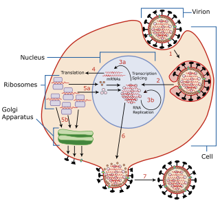
Viral entry is the earliest stage of infection in the viral life cycle, as the virus comes into contact with the host cell and introduces viral material into the cell. The major steps involved in viral entry are shown below. Despite the variation among viruses, there are several shared generalities concerning viral entry.

Pseudotyping is the process of producing viruses or viral vectors in combination with foreign viral envelope proteins. The result is a pseudotyped virus particle, also called a pseudovirus. With this method, the foreign viral envelope proteins can be used to alter host tropism or increase or decrease the stability of the virus particles. Pseudotyped particles do not carry the genetic material to produce additional viral envelope proteins, so the phenotypic changes cannot be passed on to progeny viral particles. In some cases, the inability to produce viral envelope proteins renders the pseudovirus replication incompetent. In this way, the properties of dangerous viruses can be studied in a lower risk setting.

Cedar virus, officially Cedar henipavirus, is a henipavirus known to be harboured by Pteropus spp. Infectious virus was isolated from the urine of a mixed Pteropus alecto and P. poliocephalus in Queensland, Australia in 2009. Unlike the Nipah and Hendra virus, Cedar virus infection does not lead to obvious disease in vivo. Infected animals mounted effective immune responses and seroconverted in challenge studies.

<i>Pneumoviridae</i> Family of viruses

Pneumoviridae is a family of negative-strand RNA viruses in the order Mononegavirales. Humans, cattle, and rodents serve as natural hosts. Respiratory tract infections are associated with member viruses such as human respiratory syncytial virus. There are five species in the family which are divided between the genera Metapneumovirus and Orthopneumovirus. The family used to be considered as a sub-family of Paramyxoviridae, but has been reclassified as of 2016.

Mammalian orthoreovirus (MRV) is a double-stranded RNA virus. It is a part of the family Reoviridae, as well as the subfamily Spinareovirinae. As seen in the name, the Mammalian Ortheoreovirus infects numerous mammalian species and vertebrates which serve as natural hosts. Some diseases that occur as a result of this virus or are associated with this virus include mild upper respiratory illness, and gastrointestinal illness. Examples of these are: upper respiratory tract syndromes, gastroenteritis, biliary atresia, obstructive hydrocephalus, jaundice, alopecia, conjunctivitis, and ‘oily hair’ associated with steatorrhea.

<span class="mw-page-title-main">Nipah virus infection</span> Disease caused by Nipah virus

A Nipah virus infection is a viral infection caused by the Nipah virus. Symptoms from infection vary from none to fever, cough, headache, shortness of breath, and confusion. This may worsen into a coma over a day or two, and 50 to 75% of those infected die. Complications can include inflammation of the brain and seizures following recovery.

West Caucasian bat lyssavirus (WCBL) is a member of genus Lyssavirus, family Rhabdoviridae and order Mononegavirales. This virus was first isolated from Miniopterus schreibersii, in the western Caucasus Mountains of southeastern Europe in 2002. WCBL is the most divergent form of Lyssavirus, and is found in Miniopterus bats (insectivorous), Rousettus aegyptiacus, and Eidolon helvum. The latter two are both fruit bats. The virus is fragile as it can be inactivated by UV light and chemicals, such as ether, chloroform, and bleach. WCBL has not been known to infect humans thus far.

Bat mumps orthorubulavirus, formerly Bat mumps rubulavirus (BMV), is a member of genus Orthorubulavirus, family Paramyxoviridae, and order Mononegavirales. Paramyxoviridae viruses were first isolated from bats using heminested PCR with degenerate primers. This process was then followed by Sanger sequencing. A specific location of this virus is not known because it was isolated from bats worldwide. Although multiple paramyxoviridae viruses have been isolated worldwide, BMV specifically has not been isolated thus far. However, BMV was detected in African fruit bats, but no infectious form has been isolated to date. It is known that BMV is transmitted through saliva in the respiratory system of bats. While the virus was considered its own species for a few years, phylogenetic analysis has since shown that it is a member of Mumps orthorubulavirus.

Rio Negro virus is an alphavirus that was first isolated in Argentina in 1980. The virus was first called Ag80-663 but was renamed to Rio Negro virus in 2005. It is a former member of the Venezuelan equine encephalitis complex (VEEC), which are a group of alphaviruses in the Americas that have the potential to emerge and cause disease. Río Negro virus was recently reclassified as a distinct species. Closely related viruses include Mucambo virus and Everglades virus.

<span class="mw-page-title-main">Mòjiāng virus</span> Species of virus

Mòjiāng virus(MojV), officially Mojiang henipavirus, is a virus in the family Paramyxoviridae. Based on phylogenetics, Mòjiāng virus is placed in the genus Henipavirus or described as a henipa-like virus. Antibodies raised against Mòjiāng virus glycoproteins are serologically distinct from other henipaviruses (among which higher cross-reactivity is observed).

References

  1. Stewart, Cameron R.; Deffrasnes, Celine; Foo, Chwan Hong; Bean, Andrew G. D.; Wang, Lin-Fa (2018), Tripp, Ralph A.; Tompkins, S. Mark (eds.), "A Functional Genomics Approach to Henipavirus Research: The Role of Nuclear Proteins, MicroRNAs and Immune Regulators in Infection and Disease", Roles of Host Gene and Non-coding RNA Expression in Virus Infection, Current Topics in Microbiology and Immunology, vol. 419, Springer International Publishing, pp. 191–213, doi:10.1007/82_2017_28, ISBN   978-3-030-05369-7, PMC   7122743 , PMID   28674944
  2. Behner, Laura; Zimmermann, Louisa; Ringel, Marc; Weis, Michael; Maisner, Andrea (2018-05-01). "Formation of high-order oligomers is required for functional bioactivity of an African bat henipavirus surface glycoprotein". Veterinary Microbiology. 218: 90–97. doi:10.1016/j.vetmic.2018.03.031. ISSN   0378-1135. PMID   29685227.
  3. Murray, K.; Selleck, P.; Hooper, P.; Hyatt, A.; Gould, A.; Gleeson, L.; Westbury, H.; Hiley, L.; Selvey, L.; Rodwell, B.; Et, Al (1995-04-07). "A morbillivirus that caused fatal disease in horses and humans". Science. 268 (5207): 94–97. Bibcode:1995Sci...268...94M. doi:10.1126/science.7701348. ISSN   0036-8075. PMID   7701348.
  4. Halpin, K.; Young, P. L.; Field, H. E.; Mackenzie, J. S. (2000). "Isolation of Hendra virus from pteropid bats: a natural reservoir of Hendra virus". Journal of General Virology. 81 (8): 1927–1932. doi: 10.1099/0022-1317-81-8-1927 . ISSN   0022-1317. PMID   10900029.
  5. Middleton, Deborah; Pallister, Jackie; Klein, Reuben; Feng, Yan-Ru; Haining, Jessica; Arkinstall, Rachel; Frazer, Leah; Huang, Jin-An; Edwards, Nigel; Wareing, Mark; Elhay, Martin (March 2014). "Hendra Virus Vaccine, a One Health Approach to Protecting Horse, Human, and Environmental Health". Emerging Infectious Diseases. 20 (3): 372–379. doi:10.3201/eid2003.131159. ISSN   1080-6040. PMC   3944873 . PMID   24572697.
  6. Hayman, David T. S.; Wang, Lin-Fa; Barr, Jennifer; Baker, Kate S.; Suu-Ire, Richard; Broder, Christopher C.; Cunningham, Andrew A.; Wood, James L. N. (2011-09-22). "Antibodies to Henipavirus or Henipa-Like Viruses in Domestic Pigs in Ghana, West Africa". PLOS ONE. 6 (9): e25256. Bibcode:2011PLoSO...625256H. doi: 10.1371/journal.pone.0025256 . ISSN   1932-6203. PMC   3178620 . PMID   21966471.
  7. Hayman, David T. S.; Suu-Ire, Richard; Breed, Andrew C.; McEachern, Jennifer A.; Wang, Linfa; Wood, James L. N.; Cunningham, Andrew A. (2008-07-23). "Evidence of Henipavirus Infection in West African Fruit Bats". PLOS ONE. 3 (7): e2739. Bibcode:2008PLoSO...3.2739H. doi: 10.1371/journal.pone.0002739 . ISSN   1932-6203. PMC   2453319 . PMID   18648649.
  8. 1 2 Drexler, Jan Felix; Corman, Victor Max; Gloza-Rausch, Florian; Seebens, Antje; Annan, Augustina; Ipsen, Anne; Kruppa, Thomas; Müller, Marcel A.; Kalko, Elisabeth K. V.; Adu-Sarkodie, Yaw; Oppong, Samuel (2009-07-28). "Henipavirus RNA in African Bats". PLOS ONE. 4 (7): e6367. Bibcode:2009PLoSO...4.6367D. doi: 10.1371/journal.pone.0006367 . ISSN   1932-6203. PMC   2712088 . PMID   19636378.
  9. Drexler, Jan Felix; Corman, Victor Max; Müller, Marcel Alexander; Maganga, Gael Darren; Vallo, Peter; Binger, Tabea; Gloza-Rausch, Florian; Cottontail, Veronika M.; Rasche, Andrea; Yordanov, Stoian; Seebens, Antje (2012-04-24). "Bats host major mammalian paramyxoviruses". Nature Communications. 3 (1): 796. Bibcode:2012NatCo...3..796D. doi: 10.1038/ncomms1796 . ISSN   2041-1723. PMC   3343228 . PMID   22531181.
  10. de Araujo, Jansen; Lo, Michael K.; Tamin, Azaibi; Ometto, Tatiana L.; Thomazelli, Luciano M.; Nardi, Marcello S.; Hurtado, Renata F.; Nava, Alessandra; Spiropoulou, Christina F.; Rota, Paul A.; Durigon, Edison L. (2017). "Antibodies Against Henipa-Like Viruses in Brazilian Bats". Vector-Borne and Zoonotic Diseases. 17 (4): 271–274. doi:10.1089/vbz.2016.2051. ISSN   1530-3667. PMID   28103156.
  11. 1 2 Wang, Lin-Fa; Harcourt, Brian H; Yu, Meng; Tamin, Azaibi; Rota, Paul A; Bellini, William J; Eaton, Bryan T (2001-04-01). "Molecular biology of Hendra and Nipah viruses". Microbes and Infection. 3 (4): 279–287. doi:10.1016/S1286-4579(01)01381-8. ISSN   1286-4579. PMID   11334745.
  12. Weatherman, Sarah; Feldmann, Heinz; de Wit, Emmie (February 2018). "Transmission of henipaviruses". Current Opinion in Virology. 28: 7–11. doi:10.1016/j.coviro.2017.09.004. ISSN   1879-6257. PMC   5835161 . PMID   29035743.
  13. 1 2 Voigt, Kathleen; Hoffmann, Markus; Drexler, Jan Felix; Müller, Marcel Alexander; Drosten, Christian; Herrler, Georg; Krüger, Nadine (2019-08-29). "Fusogenicity of the Ghana Virus (Henipavirus: Ghanaian bat henipavirus) Fusion Protein is Controlled by the Cytoplasmic Domain of the Attachment Glycoprotein". Viruses. 11 (9): 800. doi: 10.3390/v11090800 . ISSN   1999-4915. PMC   6784138 . PMID   31470664.
  14. 1 2 Xu, Kai; Broder, Christopher C.; Nikolov, Dimitar B. (February 2012). "Ephrin-B2 and ephrin-B3 as functional henipavirus receptors". Seminars in Cell & Developmental Biology. 23 (1): 116–123. doi:10.1016/j.semcdb.2011.12.005. ISSN   1084-9521. PMC   3327611 . PMID   22227101.
  15. Liu, Qian; Stone, Jacquelyn A.; Bradel-Tretheway, Birgit; Dabundo, Jeffrey; Benavides Montano, Javier A.; Santos-Montanez, Jennifer; Biering, Scott B.; Nicola, Anthony V.; Iorio, Ronald M.; Lu, Xiaonan; Aguilar, Hector C. (2013-11-21). "Unraveling a Three-Step Spatiotemporal Mechanism of Triggering of Receptor-Induced Nipah Virus Fusion and Cell Entry". PLOS Pathogens. 9 (11): e1003770. doi: 10.1371/journal.ppat.1003770 . ISSN   1553-7366. PMC   3837712 . PMID   24278018.
  16. Ranadheera, Charlene; Proulx, Roxanne; Chaiyakul, Mark; Jones, Shane; Grolla, Allen; Leung, Anders; Rutherford, John; Kobasa, Darwyn; Carpenter, Michael; Czub, Markus (2018-10-30). "The interaction between the Nipah virus nucleocapsid protein and phosphoprotein regulates virus replication". Scientific Reports. 8 (1): 15994. Bibcode:2018NatSR...815994R. doi:10.1038/s41598-018-34484-7. ISSN   2045-2322. PMC   6207681 . PMID   30375468.
  17. Kolakofsky, Daniel; Pelet, Thierry; Garcin, Dominique; Hausmann, Stéphane; Curran, Joseph; Roux, Laurent (February 1998). "Paramyxovirus RNA Synthesis and the Requirement for Hexamer Genome Length: the Rule of Six Revisited". Journal of Virology. 72 (2): 891–899. doi:10.1128/JVI.72.2.891-899.1998. ISSN   0022-538X. PMC   124558 . PMID   9444980.
  18. Pernet, Olivier; Lee, Benhur (2012). "Henipavirus Receptor Usage and Tropism". Current Topics in Microbiology and Immunology. 359: 59–78. doi:10.1007/82_2012_222. ISBN   978-3-642-29818-9. ISSN   0070-217X. PMC   3587688 . PMID   22695915.
  19. Wong, Kum Thong; Shieh, Wun-Ju; Kumar, Shalini; Norain, Karim; Abdullah, Wahidah; Guarner, Jeannette; Goldsmith, Cynthia S.; Chua, Kaw Bing; Lam, Sai Kit; Tan, Chong Tin; Goh, Khean Jin (December 2002). "Nipah Virus Infection". The American Journal of Pathology. 161 (6): 2153–2167. doi:10.1016/S0002-9440(10)64493-8. ISSN   0002-9440. PMC   1850894 . PMID   12466131.