Glyceraldehyde 3-phosphate dehydrogenase

Last updated

GAPDH
GAPDH with labels.png
Available structures
PDB Ortholog search: PDBe RCSB
Identifiers
Aliases GAPDH , GAPD, G3PD, HEL-S-162eP, glyceraldehyde-3-phosphate dehydrogenase
External IDs OMIM: 138400 MGI: 5434255 HomoloGene: 107053 GeneCards: GAPDH
Orthologs
SpeciesHumanMouse
Entrez
Ensembl
UniProt
RefSeq (mRNA)

XM_001476707

RefSeq (protein)

NP_001243728
NP_001276674
NP_001276675
NP_002037
NP_001344872

Contents

NP_001276655
NP_032110

Location (UCSC) Chr 12: 6.53 – 6.54 Mb n/a
PubMed search [2] [3]
Wikidata
View/Edit Human View/Edit Mouse
Glyceraldehyde 3-phosphate dehydrogenase, NAD binding domain
PDB 1cer EBI.jpg
determinants of enzyme thermostability observed in the molecular structure of thermus aquaticus d-glyceraldehyde-3-phosphate dehydrogenase at 2.5 angstroms resolution
Identifiers
SymbolGp_dh_N
Pfam PF00044
Pfam clan CL0063
InterPro IPR020828
PROSITE PDOC00069
SCOP2 1gd1 / SCOPe / SUPFAM
Available protein structures:
Pfam   structures / ECOD  
PDB RCSB PDB; PDBe; PDBj
PDBsum structure summary
Glyceraldehyde 3-phosphate dehydrogenase, C-terminal domain
PDB 2czc EBI.jpg
crystal structure of glyceraldehyde-3-phosphate dehydrogenase from pyrococcus horikoshii ot3
Identifiers
SymbolGp_dh_C
Pfam PF02800
Pfam clan CL0139
InterPro IPR020829
PROSITE PDOC00069
SCOP2 1gd1 / SCOPe / SUPFAM
Available protein structures:
Pfam   structures / ECOD  
PDB RCSB PDB; PDBe; PDBj
PDBsum structure summary

Glyceraldehyde 3-phosphate dehydrogenase (abbreviated GAPDH) (EC 1.2.1.12) is an enzyme of about 37kDa that catalyzes the sixth step of glycolysis and thus serves to break down glucose for energy and carbon molecules. In addition to this long established metabolic function, GAPDH has recently been implicated in several non-metabolic processes, including transcription activation, initiation of apoptosis, [4] ER-to-Golgi vesicle shuttling, and fast axonal, or axoplasmic transport. [5] In sperm, a testis-specific isoenzyme GAPDHS is expressed.

Structure

Under normal cellular conditions, cytoplasmic GAPDH exists primarily as a tetramer. This form is composed of four identical 37-kDa subunits containing a single catalytic thiol group each and critical to the enzyme's catalytic function. [6] [7] Nuclear GAPDH has increased isoelectric point (pI) of pH 8.3–8.7. [7] Of note, the cysteine residue C152 in the enzyme's active site is required for the induction of apoptosis by oxidative stress. [7] Notably, post-translational modifications of cytoplasmic GAPDH contribute to its functions outside of glycolysis. [6]

GAPDH is encoded by a single gene that produces a single mRNA transcript with 8 splice variants, though an isoform does exist as a separate gene that is expressed only in spermatozoa. [7]

Reaction

glyceraldehyde 3-phosphate glyceraldehyde phosphate dehydrogenase D-glycerate 1,3-bisphosphate
D-glyceraldehyde-3-phosphate.svg   D-glycerate 1,3-bisphosphate.svg
NAD+ +PiNADH + H+
Biochem reaction arrow reversible YYYY horiz med.svg
NAD+ +PiNADH + H+


Compound C00118 at KEGG Pathway Database.Enzyme 1.2.1.12 at KEGG Pathway Database.Reaction R01063 at KEGG Pathway Database.Compound C00236 at KEGG Pathway Database.

Two-step conversion of G3P

The first reaction is the oxidation of glyceraldehyde 3-phosphate (G3P) at the position-1 (in the diagram it is shown as the 4th carbon from glycolysis), in which an aldehyde is converted into a carboxylic acid (ΔG°'=-50 kJ/mol (−12kcal/mol)) and NAD+ is simultaneously reduced endergonically to NADH.

The energy released by this highly exergonic oxidation reaction drives the endergonic second reaction (ΔG°'=+50 kJ/mol (+12kcal/mol)), in which a molecule of inorganic phosphate is transferred to the GAP intermediate to form a product with high phosphoryl-transfer potential: 1,3-bisphosphoglycerate (1,3-BPG).

This is an example of phosphorylation coupled to oxidation, and the overall reaction is somewhat endergonic (ΔG°'=+6.3 kJ/mol (+1.5)). Energy coupling here is made possible by GAPDH.

Mechanism

GAPDH uses covalent catalysis and general base catalysis to decrease the very large activation energy of the second step (phosphorylation) of this reaction.

1: Oxidation

First, a cysteine residue in the active site of GAPDH attacks the carbonyl group of G3P, creating a hemithioacetal intermediate (covalent catalysis).

The hemithioacetal is deprotonated by a histidine residue in the enzyme's active site (general base catalysis). Deprotonation encourages the reformation of the carbonyl group in the subsequent thioester intermediate and ejection of a hydride ion.

Next, an adjacent, tightly bound molecule of NAD+ accepts the hydride ion, forming NADH while the hemithioacetal is oxidized to a thioester.

This thioester species is much higher in energy (less stable) than the carboxylic acid species that would result if G3P were oxidized in the absence of GAPDH (the carboxylic acid species is so low in energy that the energy barrier for the second step of the reaction (phosphorylation) would be too high, and the reaction, therefore, too slow and unfavorable for a living organism).

2: Phosphorylation

NADH leaves the active site and is replaced by another molecule of NAD+, the positive charge of which stabilizes the negatively charged carbonyl oxygen in the transition state of the next and ultimate step. Finally, a molecule of inorganic phosphate attacks the thioester and forms a tetrahedral intermediate, which then collapses to release 1,3-bisphosphoglycerate, and the thiol group of the enzyme's cysteine residue.

Regulation

This protein may use the morpheein model of allosteric regulation. [8]

Function

Metabolic

As its name indicates, glyceraldehyde 3-phosphate dehydrogenase (GAPDH) catalyses the conversion of glyceraldehyde 3-phosphate to D-glycerate 1,3-bisphosphate. This is the 6th step in the glycolytic breakdown of glucose, an important pathway of energy and carbon molecule supply which takes place in the cytosol of eukaryotic cells. The conversion occurs in two coupled steps. The first is favourable and allows the second unfavourable step to occur.

Adhesion

One of the GAPDH moonlighting functions is its role in adhesion and binding to other partners. Bacterial GAPDH from Mycoplasma and Streptococcus and fungal GAPDH from Paracoccidioides brasiliensis are known to bind with the human extracellular matrix component and act in adhesion. [9] [10] [11] GAPDH is found to be surface bound contributing in adhesion and also in competitive exclusion of harmful pathogens. [12] GAPDH from Candida albicans is found to cell-wall associated and binds to Fibronectin and Laminin. [13] GAPDH from probiotics species are known to bind human colonic mucin and ECM, resulting in enhanced colonization of probiotics in the human gut. [14] [15] [16] Patel D. et al., showed that Lactobacillus acidophilus GAPDH binds with mucin, acting in adhesion. [17]

Transcription and apoptosis

GAPDH can itself activate transcription. The OCA-S transcriptional coactivator complex contains GAPDH and lactate dehydrogenase, two proteins previously only thought to be involved in metabolism. GAPDH moves between the cytosol and the nucleus and may thus link the metabolic state to gene transcription. [18]

In 2005, Hara et al. showed that GAPDH initiates apoptosis. This is not a third function, but can be seen as an activity mediated by GAPDH binding to DNA like in transcription activation, discussed above. The study demonstrated that GAPDH is S-nitrosylated by NO in response to cell stress, which causes it to bind to the protein SIAH1, a ubiquitin ligase. The complex moves into the nucleus where Siah1 targets nuclear proteins for degradation, thus initiating controlled cell shutdown. [19] In subsequent study the group demonstrated that deprenyl, which has been used clinically to treat Parkinson's disease, strongly reduces the apoptotic action of GAPDH by preventing its S-nitrosylation and might thus be used as a drug. [20]

Metabolic switch

GAPDH acts as a reversible metabolic switch under oxidative stress. [21] When cells are exposed to oxidants, they need excessive amounts of the antioxidant cofactor NADPH. In the cytosol, NADPH is reduced from NADP+ by several enzymes, three of them catalyze the first steps of the pentose phosphate pathway. Oxidant-treatments cause an inactivation of GAPDH. This inactivation re-routes temporally the metabolic flux from glycolysis to the pentose phosphate pathway, allowing the cell to generate more NADPH. [22] Under stress conditions, NADPH is needed by some antioxidant-systems including glutaredoxin and thioredoxin as well as being essential for the recycling of gluthathione.

ER-to-Golgi transport

GAPDH also appears to be involved in the vesicle transport from the endoplasmic reticulum (ER) to the Golgi apparatus which is part of shipping route for secreted proteins. It was found that GAPDH is recruited by rab2 to the vesicular-tubular clusters of the ER where it helps to form COP 1 vesicles. GAPDH is activated via tyrosine phosphorylation by Src. [23]

Additional functions

GAPDH, like many other enzymes, has multiple functions. In addition to catalysing the 6th step of glycolysis, recent evidence implicates GAPDH in other cellular processes. GAPDH has been described to exhibit higher order multifunctionality in the context of maintaining cellular iron homeostasis, [24] specifically as a chaperone protein for labile heme within cells. [25] This came as a surprise to researchers but it makes evolutionary sense to re-use and adapt existing proteins instead of evolving a novel protein from scratch.

Use as loading control

Because the GAPDH gene is often stably and constitutively expressed at high levels in most tissues and cells, it is considered a housekeeping gene. For this reason, GAPDH is commonly used by biological researchers as a loading control for western blot and as a control for qPCR. However, researchers have reported different regulation of GAPDH under specific conditions. [26] For example, the transcription factor MZF-1 has been shown to regulate the GAPDH gene. [27] Hypoxia also strongly upregulates GAPDH. [28] Therefore, the use of GAPDH as loading control has to be considered carefully.

Cellular distribution

All steps of glycolysis take place in the cytosol and so does the reaction catalysed by GAPDH. In red blood cells, GAPDH and several other glycolytic enzymes assemble in complexes on the inside of the cell membrane. The process appears to be regulated by phosphorylation and oxygenation. [29] Bringing several glycolytic enzymes close to each other is expected to greatly increase the overall speed of glucose breakdown. Recent studies have also revealed that GAPDH is expressed in an iron dependent fashion on the exterior of the cell membrane a where it plays a role in maintenance of cellular iron homeostasis. [30] [31]

Clinical significance

Cancer

GAPDH is overexpressed in multiple human cancers, such as cutaneous melanoma, and its expression is positively correlated with tumor progression. [32] [33] Its glycolytic and antiapoptotic functions contribute to proliferation and protection of tumor cells, promoting tumorigenesis. Notably, GAPDH protects against telomere shortening induced by chemotherapeutic drugs that stimulate the sphingolipid ceramide. Meanwhile, conditions like oxidative stress impair GAPDH function, leading to cellular aging and death. [7] Moreover, depletion of GAPDH has managed to induce senescence in tumor cells, thus presenting a novel therapeutic strategy for controlling tumor growth. [34]

Neurodegeneration

GAPDH has been implicated in several neurodegenerative diseases and disorders, largely through interactions with other proteins specific to that disease or disorder. These interactions may affect not only energy metabolism but also other GAPDH functions. [6] For example, GAPDH interactions with beta-amyloid precursor protein (betaAPP) could interfere with its function regarding the cytoskeleton or membrane transport, while interactions with huntingtin could interfere with its function regarding apoptosis, nuclear tRNA transport, DNA replication, and DNA repair. In addition, nuclear translocation of GAPDH has been reported in Parkinson's disease (PD), and several anti-apoptotic PD drugs, such as rasagiline, function by preventing the nuclear translocation of GAPDH. It is proposed that hypometabolism may be one contributor to PD, but the exact mechanisms underlying GAPDH involvement in neurodegenerative disease remains to be clarified. [35] The SNP rs3741916 in the 5' UTR of the GAPDH gene may be associated with late onset Alzheimer's disease. [36]

Interactions

Protein binding partners

GAPDH participates in a number of biological functions through its protein–protein interactions with:

Nucleic acid binding partners

GAPDH binds to single-stranded RNA [39] and DNA and a number of nucleic acid binding partners have been identified: [7]

Inhibitors

Interactive pathway map

Click on genes, proteins and metabolites below to link to respective articles. [§ 1]

[[File:
WP534.png go to articlego to articlego to articlego to articlego to articlego to articlego to articlego to articlego to articlego to articlego to articlego to articlego to articlego to articlego to articlego to articlego to articlego to articlego to articlego to articlego to articlego to articlego to articlego to articlego to articlego to articlego to articlego to articlego to articlego to articlego to articlego to articlego to articlego to articlego to articlego to articlego to articlego to articlego to articlego to articlego to articlego to articlego to articlego to articlego to articlego to articlego to articlego to articlego to articlego to articlego to articlego to articlego to articlego to articlego to articlego to articlego to articlego to articlego to articlego to articlego to articlego to articlego to articlego to articlego to articlego to articlego to articlego to articlego to articlego to articlego to WikiPathwaysgo to articlego to Entrezgo to article
[[
]]
[[
]]
[[
]]
[[
]]
[[
]]
[[
]]
[[
]]
[[
]]
[[
]]
[[
]]
[[
]]
[[
]]
[[
]]
[[
]]
[[
]]
[[
]]
[[
]]
[[
]]
[[
]]
[[
]]
[[
]]
[[
]]
[[
]]
[[
]]
[[
]]
[[
]]
[[
]]
[[
]]
[[
]]
[[
]]
[[
]]
[[
]]
[[
]]
[[
]]
[[
]]
[[
]]
[[
]]
[[
]]
[[
]]
[[
]]
[[
]]
[[
]]
[[
]]
[[
]]
[[
]]
[[
]]
[[
]]
[[
]]
[[
]]
[[
]]
[[
]]
[[
]]
[[
]]
[[
]]
[[
]]
[[
]]
[[
]]
[[
]]
[[
]]
[[
]]
[[
]]
[[
]]
[[
]]
[[
]]
[[
]]
[[
]]
[[
]]
[[
]]
[[
]]
[[
]]
[[
]]
[[
]]
[[
]]
[[
]]
[[
]]
WP534.png go to articlego to articlego to articlego to articlego to articlego to articlego to articlego to articlego to articlego to articlego to articlego to articlego to articlego to articlego to articlego to articlego to articlego to articlego to articlego to articlego to articlego to articlego to articlego to articlego to articlego to articlego to articlego to articlego to articlego to articlego to articlego to articlego to articlego to articlego to articlego to articlego to articlego to articlego to articlego to articlego to articlego to articlego to articlego to articlego to articlego to articlego to articlego to articlego to articlego to articlego to articlego to articlego to articlego to articlego to articlego to articlego to articlego to articlego to articlego to articlego to articlego to articlego to articlego to articlego to articlego to articlego to articlego to articlego to articlego to articlego to WikiPathwaysgo to articlego to Entrezgo to article
|alt=Glycolysis and Gluconeogenesis edit]]
Glycolysis and Gluconeogenesis edit
  1. The interactive pathway map can be edited at WikiPathways: "GlycolysisGluconeogenesis_WP534".

Related Research Articles

<span class="mw-page-title-main">Adenosine triphosphate</span> Energy-carrying molecule in living cells

Adenosine triphosphate (ATP) is a nucleotide that provides energy to drive and support many processes in living cells, such as muscle contraction, nerve impulse propagation, condensate dissolution, and chemical synthesis. Found in all known forms of life, it is often referred to as the "molecular unit of currency" of intracellular energy transfer.

<span class="mw-page-title-main">Glycolysis</span> Series of interconnected biochemical reactions

Glycolysis is the metabolic pathway that converts glucose into pyruvate and, in most organisms, occurs in the liquid part of cells. The free energy released in this process is used to form the high-energy molecules adenosine triphosphate (ATP) and reduced nicotinamide adenine dinucleotide (NADH). Glycolysis is a sequence of ten reactions catalyzed by enzymes.

<span class="mw-page-title-main">Phosphorylation</span> Chemical process of introducing a phosphate

In biochemistry, phosphorylation is the attachment of a phosphate group to a molecule or an ion. This process and its inverse, dephosphorylation, are common in biology. Protein phosphorylation often activates many enzymes.

<span class="mw-page-title-main">Tumor hypoxia</span> Situation where tumor cells have been deprived of oxygen

Tumor hypoxia is the situation where tumor cells have been deprived of oxygen. As a tumor grows, it rapidly outgrows its blood supply, leaving portions of the tumor with regions where the oxygen concentration is significantly lower than in healthy tissues. Hypoxic microenvironments in solid tumors are a result of available oxygen being consumed within 70 to 150 μm of tumor vasculature by rapidly proliferating tumor cells thus limiting the amount of oxygen available to diffuse further into the tumor tissue. In order to support continuous growth and proliferation in challenging hypoxic environments, cancer cells are found to alter their metabolism. Furthermore, hypoxia is known to change cell behavior and is associated with extracellular matrix remodeling and increased migratory and metastatic behavior.

<span class="mw-page-title-main">Aldolase A</span> Mammalian protein found in Homo sapiens

Aldolase A, also known as fructose-bisphosphate aldolase, is an enzyme that in humans is encoded by the ALDOA gene on chromosome 16.

<span class="mw-page-title-main">Human iron metabolism</span> Iron metabolism in the body

Human iron metabolism is the set of chemical reactions that maintain human homeostasis of iron at the systemic and cellular level. Iron is both necessary to the body and potentially toxic. Controlling iron levels in the body is a critically important part of many aspects of human health and disease. Hematologists have been especially interested in systemic iron metabolism, because iron is essential for red blood cells, where most of the human body's iron is contained. Understanding iron metabolism is also important for understanding diseases of iron overload, such as hereditary hemochromatosis, and iron deficiency, such as iron-deficiency anemia.

<span class="mw-page-title-main">Tumor metabolome</span>

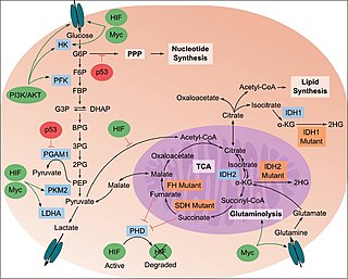
The study of the tumor metabolism, also known as tumor metabolome describes the different characteristic metabolic changes in tumor cells. The characteristic attributes of the tumor metabolome are high glycolytic enzyme activities, the expression of the pyruvate kinase isoenzyme type M2, increased channeling of glucose carbons into synthetic processes, such as nucleic acid, amino acid and phospholipid synthesis, a high rate of pyrimidine and purine de novo synthesis, a low ratio of Adenosine triphosphate and Guanosine triphosphate to Cytidine triphosphate and Uridine triphosphate, low Adenosine monophosphate levels, high glutaminolytic capacities, release of immunosuppressive substances and dependency on methionine.

Glyceraldehyde-3-phosphate dehydrogenase (NADP+) (GAPN) is an enzyme that irreversibly catalyzes the oxidation of glyceraldehyde-3-phosphate (GAP) to 3-phosphoglycerate using the reduction of NADP+ to NADPH. GAPN is used in a variant of glycolysis that conserves energy as NADPH rather than as ATP. The NADPH and 3-PG can then be used for synthesis. The most familiar variant of glycolysis uses glyceraldehyde-3-phosphate dehydrogenase (GAPDH) and phosphoglycerate kinase to produce ATP. GAPDH is phosphorylating. GAPN is non-phosphorylating.

<span class="mw-page-title-main">Transferrin receptor</span> Family of transport proteins

Transferrin receptor (TfR) is a carrier protein for transferrin. It is needed for the import of iron into cells and is regulated in response to intracellular iron concentration. It imports iron by internalizing the transferrin-iron complex through receptor-mediated endocytosis. The existence of a receptor for transferrin iron uptake has been recognized since the late 1950s. Earlier two transferrin receptors in humans, transferrin receptor 1 and transferrin receptor 2 had been characterized and until recently cellular iron uptake was believed to occur chiefly via these two well documented transferrin receptors. Both these receptors are transmembrane glycoproteins. TfR1 is a high affinity ubiquitously expressed receptor while expression of TfR2 is restricted to certain cell types and is unaffected by intracellular iron concentrations. TfR2 binds to transferrin with a 25-30 fold lower affinity than TfR1. Although TfR1 mediated iron uptake is the major pathway for iron acquisition by most cells and especially developing erythrocytes, several studies have indicated that the uptake mechanism varies depending upon the cell type. It is also reported that Tf uptake exists independent of these TfRs although the mechanisms are not well characterized. The multifunctional glycolytic enzyme glyceraldehyde 3-phosphate dehydrogenase has been shown to utilize post translational modifications to exhibit higher order moonlighting behavior wherein it switches its function as a holo or apo transferrin receptor leading to either iron delivery or iron export respectively.

<span class="mw-page-title-main">Glycerol phosphate shuttle</span>

The glycerol-3-phosphate shuttle is a mechanism used in skeletal muscle and the brain that regenerates NAD+ from NADH, a by-product of glycolysis. The NADH generated during glycolysis is found in the cytoplasm and must be transported into the mitochondria to enter the oxidative phosphorylation pathway. However, the inner mitochondrial membrane is impermeable to NADH and NAD+ and does not contain a transport system for these electron carriers. Either the glycerol-3-phosphate shuttle pathway or the malate-aspartate shuttle pathway, depending on the tissue of the organism, must be taken to transport electrons from cytoplasmic NADH into the mitochondria. The shuttle consists of the sequential activity of two proteins; Cytoplasmic glycerol-3-phosphate dehydrogenase (cGPD) transfers an electron pair from NADH to dihydroxyacetone phosphate (DHAP), forming glycerol-3-phosphate (G3P) and regenerating NAD+ needed to generate energy via glycolysis. The other protein, mitochondrial glycerol-3-phosphate dehydrogenase (mGPD) catalyzes the oxidation of G3P by FAD, regenerating DHAP in the cytosol and forming FADH2 in the mitochondrial matrix. In mammals, its activity in transporting reducing equivalents across the mitochondrial membrane is considered secondary to the malate-aspartate shuttle.

<span class="mw-page-title-main">Fructose-bisphosphate aldolase</span>

Fructose-bisphosphate aldolase, often just aldolase, is an enzyme catalyzing a reversible reaction that splits the aldol, fructose 1,6-bisphosphate, into the triose phosphates dihydroxyacetone phosphate (DHAP) and glyceraldehyde 3-phosphate (G3P). Aldolase can also produce DHAP from other (3S,4R)-ketose 1-phosphates such as fructose 1-phosphate and sedoheptulose 1,7-bisphosphate. Gluconeogenesis and the Calvin cycle, which are anabolic pathways, use the reverse reaction. Glycolysis, a catabolic pathway, uses the forward reaction. Aldolase is divided into two classes by mechanism.

<span class="mw-page-title-main">Glycerol-3-phosphate dehydrogenase</span> Class of enzymes

Glycerol-3-phosphate dehydrogenase (GPDH) is an enzyme that catalyzes the reversible redox conversion of dihydroxyacetone phosphate to sn-glycerol 3-phosphate.

<span class="mw-page-title-main">Hepatitis A virus internal ribosome entry site (IRES)</span>

This family represents the internal ribosome entry site (IRES) of the hepatitis A virus. HAV IRES is a 450 nucleotide long sequence located in the 735 nt long 5’ UTR of Hepatitis A viral RNA genome. IRES elements allow cap and end-independent translation of mRNA in the host cell. The IRES achieves this by mediating the internal initiation of translation by recruiting a ribosomal 40S pre-initiation complex directly to the initiation codon and eliminates the requirement for eukaryotic initiation factor, eIF4F.

<span class="mw-page-title-main">Lactate dehydrogenase A</span> Type of enzyme

Lactate dehydrogenase A (LDHA) is an enzyme which in humans is encoded by the LDHA gene. It is a monomer of Lactate dehydrogenase, which exists as a tetramer. The other main subunit is lactate dehydrogenase B (LDHB).

<span class="mw-page-title-main">PRKCI</span> Protein-coding gene in the species Homo sapiens

Protein kinase C iota type is an enzyme that in humans is encoded by the PRKCI gene.

<span class="mw-page-title-main">GAPDHS</span> Protein-coding gene in the species Homo sapiens

Glyceraldehyde-3-phosphate dehydrogenase, spermatogenic or glyceraldehyde-3-phosphate dehydrogenase, testis-specific is an enzyme that in humans is encoded by the GAPDHS gene.

<span class="mw-page-title-main">Aldolase C</span> Protein-coding gene in the species Homo sapiens

Aldolase C, fructose-bisphosphate, is an enzyme that, in humans, is encoded by the ALDOC gene on chromosome 17. This gene encodes a member of the class I fructose-bisphosphate aldolase gene family. Expressed specifically in the hippocampus and Purkinje cells of the brain, the encoded protein is a glycolytic enzyme that catalyzes the reversible aldol cleavage of fructose 1,6-bisphosphate and fructose-1-phosphate to dihydroxyacetone phosphate and either glyceraldehyde 3-phosphate or glyceraldehyde, respectively.[provided by RefSeq, Jul 2008]

<span class="mw-page-title-main">Protein moonlighting</span> Proteins performing more than one function

Protein moonlighting is a phenomenon by which a protein can perform more than one function. It is an excellent example of gene sharing.

<span class="mw-page-title-main">TP53-inducible glycolysis and apoptosis regulator</span> Protein-coding gene in the species Homo sapiens

The TP53-inducible glycolysis and apoptosis regulator (TIGAR) also known as fructose-2,6-bisphosphatase TIGAR is an enzyme that in humans is encoded by the C12orf5 gene.

<span class="mw-page-title-main">1-Arseno-3-phosphoglycerate</span>

1-Arseno-3-phosphoglycerate is a compound produced by the enzyme glyceraldehyde 3-phosphate dehydrogenase, present in high concentrations in many organisms, from glyceraldehyde 3-phosphate and arsenate in the glycolysis pathway. The compound is unstable and hydrolyzes spontaneously to 3-phosphoglycerate, bypassing the energy producing step of glycolysis.

References

  1. 1 2 3 GRCh38: Ensembl release 89: ENSG00000111640 - Ensembl, May 2017
  2. "Human PubMed Reference:". National Center for Biotechnology Information, U.S. National Library of Medicine.
  3. "Mouse PubMed Reference:". National Center for Biotechnology Information, U.S. National Library of Medicine.
  4. Tarze A, Deniaud A, Le Bras M, Maillier E, Molle D, Larochette N, Zamzami N, Jan G, Kroemer G, Brenner C (April 2007). "GAPDH, a novel regulator of the pro-apoptotic mitochondrial membrane permeabilization". Oncogene. 26 (18): 2606–2620. doi:10.1038/sj.onc.1210074. PMID   17072346. S2CID   20291542.
  5. Zala D, Hinckelmann MV, Yu H, Lyra da Cunha MM, Liot G, Cordelières FP, Marco S, Saudou F (January 2013). "Vesicular glycolysis provides on-board energy for fast axonal transport". Cell. 152 (3): 479–491. doi: 10.1016/j.cell.2012.12.029 . PMID   23374344.
  6. 1 2 3 4 5 6 7 Tristan C, Shahani N, Sedlak TW, Sawa A (February 2011). "The diverse functions of GAPDH: views from different subcellular compartments". Cellular Signalling. 23 (2): 317–323. doi:10.1016/j.cellsig.2010.08.003. PMC   3084531 . PMID   20727968.
  7. 1 2 3 4 5 6 7 8 9 10 11 12 13 14 15 16 17 18 19 Nicholls C, Li H, Liu JP (August 2012). "GAPDH: a common enzyme with uncommon functions". Clinical and Experimental Pharmacology & Physiology. 39 (8): 674–679. doi:10.1111/j.1440-1681.2011.05599.x. PMID   21895736. S2CID   23499684.
  8. Selwood T, Jaffe EK (March 2012). "Dynamic dissociating homo-oligomers and the control of protein function". Archives of Biochemistry and Biophysics. 519 (2): 131–143. doi:10.1016/j.abb.2011.11.020. PMC   3298769 . PMID   22182754.
  9. Dumke R, Hausner M, Jacobs E (August 2011). "Role of Mycoplasma pneumoniae glyceraldehyde-3-phosphate dehydrogenase (GAPDH) in mediating interactions with the human extracellular matrix". Microbiology. 157 (Pt 8): 2328–2338. doi: 10.1099/mic.0.048298-0 . PMID   21546586.
  10. Brassard J, Gottschalk M, Quessy S (August 2004). "Cloning and purification of the Streptococcus suis serotype 2 glyceraldehyde-3-phosphate dehydrogenase and its involvement as an adhesin". Veterinary Microbiology. 102 (1–2): 87–94. doi:10.1016/j.vetmic.2004.05.008. PMID   15288930.
  11. Barbosa MS, Báo SN, Andreotti PF, de Faria FP, Felipe MS, dos Santos Feitosa L, Mendes-Giannini MJ, Soares CM (January 2006). "Glyceraldehyde-3-phosphate dehydrogenase of Paracoccidioides brasiliensis is a cell surface protein involved in fungal adhesion to extracellular matrix proteins and interaction with cells". Infection and Immunity. 74 (1): 382–389. doi:10.1128/IAI.74.1.382-389.2006. PMC   1346668 . PMID   16368993.
  12. Ramiah K, van Reenen CA, Dicks LM (1 July 2008). "Surface-bound proteins of Lactobacillus plantarum 423 that contribute to adhesion of Caco-2 cells and their role in competitive exclusion and displacement of Clostridium sporogenes and Enterococcus faecalis". Research in Microbiology. 159 (6): 470–475. doi: 10.1016/j.resmic.2008.06.002 . PMID   18619532.
  13. Gozalbo D, Gil-Navarro I, Azorín I, Renau-Piqueras J, Martínez JP, Gil ML (May 1998). "The cell wall-associated glyceraldehyde-3-phosphate dehydrogenase of Candida albicans is also a fibronectin and laminin binding protein". Infection and Immunity. 66 (5): 2052–2059. doi:10.1128/IAI.66.5.2052-2059.1998. PMC   108162 . PMID   9573088.
  14. Deng Z, Dai T, Zhang W, Zhu J, Luo XM, Fu D, Liu J, Wang H (December 2020). "Glyceraldehyde-3-Phosphate Dehydrogenase Increases the Adhesion of Lactobacillus reuteri to Host Mucin to Enhance Probiotic Effects". International Journal of Molecular Sciences. 21 (24): 9756. doi: 10.3390/ijms21249756 . PMC   7766874 . PMID   33371288.
  15. Kinoshita H, Uchida H, Kawai Y, Kawasaki T, Wakahara N, Matsuo H, Watanabe M, Kitazawa H, Ohnuma S, Miura K, Horii A, Saito T (June 2008). "Cell surface Lactobacillus plantarum LA 318 glyceraldehyde-3-phosphate dehydrogenase (GAPDH) adheres to human colonic mucin". Journal of Applied Microbiology. 104 (6): 1667–1674. doi:10.1111/j.1365-2672.2007.03679.x. PMID   18194256. S2CID   22346488.
  16. Kinoshita H, Wakahara N, Watanabe M, Kawasaki T, Matsuo H, Kawai Y, Kitazawa H, Ohnuma S, Miura K, Horii A, Saito T (1 November 2008). "Cell surface glyceraldehyde-3-phosphate dehydrogenase (GAPDH) of Lactobacillus plantarum LA 318 recognizes human A and B blood group antigens". Research in Microbiology. 159 (9–10): 685–691. doi: 10.1016/j.resmic.2008.07.005 . PMID   18790050.
  17. Patel DK, Shah KR, Pappachan A, Gupta S, Singh DD (October 2016). "Cloning, expression and characterization of a mucin-binding GAPDH from Lactobacillus acidophilus". International Journal of Biological Macromolecules. 91: 338–346. doi:10.1016/j.ijbiomac.2016.04.041. PMID   27180300.
  18. Zheng L, Roeder RG, Luo Y (July 2003). "S phase activation of the histone H2B promoter by OCA-S, a coactivator complex that contains GAPDH as a key component". Cell. 114 (2): 255–266. doi: 10.1016/S0092-8674(03)00552-X . PMID   12887926. S2CID   5543647.
  19. Hara MR, Agrawal N, Kim SF, Cascio MB, Fujimuro M, Ozeki Y, Takahashi M, Cheah JH, Tankou SK, Hester LD, Ferris CD, Hayward SD, Snyder SH, Sawa A (July 2005). "S-nitrosylated GAPDH initiates apoptotic cell death by nuclear translocation following Siah1 binding". Nature Cell Biology. 7 (7): 665–674. doi:10.1038/ncb1268. PMID   15951807. S2CID   1922911.
  20. Hara MR, Thomas B, Cascio MB, Bae BI, Hester LD, Dawson VL, Dawson TM, Sawa A, Snyder SH (March 2006). "Neuroprotection by pharmacologic blockade of the GAPDH death cascade". Proceedings of the National Academy of Sciences of the United States of America. 103 (10): 3887–3889. Bibcode:2006PNAS..103.3887H. doi: 10.1073/pnas.0511321103 . PMC   1450161 . PMID   16505364.
  21. Agarwal AR, Zhao L, Sancheti H, Sundar IK, Rahman I, Cadenas E (November 2012). "Short-term cigarette smoke exposure induces reversible changes in energy metabolism and cellular redox status independent of inflammatory responses in mouse lungs". American Journal of Physiology. Lung Cellular and Molecular Physiology. 303 (10): L889–L898. doi:10.1152/ajplung.00219.2012. PMID   23064950.
  22. Ralser M, Wamelink MM, Kowald A, Gerisch B, Heeren G, Struys EA, Klipp E, Jakobs C, Breitenbach M, Lehrach H, Krobitsch S (December 2007). "Dynamic rerouting of the carbohydrate flux is key to counteracting oxidative stress". Journal of Biology. 6 (4): 10. doi: 10.1186/jbiol61 . PMC   2373902 . PMID   18154684.
  23. Tisdale EJ, Artalejo CR (June 2007). "A GAPDH mutant defective in Src-dependent tyrosine phosphorylation impedes Rab2-mediated events". Traffic. 8 (6): 733–741. doi:10.1111/j.1600-0854.2007.00569.x. PMC   3775588 . PMID   17488287.
  24. Boradia VM, Raje M, Raje CI (December 2014). "Protein moonlighting in iron metabolism: glyceraldehyde-3-phosphate dehydrogenase (GAPDH)". Biochemical Society Transactions. 42 (6): 1796–1801. doi:10.1042/BST20140220. PMID   25399609.
  25. Sweeny EA, Singh AB, Chakravarti R, Martinez-Guzman O, Saini A, Haque MM, Garee G, Dans PD, Hannibal L, Reddi AR, Stuehr DJ (September 2018). "Glyceraldehyde-3-phosphate dehydrogenase is a chaperone that allocates labile heme in cells". The Journal of Biological Chemistry. 293 (37): 14557–14568. doi: 10.1074/jbc.RA118.004169 . PMC   6139559 . PMID   30012884.
  26. Barber RD, Harmer DW, Coleman RA, Clark BJ (May 2005). "GAPDH as a housekeeping gene: analysis of GAPDH mRNA expression in a panel of 72 human tissues". Physiological Genomics. 21 (3): 389–395. CiteSeerX   10.1.1.459.7039 . doi:10.1152/physiolgenomics.00025.2005. PMID   15769908.
  27. Piszczatowski RT, Rafferty BJ, Rozado A, Tobak S, Lents NH (August 2014). "The glyceraldehyde 3-phosphate dehydrogenase gene (GAPDH) is regulated by myeloid zinc finger 1 (MZF-1) and is induced by calcitriol". Biochemical and Biophysical Research Communications. 451 (1): 137–141. doi:10.1016/j.bbrc.2014.07.082. PMID   25065746.
  28. Saygin D, Tabib T, Bittar HE, Valenzi E, Sembrat J, Chan SY, Rojas M, Lafyatis R (2008). "Transcriptional profiling of lung cell populations in idiopathic pulmonary arterial hypertension". Pulmonary Circulation. 10 (1): 239–243. doi:10.1007/s11684-008-0045-7. PMC   7052475 . PMID   32166015. S2CID   85327763.
  29. Campanella ME, Chu H, Low PS (February 2005). "Assembly and regulation of a glycolytic enzyme complex on the human erythrocyte membrane". Proceedings of the National Academy of Sciences of the United States of America. 102 (7): 2402–2407. Bibcode:2005PNAS..102.2402C. doi: 10.1073/pnas.0409741102 . PMC   549020 . PMID   15701694.
  30. Sirover MA (December 2014). "Structural analysis of glyceraldehyde-3-phosphate dehydrogenase functional diversity". The International Journal of Biochemistry & Cell Biology. 57: 20–26. doi:10.1016/j.biocel.2014.09.026. PMC   4268148 . PMID   25286305.
  31. 1 2 Kumar S, Sheokand N, Mhadeshwar MA, Raje CI, Raje M (January 2012). "Characterization of glyceraldehyde-3-phosphate dehydrogenase as a novel transferrin receptor". The International Journal of Biochemistry & Cell Biology. 44 (1): 189–199. doi:10.1016/j.biocel.2011.10.016. PMID   22062951.
  32. Ramos D, Pellín-Carcelén A, Agustí J, Murgui A, Jordá E, Pellín A, Monteagudo C (January 2015). "Deregulation of glyceraldehyde-3-phosphate dehydrogenase expression during tumor progression of human cutaneous melanoma". Anticancer Research. 35 (1): 439–444. PMID   25550585.
  33. Wang D, Moothart DR, Lowy DR, Qian X (2013). "The expression of glyceraldehyde-3-phosphate dehydrogenase associated cell cycle (GACC) genes correlates with cancer stage and poor survival in patients with solid tumors". PLOS ONE. 8 (4): e61262. Bibcode:2013PLoSO...861262W. doi: 10.1371/journal.pone.0061262 . PMC   3631177 . PMID   23620736.
  34. Phadke M, Krynetskaia N, Mishra A, Krynetskiy E (July 2011). "Accelerated cellular senescence phenotype of GAPDH-depleted human lung carcinoma cells". Biochemical and Biophysical Research Communications. 411 (2): 409–415. doi:10.1016/j.bbrc.2011.06.165. PMC   3154080 . PMID   21749859.
  35. 1 2 3 Mazzola JL, Sirover MA (October 2002). "Alteration of intracellular structure and function of glyceraldehyde-3-phosphate dehydrogenase: a common phenotype of neurodegenerative disorders?". Neurotoxicology. 23 (4–5): 603–609. doi:10.1016/s0161-813x(02)00062-1. PMID   12428732.
  36. Allen M, Cox C, Belbin O, Ma L, Bisceglio GD, Wilcox SL, Howell CC, Hunter TA, Culley O, Walker LP, Carrasquillo MM, Dickson DW, Petersen RC, Graff-Radford NR, Younkin SG, Ertekin-Taner N (January 2012). "Association and heterogeneity at the GAPDH locus in Alzheimer's disease". Neurobiology of Aging. 33 (1): 203.e25–203.e33. doi:10.1016/j.neurobiolaging.2010.08.002. PMC   3017231 . PMID   20864222.
  37. Raje CI, Kumar S, Harle A, Nanda JS, Raje M (February 2007). "The macrophage cell surface glyceraldehyde-3-phosphate dehydrogenase is a novel transferrin receptor". The Journal of Biological Chemistry. 282 (5): 3252–3261. doi: 10.1074/jbc.M608328200 . PMID   17121833.
  38. Chauhan AS, Rawat P, Malhotra H, Sheokand N, Kumar M, Patidar A, Chaudhary S, Jakhar P, Raje CI, Raje M (December 2015). "Secreted multifunctional Glyceraldehyde-3-phosphate dehydrogenase sequesters lactoferrin and iron into cells via a non-canonical pathway". Scientific Reports. 5: 18465. Bibcode:2015NatSR...518465C. doi:10.1038/srep18465. PMC   4682080 . PMID   26672975.
  39. White MR, Khan MM, Deredge D, Ross CR, Quintyn R, Zucconi BE, Wysocki VH, Wintrode PL, Wilson GM, Garcin ED (January 2015). "A dimer interface mutation in glyceraldehyde-3-phosphate dehydrogenase regulates its binding to AU-rich RNA". The Journal of Biological Chemistry. 290 (3): 1770–1785. doi: 10.1074/jbc.M114.618165 . PMC   4340419 . PMID   25451934.

Further reading