
Histology, also known as microscopic anatomy or microanatomy, is the branch of biology which studies the microscopic anatomy of biological tissues. Histology is the microscopic counterpart to gross anatomy, which looks at larger structures visible without a microscope. Although one may divide microscopic anatomy into organology, the study of organs, histology, the study of tissues, and cytology, the study of cells, modern usage places these topics under the field of histology. In medicine, histopathology is the branch of histology that includes the microscopic identification and study of diseased tissue. In the field of paleontology, the term paleohistology refers to the histology of fossil organisms.

Eosin is the name of several fluorescent acidic compounds which bind to and form salts with basic, or eosinophilic, compounds like proteins containing amino acid residues such as arginine and lysine, and stains them dark red or pink as a result of the actions of bromine on fluorescein. In addition to staining proteins in the cytoplasm, it can be used to stain collagen and muscle fibers for examination under the microscope. Structures that stain readily with eosin are termed eosinophilic. In the field of histology, Eosin Y is the form of eosin used most often as a histologic stain.

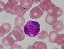
Staining is a technique used to enhance contrast in samples, generally at the microscopic level. Stains and dyes are frequently used in histology and in the medical fields of histopathology, hematology, and cytopathology that focus on the study and diagnoses disease at a microscopic level. Stains may be used to define biological tissues, cell populations, or organelles within individual cells.

In biochemistry, immunostaining is any use of an antibody-based method to detect a specific protein in a sample. The term "immunostaining" was originally used to refer to the immunohistochemical staining of tissue sections, as first described by Albert Coons in 1941. However, immunostaining now encompasses a broad range of techniques used in histology, cell biology, and molecular biology that use antibody-based staining methods.

Histopathology refers to the microscopic examination of tissue in order to study the manifestations of disease. Specifically, in clinical medicine, histopathology refers to the examination of a biopsy or surgical specimen by a pathologist, after the specimen has been processed and histological sections have been placed onto glass slides. In contrast, cytopathology examines free cells or tissue micro-fragments.
Eosinophilic refers to the staining of certain tissues, cells, or organelles after they have been washed with eosin, a dye.
Basophilic is a technical term used by histologists. It describes the microscopic appearance of cells and tissues, as seen down the microscope, after a histological section has been stained with a basic dye. The most common such dye is haematoxylin.

Sex cord–gonadal stromal tumour is a group of tumors derived from the stromal component of the ovary and testis, which comprises the granulosa, thecal cells and fibrocytes. In contrast, the epithelial cells originate from the outer epithelial lining surrounding the gonad while the germ cell tumors arise from the precursor cells of the gametes, hence the name germ cell. In humans, this group accounts for 8% of ovarian cancers and under 5% of testicular cancers. Their diagnosis is histological: only a biopsy of the tumour can make an exact diagnosis. They are often suspected of being malignant prior to operation, being solid ovarian tumours that tend to occur most commonly in post menopausal women.
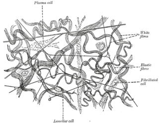
Elastic fibers are bundles of proteins (elastin) found in extracellular matrix of connective tissue and produced by fibroblasts and smooth muscle cells in arteries. These fibers can stretch up to 1.5 times their length, and snap back to their original length when relaxed. Elastic fibers include elastin, elaunin and oxytalan.

Invasive carcinoma of no special type (NST) also known as invasive ductal carcinoma or ductal NOS and previously known as invasive ductal carcinoma, not otherwise specified (NOS) is a group of breast cancers that do not have the "specific differentiating features". Those that have these features belong to other types.
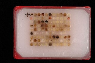
Tissue microarrays consist of paraffin blocks in which up to 1000 separate tissue cores are assembled in array fashion to allow multiplex histological analysis.

Hematoxylin and eosin stain or haematoxylin and eosin stain is one of the principal tissue stains used in histology. It is the most widely used stain in medical diagnosis and is often the gold standard; for example, when a pathologist looks at a biopsy of a suspected cancer, the histological section is likely to be stained with H&E.
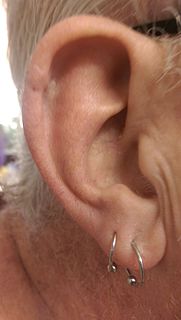
Mohs surgery, developed in 1938 by a general surgeon, Frederic E. Mohs, is microscopically controlled surgery used to treat common types of skin cancer. During the surgery, after each removal of tissue and while the patient waits, the tissue is examined for cancer cells. That examination informs the decision for additional tissue removal. Mohs surgery is the gold standard method for obtaining complete margin control during removal of a skin cancer using frozen section histology. CCPDMA or Mohs surgery allows for the removal of a skin cancer with very narrow surgical margin and a high cure rate.

Toluidine blue, also known as TBO or tolonium chloride (INN) is a blue cationic (basic) dye used in histology and sometimes clinically.
In histology, the HOPS stain is a way of marking tissue for microscopic examination. HOPS is an acronym for haematoxylin, orcein, phyloxin and saffron.

An inverted papilloma is a type of tumor in which surface epithelial cells grow downward into the underlying supportive tissue. It may occur in the nose and/or sinuses or in the urinary tract. When it occurs in the nose or sinuses, it may cause symptoms similar to those caused by sinusitis, such as nasal congestion. When it occurs in the urinary tract, it may cause blood in the urine.
Movat's stain is a pentachrome stain originally developed by Henry Zoltan Movat (1923–1995), a Hungarian-Canadian Pathologist in Toronto in 1955 to highlight the various constituents of connective tissue, especially cardiovascular tissue, by five colors in a single stained slide. In 1972, H. K. Russell, Jr. modified the technique so as to reduce the time for staining and to increase the consistency and reliability of the staining.
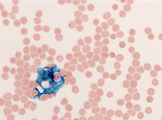
Perls' Prussian blue is a commonly used method in histology, histopathology, and clinical pathology to detect the presence of iron in tissue or cell samples. Perls' Prussian Blue derives its name from the German pathologist Max Perls (1843-1881), who described the technique in 1867. The method does not involve the application of a dye, but rather causes the pigment Prussian blue to form directly within the tissue. The method stains mostly iron in the ferric state which includes ferritin and hemosiderin, rather than iron in the ferrous state.

In pathology, the reticulin stain, is a popular staining method in histology. It is used to visualize reticular fiber and used extensively in liver histopathology.
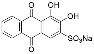
Alizarin Red S (also known as C.I. Mordant Red 3, Alizarin Carmine, and C.I 58005. It is a water-soluble sodium salt of Alizarin sulfonic acid with a chemical formula of C
14H
7NaO
7S. Alizarin Red S was discovered by Graebe and Libermann in 1871. In the field of histology alizarin Red S is been used to stain calcium deposits in tissues, and in geology to stain and differentiate carbonate minerals.
