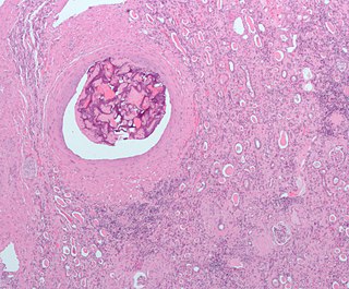
Hemorrhagic infarct

Last updated
Hemorrhagic infarct
Recent hemorrhagic infarcts.jpg
Recent hemorrhagic infarcts.
Specialty Vascular surgery

A hemorrhagic infarct is determined when hemorrhage is present around an area of infarction. Simply stated, an infarction is an area of dead tissue or necrosis. [1] When blood escapes outside of the vessel (extravasation) and re-perfuses back into the tissue surrounding the infarction, the infarction is then termed a hemorrhagic infarct (infarction). [1] Hemorrhagic infarcts can occur in any region of the body, such as the head, trunk and abdomen-pelvic regions, typically arising from their arterial blood supply being interrupted by a blockage or compression of an artery.

Contents

Causes

Infarcts typically occur due to one of two reasons. One is due to hypoxia or a lack of oxygen to tissue. The other cause is called ischemia, which occurs when there is a lack of blood flow to the tissues. Ischemia is more serious and can be caused by several factors. These factors include atherosclerosis, thrombus, embolism, trauma to vessels, neoplasms, cytomegalovirus, encapsulation or external compression of a vessel, or if a vessel becomes twisted, such as in torsion. [1] [2] [3]

Classification

After an infarction, the color of the tissue is used to classify the infarctions into one of two type of infarcts:

  1. White infarcts (anemic infarct or pale infarcts) are used to describe a soft, pale area of tissue and occur in organs that have a single supply of blood and solid parenchyma. [4] Typical organs that experience an anemic infarct include the heart, kidneys and spleen. [3]
  2. Red (hemorrhagic) infarcts have a dual blood supply and appear red to brown in color due to the hemorrhage of blood perfusing back into the tissue from a collateral or secondary vessel. These organs have a loose parenchyma, such as the lungs, brain and gastrointestinal tract. [3]

Common red infarcts

Cerebral

Intercerebral hemorrhage Intracerebral hemorrage (CT scan).jpg
Intercerebral hemorrhage

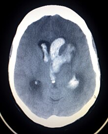
Otherwise known as hemorrhagic stroke, these account for approximately 15% of all strokes. [5] Classified according to one of four origination sites, intracerebral (ICH), subarachnoid, subdural, or epidural. Both subdural and epidural hemorrhages are most always caused by trauma, therefore, they are not considered hemorrhagic strokes. [5]

  • Hypertension is the predominant cause of intracerebral hemorrhage. The clinical presentation and signs and symptoms depend on the location of affected structures in the brain and which vessel is involved. A non-contrast Cat Scan of the head should be utilized as the primary imaging modality during initial diagnosis. Prognosis and treatment are determined by location and size of the hemorrhage. [5]
  • Subarachnoid hemorrhage is most commonly caused by the rupture of an aneurysm, usually at the base of the brain. Prior to rupture,
    Diffuse hemorrhage from the alveoli in the lungs Diffuse alveolaere Haemorrhagie unter INR Entgleisung 78W - CT - 001.jpg
    Diffuse hemorrhage from the alveoli in the lungs

symptoms include headache, diplopia or blurred vision. After rupture, the headache is typically described as the "worst headache of my life". Other symptoms include nausea, vomiting and loss of consciousness. Neurological signs can develop as the hemorrhage flows into surrounding tissues. Initial imaging will include a non-contrast CT head possibly followed by the use of contrast media to perform CT angiography of the vessels in the brain to detect the aneurysm site if the non-contrast CT head is negative. Treatment can include surgical or endovascular intervention with clips or coils to prevent re-rupture or ventricular drainage or shunting due to hydrocephalus. Cerebral vasospasm can occur up to two weeks after the subarachnoid hemorrhage, which can cause further ischemia because of the vessel narrowing during the spasm. [5]

Pulmonary

This commonly occurs when a distal pulmonary artery becomes occluded following a pulmonary embolism or other lung pathologies that can include malignancy, infection, or vasculitis. A large percentage (77%- 87%) of lung infarctions occur unilaterally, and typically in the lower lobes compared to the upper lobes. Symptoms of pulmonary infarction and pulmonary embolism are similar. Dyspnea, chest pain, pain and swelling in a lower extremity, and hemoptysis are common symptoms in both. Chest X-Ray can demonstrate a "Hampton's Hump" when pulmonary infarction is present, but Cat Scan is more commonly used. Treatment is dictated by the underlying cause of the pulmonary infarction which has a broad differential diagnosis that should be considered during treatment. The underlying cause will assist in a treatment plan to include level of care during initial management and the involvement of various specialists as pulmonary infarction can be deadly. [6]

Gastrointestinal

The most frequent type of gastrointestinal ischemia is ischemic colitis. The disruption to the blood supply can be chronic or acute. Its effects can vary from ulceration, hemorrhage or necrosis. A surgical repair is typically emergent as the ischemia is associated with high morbidity and mortality rates. [7] Ischemic colitis is the second-leading cause of lower gastrointestinal bleeding. The risk of ischemic colitis is directly proportional to age and is more common in the older population but can occur in younger individuals. [7] A Cat Scan of the Abdomen and Pelvis with IV contrast is considered the gold standard in imaging for diagnosis, but other imaging modalities can be used if it is contraindicated for a patient to have a Cat Scan with contrast. [7] Presentation of patients typically include complaints of "abdominal pain, nausea, vomiting, diarrhea, abdominal distention and rectal bleeding". [7]

See also

Related Research Articles

<span class="mw-page-title-main">Embolism</span> Disease of arteries, arterioles and capillaries

An embolism is the lodging of an embolus, a blockage-causing piece of material, inside a blood vessel. The embolus may be a blood clot (thrombus), a fat globule, a bubble of air or other gas, amniotic fluid, or foreign material.

<span class="mw-page-title-main">Thrombosis</span> Medical condition caused by blood clots

Thrombosis is the formation of a blood clot inside a blood vessel, obstructing the flow of blood through the circulatory system. When a blood vessel is injured, the body uses platelets (thrombocytes) and fibrin to form a blood clot to prevent blood loss. Even when a blood vessel is not injured, blood clots may form in the body under certain conditions. A clot, or a piece of the clot, that breaks free and begins to travel around the body is known as an embolus.

<span class="mw-page-title-main">Cerebrovascular disease</span> Condition that affects the arteries that supply the brain

Cerebrovascular disease includes a variety of medical conditions that affect the blood vessels of the brain and the cerebral circulation. Arteries supplying oxygen and nutrients to the brain are often damaged or deformed in these disorders. The most common presentation of cerebrovascular disease is an ischemic stroke or mini-stroke and sometimes a hemorrhagic stroke. Hypertension is the most important contributing risk factor for stroke and cerebrovascular diseases as it can change the structure of blood vessels and result in atherosclerosis. Atherosclerosis narrows blood vessels in the brain, resulting in decreased cerebral perfusion. Other risk factors that contribute to stroke include smoking and diabetes. Narrowed cerebral arteries can lead to ischemic stroke, but continually elevated blood pressure can also cause tearing of vessels, leading to a hemorrhagic stroke.

<span class="mw-page-title-main">Ischemia</span> Restriction in blood supply to tissues

Ischemia or ischaemia is a restriction in blood supply to any tissue, muscle group, or organ of the body, causing a shortage of oxygen that is needed for cellular metabolism. Ischemia is generally caused by problems with blood vessels, with resultant damage to or dysfunction of tissue i.e. hypoxia and microvascular dysfunction. It also implies local hypoxia in a part of a body resulting from constriction.
<span class="mw-page-title-main">Thrombolysis</span> Breakdown (lysis) of blood clots formed in blood vessels, using medication

Thrombolysis, also called fibrinolytic therapy, is the breakdown (lysis) of blood clots formed in blood vessels, using medication. It is used in ST elevation myocardial infarction, stroke, and in cases of severe venous thromboembolism.
<span class="mw-page-title-main">Infarction</span> Tissue death due to inadequate blood supply

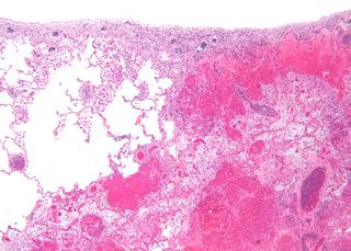
Infarction is tissue death (necrosis) due to inadequate blood supply to the affected area. It may be caused by artery blockages, rupture, mechanical compression, or vasoconstriction. The resulting lesion is referred to as an infarct (from the Latin infarctus, "stuffed into").
<span class="mw-page-title-main">Stroke</span> Death of a region of brain cells due to poor blood flow

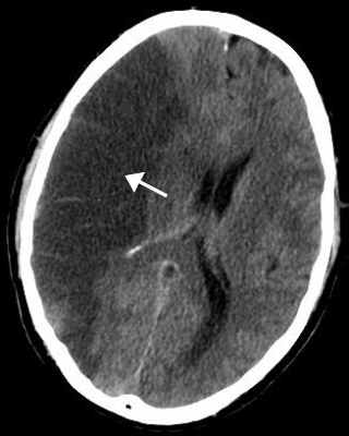
Stroke is a medical condition in which poor blood flow to the brain causes cell death. There are two main types of stroke: ischemic, due to lack of blood flow, and hemorrhagic, due to bleeding. Both cause parts of the brain to stop functioning properly.
<span class="mw-page-title-main">Subarachnoid hemorrhage</span> Bleeding into the subarachnoid space

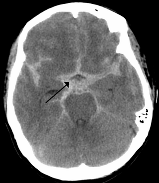
Subarachnoid hemorrhage (SAH) is bleeding into the subarachnoid space—the area between the arachnoid membrane and the pia mater surrounding the brain. Symptoms may include a severe headache of rapid onset, vomiting, decreased level of consciousness, fever, weakness, numbness, and sometimes seizures. Neck stiffness or neck pain are also relatively common. In about a quarter of people a small bleed with resolving symptoms occurs within a month of a larger bleed.

<span class="mw-page-title-main">Intracranial hemorrhage</span> Hemorrhage, or bleeding, within the skull

Intracranial hemorrhage (ICH), also known as intracranial bleed, is bleeding within the skull. Subtypes are intracerebral bleeds, subarachnoid bleeds, epidural bleeds, and subdural bleeds.

<span class="mw-page-title-main">Cerebral hypoxia</span> Oxygen shortage of the brain

Cerebral hypoxia is a form of hypoxia, specifically involving the brain; when the brain is completely deprived of oxygen, it is called cerebral anoxia. There are four categories of cerebral hypoxia; they are, in order of increasing severity: diffuse cerebral hypoxia (DCH), focal cerebral ischemia, cerebral infarction, and global cerebral ischemia. Prolonged hypoxia induces neuronal cell death via apoptosis, resulting in a hypoxic brain injury.

<span class="mw-page-title-main">Intraparenchymal hemorrhage</span> Medical condition

Intraparenchymal hemorrhage (IPH) is one form of intracerebral bleeding in which there is bleeding within brain parenchyma. The other form is intraventricular hemorrhage (IVH).

<span class="mw-page-title-main">Cerebral infarction</span> Stroke resulting from lack of blood flow

Cerebral infarction, also known as an ischemic stroke, is the pathologic process that results in an area of necrotic tissue in the brain. In mid to high income countries, a stroke is the main reason for disability among people and the 2nd cause of death. It is caused by disrupted blood supply (ischemia) and restricted oxygen supply (hypoxia). This is most commonly due to a thrombotic occlusion, or an embolic occlusion of major vessels which leads to a cerebral infarct. In response to ischemia, the brain degenerates by the process of liquefactive necrosis.

An embolus, is described as a free-floating mass, located inside blood vessels that can travel from one site in the blood stream to another. An embolus can be made up of solid, liquid, or gas. Once these masses get "stuck" in a different blood vessel, it is then known as an "embolism." An embolism can cause ischemia—damage to an organ from lack of oxygen. A paradoxical embolism is a specific type of embolism in which the embolus travels from the right side of the heart to the left side of the heart and lodges itself in a blood vessel known as an artery. Thus, it is termed "paradoxical" because the embolus lands in an artery, rather than a vein.

<span class="mw-page-title-main">Watershed stroke</span> Medical condition

A watershed stroke is defined as a brain ischemia that is localized to the vulnerable border zones between the tissues supplied by the anterior, posterior and middle cerebral arteries. The actual blood stream blockage/restriction site can be located far away from the infarcts. Watershed locations are those border-zone regions in the brain supplied by the major cerebral arteries where blood supply is decreased. Watershed strokes are a concern because they comprise approximately 10% of all ischemic stroke cases. The watershed zones themselves are particularly susceptible to infarction from global ischemia as the distal nature of the vasculature predisposes these areas to be most sensitive to profound hypoperfusion.
<span class="mw-page-title-main">Brain ischemia</span> Medical condition

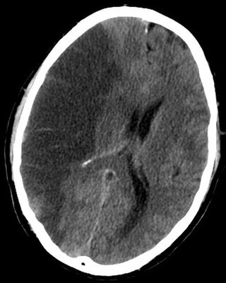
Brain ischemia is a condition in which there is insufficient bloodflow to the brain to meet metabolic demand. This leads to poor oxygen supply or cerebral hypoxia and thus leads to the death of brain tissue or cerebral infarction/ischemic stroke. It is a sub-type of stroke along with subarachnoid hemorrhage and intracerebral hemorrhage.

Animal models of ischemic stroke are procedures inducing cerebral ischemia. The aim is the study of basic processes or potential therapeutic interventions in this disease, and the extension of the pathophysiological knowledge on and/or the improvement of medical treatment of human ischemic stroke. Ischemic stroke has a complex pathophysiology involving the interplay of many different cells and tissues such as neurons, glia, endothelium, and the immune system. These events cannot be mimicked satisfactorily in vitro yet. Thus a large portion of stroke research is conducted on animals.

<span class="mw-page-title-main">Ischemic colitis</span> Medical condition

Ischemic colitis is a medical condition in which inflammation and injury of the large intestine result from inadequate blood supply. Although uncommon in the general population, ischemic colitis occurs with greater frequency in the elderly, and is the most common form of bowel ischemia. Causes of the reduced blood flow can include changes in the systemic circulation or local factors such as constriction of blood vessels or a blood clot. In most cases, no specific cause can be identified.
<span class="mw-page-title-main">Intestinal ischemia</span> Restriction of blood flow to the small intestine resulting in injury

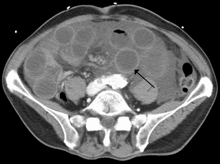
Intestinal ischemia is a medical condition in which injury to the large or small intestine occurs due to not enough blood supply. It can come on suddenly, known as acute intestinal ischemia, or gradually, known as chronic intestinal ischemia. The acute form of the disease often presents with sudden severe abdominal pain and is associated with a high risk of death. The chronic form typically presents more gradually with abdominal pain after eating, unintentional weight loss, vomiting, and fear of eating.

<span class="mw-page-title-main">Arterial embolism</span> Interruption of blood flow to an organ

Arterial embolism is a sudden interruption of blood flow to an organ or body part due to an embolus adhering to the wall of an artery blocking the flow of blood, the major type of embolus being a blood clot (thromboembolism). Sometimes, pulmonary embolism is classified as arterial embolism as well, in the sense that the clot follows the pulmonary artery carrying deoxygenated blood away from the heart. However, pulmonary embolism is generally classified as a form of venous embolism, because the embolus forms in veins. Arterial embolism is the major cause of infarction.

<span class="mw-page-title-main">Arterial occlusion</span>

Arterial occlusion is a condition involving partial or complete blockage of blood flow through an artery. Arteries are blood vessels that carry oxygenated blood to body tissues. An occlusion of arteries disrupts oxygen and blood supply to tissues, leading to ischemia. Depending on the extent of ischemia, symptoms of arterial occlusion range from simple soreness and pain that can be relieved with rest, to a lack of sensation or paralysis that could require amputation.

References

  1. 1 2 3 Dennis M, Mead G, Forbes J, Graham C, Hackett M, Hankey GJ, et al. (FOCUS Trial Collaboration) (January 2019). "Effects of fluoxetine on functional outcomes after acute stroke (FOCUS): a pragmatic, double-blind, randomised, controlled trial". Lancet. 393 (10168): 265–274. doi:10.1016/S0140-6736(18)32823-X. PMC   6336936 . PMID   30528472.
  2. Goljan E (2011). Rapid Review Pathology. Philadelphia: Elsevier Mosby. p. 425. ISBN   9780323084383.
  3. 1 2 3 Kumar V, Abbas AK, Aster JC, Deyrup AT (2023). Robbins & Kumar basic pathology (Eleventh ed.). Philadelphia, PA: Elsevier. ISBN   978-0-323-79018-5. OCLC   1351696609.
  4. Doyle D, Herrington S. "Cardiovascular System: Infarction". Pathologia. College of Medicine & Veterinary Medicine, University of Edinburgh.
  5. 1 2 3 4 Halter JB, Ouslander JG, Studenski S, High KP, Asthana S, Supiano MA, Ritchie CS, Schmader K, eds. (2022). Hazzard's geriatric medicine and gerontology (Eighth ed.). New York, Chicago, San Francisco, Athens, London: McGraw Hill. ISBN   978-1-260-46446-7.
  6. Emerling A, Cook J (2024). "Pulmonary Infarction". StatPearls. Treasure Island (FL): StatPearls Publishing. PMID   30725874 . Retrieved 2024-04-27.
  7. 1 2 3 4 Iacobellis F, Narese D, Berritto D, Brillantino A, Di Serafino M, Guerrini S, et al. (May 2021). "Large Bowel Ischemia/Infarction: How to Recognize It and Make Differential Diagnosis? A Review". Diagnostics. 11 (6): 998. doi: 10.3390/diagnostics11060998 . PMC   8230100 . PMID   34070924.