Hoof wall separation disease

Last updated
The hoof of a foal with HWSD Hoof wall separation disease in Connemara pony - HWSD2015.jpg
The hoof of a foal with HWSD
HWSD causes the layers within the dorsal hoof wall (brown, far right) to separate from each other Horse hoof wild bare sagittal.jpg
HWSD causes the layers within the dorsal hoof wall (brown, far right) to separate from each other

Hoof wall separation disease (HWSD) is an autosomal recessive genetic hoof disease in horses. [1] Research is being carried out at, among others, UC Davis School of Veterinary Medicine in Davis, California. [2] The disease has been found in Connemara ponies and was earlier referred to as hoof wall separation syndrome, HWSS.

Contents

Symptoms

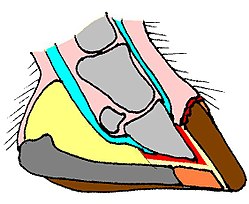
The disease develops among foals from the age of one to six months and typically occurs during their first year of life. [3] The frontal edge of the hoof cracks and splits. Pieces of hoof wall chip off in bits. This cracking, chipping and peeling may involve the whole of the dorsal hoof wall. The deficit means that when the hoof wall reaches the ground (and weightbearing load) the wall breaks away from above the level of the sole. Thus the affected pony has to walk on the sole of the hoof with no support provided by the dorsal hoof wall. All four feet are similarly affected. This can be very painful for the animal, which may lead to the necessity of having the horse put down. [3] HWSD may be confused with white line disease (onychomycosis)

Origin and cause

The disease is an autosomal recessive condition, which means that both parents of the foal must carry the gene in order for the disease to hit their offspring. [2] The disease is associated with a frameshift mutation in the gene coding for the protein serpin B11. [3]

Distribution [4]
Normal stallionStallion with HWSD genesStallion with fully developed HWSD
Normal mareNormal foals50% normal foals, 50% carriers100% carriers
Mare with HWSD genes50% normal foals, 50% carriers25% normal foals, 50% carriers, 25% with fully developed HWSD50% carriers, 50% with fully developed HWSD
Mare with fully developed HWSD100% carriers50% carriers, 50% with fully developed HWSD100% with fully developed HWSD

Tests and treatment

Genetic predisposition may be confirmed through a DNA test. [1] There is no treatment as such, owners can attempt to manage the condition though various types of hoof care, and/or the use of special shoes. Such treatment is expensive and labour-intensive. Environmental conditions can make the condition worse (alternating wet/dry hooves). Protecting the hooves from such variations may reduce the effect of the disease. [4]

Related Research Articles

<span class="mw-page-title-main">Genetic disorder</span> Health problem caused by one or more abnormalities in the genome

A genetic disorder is a health problem caused by one or more abnormalities in the genome. It can be caused by a mutation in a single gene (monogenic) or multiple genes (polygenic) or by a chromosomal abnormality. Although polygenic disorders are the most common, the term is mostly used when discussing disorders with a single genetic cause, either in a gene or chromosome. The mutation responsible can occur spontaneously before embryonic development, or it can be inherited from two parents who are carriers of a faulty gene or from a parent with the disorder. When the genetic disorder is inherited from one or both parents, it is also classified as a hereditary disease. Some disorders are caused by a mutation on the X chromosome and have X-linked inheritance. Very few disorders are inherited on the Y chromosome or mitochondrial DNA.

<span class="mw-page-title-main">Lethal white syndrome</span> Genetic disorder in horses

Lethal white syndrome (LWS), also called overo lethal white syndrome (OLWS), lethal white overo (LWO), and overo lethal white foal syndrome (OLWFS), is an autosomal genetic disorder most prevalent in the American Paint Horse. Affected foals are born after the full 11-month gestation and externally appear normal, though they have all-white or nearly all-white coats and blue eyes. However, internally, these foals have a nonfunctioning colon. Within a few hours, signs of colic appear; affected foals die within a few days. Because the death is often painful, such foals are often humanely euthanized once identified. The disease is particularly devastating because foals are born seemingly healthy after being carried to full term.

<span class="mw-page-title-main">Bay (horse)</span> Hair coat color of horses

Bay is a hair coat color of horses, characterized by a reddish-brown or brown body color with a black point coloration on the mane, tail, ear edges, and lower legs. Bay is one of the most common coat colors in many horse breeds.

<span class="mw-page-title-main">Connemara pony</span> Breed of horse

The Connemara pony is a pony breed originating in Ireland. They are known for their athleticism, versatility and good disposition. The breed makes excellent show ponies.

<span class="mw-page-title-main">Laminitis</span> Disease of the feet of hooved animals

Laminitis is a disease that affects the feet of ungulates and is found mostly in horses and cattle. Clinical signs include foot tenderness progressing to inability to walk, increased digital pulses, and increased temperature in the hooves. Severe cases with outwardly visible clinical signs are known by the colloquial term founder, and progression of the disease will lead to perforation of the coffin bone through the sole of the hoof or being unable to stand up, requiring euthanasia.

Glycogen-branching enzyme deficiency (GBED) is an inheritable glycogen storage disease affecting American Quarter Horses and American Paint Horses. It leads to abortion, stillbirths, or early death of affected animals. The human form of the disease is known as glycogen storage disease type IV.

<span class="mw-page-title-main">Robinow syndrome</span> Rare genetic disorder characterized by a fetal face

Robinow syndrome is an extremely rare genetic disorder characterized by short-limbed dwarfism, abnormalities in the head, face, and external genitalia, as well as vertebral segmentation. The disorder was first described in 1969 by human geneticist Meinhard Robinow, along with physicians Frederic N. Silverman and Hugo D. Smith, in the American Journal of Diseases of Children. By 2002, over 100 cases had been documented and introduced into medical literature.

<span class="mw-page-title-main">Horse hoof</span> Hardened, weight bearing structure of a horses limbs

A horse hoof is the lower extremity of each leg of a horse, the part that makes contact with the ground and carries the weight of the animal. It is both hard and flexible. It is a complex structure surrounding the distal phalanx of the 3rd digit of each of the four limbs, which is covered by soft tissue and keratinised (cornified) matter. The arteries that supply the hoof with blood are, the vena plantaris externa and vena plantaris interna, which branch off the tibialis posterior. The horse hoof encapsules one of the three metatarsus bones that are found in the hoof and heel area.

<span class="mw-page-title-main">Cerebellar abiotrophy</span> Genetic condition in animals

Cerebellar abiotrophy (CA), also called cerebellar cortical abiotrophy (CCA), is a genetic neurological disease in animals, best known to affect certain breeds of horses, dogs and cats. It can also develop in humans. It develops when the neurons known as Purkinje cells, located in the cerebellum of the brain, begin to die off. These cells affect balance and coordination. They have a critical role to play in the brain. The Purkinje layer allows communication between the granular and molecular cortical layers in the cerebellum. Put simply, without Purkinje cells, an animal loses its sense of space and distance, making balance and coordination difficult. People with damage to the cerebellum can experience symptoms like unsteady gait, poor muscle control, and trouble speaking or swallowing.

<span class="mw-page-title-main">Black horse</span> Horse coat color

Black is a hair coat color of horses in which the entire hair coat is black. Black is a relatively uncommon coat color, and it is not uncommon to mistake dark chestnuts or bays for black.

<span class="mw-page-title-main">Dermatopathia pigmentosa reticularis</span> Medical condition

Dermatopathia pigmentosa reticularis(DPR) is a rare, autosomal dominant congenital disorder that is a form of ectodermal dysplasia. Dermatopathia pigmentosa reticularis is composed of the triad of generalized reticulate hyperpigmentation, noncicatricial alopecia, and onychodystrophy. DPR is a non life-threatening disease that largely affects the skin, hair, and nails. It has also been identified as a keratin disorder. Historically, as of 1992, only 10 cases had been described in world literature; however, due to recent advances in genetic analysis, five additional families studied in 2006 have been added to the short list of confirmed cases.

Hereditary equine regional dermal asthenia (HERDA), also known as hyperelastosis cutis (HC), is an inherited autosomal recessive connective tissue disorder. It develops from a homozygous recessive mutation that weakens collagen fibers that allow the skin of the animal to stay connected to the rest of the animal. Affected horses have extremely fragile skin that tears easily and exhibits impaired healing. In horses with HC, the skin separates between the deep and superficial dermis. There is no cure. Most affected individuals receive an injury they cannot heal, and are euthanized. Managed breeding strategy is currently the only option for reducing the incidence of the disease.

<span class="mw-page-title-main">Junctional epidermolysis bullosa (veterinary medicine)</span> Genetic disorder of horses

Junctional epidermolysis bullosa (JEB) is an inherited disorder that is also known as red foot disease or hairless foal syndrome. JEB is the result of a genetic mutation that inhibits protein production that is essential for skin adhesion. Therefore, tissues, such as skin and mouth epithelia, are affected. Blisters form over the entire body causing pain and discomfort, and open sores leave newborn foals highly susceptible to secondary infection. The condition can be categorized into two types of mutations: JEB1 and JEB2. JEB1 is found in Belgian Draft horses, as well as other related Draft breeds. In contrast, JEB2 is found in American Saddlebred horses.

<span class="mw-page-title-main">Lavender foal syndrome</span> Genetic disease in horses

Lavender foal syndrome (LFS), also called coat color dilution lethal (CCDL), is an autosomal recessive genetic disease that affects newborn foals of certain Arabian horse bloodlines. Affected LFS foals have severe neurological abnormalities, cannot stand, and require euthanasia shortly after birth. The popular name originates due to a diluted color of the foal's coat, that in some cases appears to have a purple or lavender hue. However, not all foals possess the lavender coat colour, and colouring can range from silver to light chestnut to a pale pink. Carrier horses have no clinical signs and DNA testing can determine if a horse carries the gene.

The severe combined immunodeficiency (SCID) is a severe immunodeficiency genetic disorder that is characterized by the complete inability of the adaptive immune system to mount, coordinate, and sustain an appropriate immune response, usually due to absent or atypical T and B lymphocytes. In humans, SCID is colloquially known as "bubble boy" disease, as victims may require complete clinical isolation to prevent lethal infection from environmental microbes.

<span class="mw-page-title-main">DOCK8 deficiency</span> Medical condition

DOCK8 deficiency, also called DOCK8 immunodeficiency syndrome, is the autosomal recessive form of hyperimmunoglobulin E syndrome, a genetic disorder characterized by elevated immunoglobulin E levels, eosinophilia, and recurrent infections with staphylococcus and viruses. It is caused by a mutation in the DOCK8 gene.

<span class="mw-page-title-main">Mushroom gene</span>

Identified in 2014, the mushroom gene is a recessive dilution gene that affects red pigment in horses.

<span class="mw-page-title-main">Ann T. Bowling</span> American geneticist (1943–2000)

Ann Trommershausen Bowling was an American scientist who was one of the world's leading geneticists in the study of horses, conducting research in the areas of molecular genetics and cytogenetics. She was a major figure in the development of testing to determine animal parentage, first with blood typing in the 1980s and then DNA testing in the 1990s. She later became known for her studies of hereditary diseases in horses and equine coat color genetics, as well as research on horse evolution and the development of horse breeds. She studied the population genetics of feral horses, did considerable work to help preserve the Przewalski's horse, and was one of the founding members of the international project to map the horse genome. She was an adjunct professor at the University of California, Davis (UCD), and at the time of her death in 2000 was the executive associate director of the Veterinary Genetics Laboratory (VGL) there. Her unexpected death on December 8, 2000, at age 57 was attributed to a massive stroke.

Foal immunodeficiency syndrome (FIS), originally known as Fell pony syndrome, mainly affects two breeds of ponies: the Fell and Dales. FIS is a recessive genetic disease. Affected foals appear normal at birth, but become weak, and either die or are euthanized by three months of age due to persistent infections caused by immunodeficiency.

<span class="mw-page-title-main">Salt and pepper syndrome</span> Medical condition

Salt and pepper developmental regression syndrome, also known as Amish infantile epileptic syndrome or GM3 deficiency syndrome, is a rare autosomal recessive progressive neurological disorder characterized by developmental delay, severe intellectual disability, seizures, and skin pigmentation irregularities. The clinical symptoms of this condition start manifesting soon after birth, during the newborn/neo-natal stage of life.

References

  1. 1 2 UC Davis Veterinary medicine: Connemara Pony Hoof Wall Separation Disease, accessed April 25, 2015
  2. 1 2 Hoof Wall Separation Syndrome in Connemara Ponies Archived 2014-11-04 at the Wayback Machine , Accessed November 4, 2014
  3. 1 2 3 Finno, CJ; Stevens, C; Young, A; Affolter, V; Joshi, NA; Ramsay, S; Bannasch, DL (April 2015). "SERPINB11 frameshift variant associated with novel hoof specific phenotype in Connemara ponies". PLOS Genetics. 11 (4): e1005122. doi: 10.1371/journal.pgen.1005122 . PMC   4395385 . PMID   25875171.
  4. 1 2 Dansk Connemara Pony Selskab: Gode nyheder for Connemaraavlen – Gentest for hovdefekten HWSD er klar til brug! Archived 2015-05-18 at the Wayback Machine , Accessed April 25, 2015