Most cases of feline hypersomatotropism are caused by acidophilic pituitary tumours that predominantly secrete growth hormone. In some cases low levels of other pituitary hormones are secreted. Rarely a cat may have double adenomas. In a few cases the diagnosis has been pituitary acidophilic hyperplasia. In dogs nearly all cases of acromegaly are caused by endogenous or exogenousprogestogens, this causes a hypersecretion of growth hormones from the mammary gland. Pituitary tumours causing excess growth hormone secretion are very rare in dogs.[1]
Signs and symptoms
Cats suffering from hypersomatotropism show a multitude of symptoms, the majority of these symptoms are not useful for identifying hypersomatotropism instead of just diabetes mellitus. The presence and level of severity of symptoms vary based on levels of growth hormone excess and duration of excess secretion. In dogs the manifestation of symptoms varies much more than with cats, some dogs may only show signs of acromegaly whilst others show mostly symptoms of diabetes mellitus. It is possible that this variability is related to breed. Diabetes however, still occurs in a substantial amount of dogs.[1]
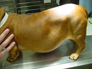
Most symptoms result from the diabetogenic effect of growth hormone and the acral enlargement effects of growth hormone and IGF-1. Neurological signs may be observed in some cats due to expansion of the tumour, this can occur in dogs with pituiary tumours too. The most common symptoms in cats are polyuria, polydipsia, and polyphagia due to diabetes mellitus; however polyphagia can be the result of growth hormone excess itself. Other symptoms in both cats and dogs include ataxia, asthenia, hepatomegaly, visceromegaly, enlargement of head and distal extremities, heart murmur, degenerative atrophy, thickening of skin and fur, stridor and a plantigrade stance in cats. Disruption of the central nervous system occurs in 10-15% of cases, potential signs of this include: adipsia, lethargy, behavioural change, vision impairment, anorexia, temperature dysregulation, somnolence, stupor, and seizures. Less common but occasional symptoms include erythrocytosis, leukocytosis, and proteinuria.[1]
The physical changes of hypersomatotropism are a result of anabolic effects of growth hormone and IGF-1; these have a gradual onset and progress slowly. The overgrowth of soft tissue and bone changes cause weight gain, broadening of the skull, prognathia inferior, and an enlarged tongue. Thickening of the oropharyngeal tissue may lead to respiratory distress and stridor. Degenerative anthropathy is caused by proliferation of chondrocytes and changes to the joint geometry.[1]
Heart murmur occurs in most feline cases, further diagnostics may reveal cardiomegaly and other heart abnormalities. Congestive heart failure can occur during later stages of the disease.[1]
A 1990 study reported 50% of cats diagnosed with hypersomatotropism had developed renal failure within 8-36 months of initial examination;[2] however, another study from 2007 found a 12% incidence of azotaemia.[3] Renal failure is a very common disease in elderly cats and it is controversial as to whether or not feline hypersomatotropism affects the development of renal failure.[1]
In dogs the symptoms of diabetes mellitus commonly overshadows acromegalic symptoms.[1]
Risk factors
There is no known breed predilection. Approximately 88% of described cats were male; male cats also have a predisposition to diabetes mellitus. Growth hormone secreting pituitary tumours in dogs have only been observed in male dogs of large breeds. Excessive growth hormone secretion caused by progestone use has been seen in a variety of breeds of varying size, as well as in mixed breed dogs, the same is true of growth hormone excess brought on by the oestrous cycle.[1]
Co-morbidities
The vast majority of cats with hypersomatotropism also have diabetes mellitus.[1]
Age of onset
Feline hypersomatotropism is usually diagnosed in elderly cats. The average age of diagnosis is between 10-11 years with a range of 4-17 years.[1]
The age range of dogs reported with growth hormone secreting tumours is between 7-10 years. The age range of bitches with progestone induced hypersomatotropism is 4-11 years, for bitches with hypersomatotropism resulting from the estrous cycle the age range is between 6-13 years.[1]
History
In 1976, two cats were described with diabetes mellitus and acidophilicpituitary adenomas. Whilst growth hormone levels were not measured it was proposed that growth hormone excess from the tumours was the cause of the diabetes. In the following three decades further cases would only be written about infrequently and feline hypersomatotropism was considered a rare disease. This was challenged in 2007 after a study looking at the IGF-1 levels in cats with diabetes found a marked increase in 32% of cats.[1][3]
Growth hormone excess induced by progestogens in dogs was first described in the 1970s and 1980s.[1] In 1980 a crossbred Belgian Shepherd bitch with acromegaly that had been administered excessive amounts of medroxyprogesterone acetate (MPA); following cessation of MPA administration symptoms improved and hormone levels returned to normal.[4] In 1981 it was reported that fifteen bitches receiving MPA injections to prevent oestrus. All fifteen showed signs of acromegaly and thirteen showed hyperglycaemia. Clinical signs were improved after cessation of MPA. Later studies would confirm an association between progestogen administration and acromegaly, glucose intolerance, and diabetes mellitus.[1]
Epidemiology
Studies have found the prevalence of heightened IGF-1 levels in cats with diabetes mellitus to range between 17.8% 27.3%.[5][1] According to Claudia Reusch, a professor at the University of Zurich, the prevalence of hypersomatotropism in diabetic cats is 10-15%, in cases that are hard to regulate it rises to 30% or higher.[1]
Cause
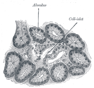
Growth hormone-producing acidophilic pituitary adenomas are the cause of hypersomatotropism in the vast majority of cats. Both dense and sparse granulated cells has been observed in the adenomas of cats with diabetes mellitus and elevated IGF-1 levels; however it is not known whether this is because of different variants of the disease or not. It is also not known what percentage of tumours are mixed or plurihormonal tumours. This information has only been reported in studies with a small number of cases.[1]
In dogs growth hormone is not just derived from the anterior pituitary gland, it also derives from the mammary gland. Mammary gland production of growth hormone is stimulated by progestogen, this occurs naturally in intact bitches. Synthetic progestogens can cause excessive growth hormone secretion and higher IGF-1 levels which may lead to acromegaly and diabetes mellitus. Progestogens are used to prevent oestrus in bitches and to treat certain hyperplasias. Growth hormone excess can occur in the luteal phase of the oestrous cycle in older bitches.[1]
Somatotrophic adenomas have only been described in two dogs.[1] In 1996 a 9 year old male Doberman Pinscher with difficult to manage diabetes mellitus but no signs of acromegaly was euthanised. Necropsy later revealed an acidophilic adenoma with immunohistochemical staining for growth hormone, adrenocorticotropic hormone, and prolactin.[6] In 2007 a 10 year old male Dalmatian dog was described with acromegalic signs such as an enlarged tongue and head, widened interdental space, and thickening of skin; along with other signs of hypersomatotropism such as polyphagia, obesity, and stridor. Insulin levels were heightened and the dog had glucose intolerance; however, the dog was not diabetic as blood glucose levels were found to be normal after multiple tests. Levels of IGF-1 and growth hormone were heightened. CT imaging showed a pituitary mass which was later confirmed to be an acidophilic adenoma stained for growth hormone;[7] however, mammary tumour-induced acromegaly is rare with few reported cases.[1]
Hypothyroidism can increase levels of growth hormone and IGF-1 in dogs. Some of the physical changes in dogs with hypothyroidism may be due to increased growth hormone concentration.[1]
Pathophysiology
In cases of hypersomatotropism the growth hormone concentrations that circulate are chronically increased, however the secretion of growth hormone remains the same. Growth hormone directly and indirectly affects the metabolic system; stimulation of IGF-1 synthesis is the indirect cause. Increased levels of growth hormone and IGF-1 result in proliferation of bone, cartilage, soft tissue, and increases the size of organs. These changes are responsible for the physical changes of hypersomatotropism that are characteristic to the condition.[1]
Both growth hormone and IGF-1 can impact insulin in different manners. Chronic growth hormone excess has been linked to defects in hepatic and extrahepatic insulin actions. Growth hormone increases hepatic glucose production and decreases glucose uptake in extrahepatic tissue.[1] Studies have suggested that growth hormone excess reduces insulin sensitivity.[1][8][9] IGF-1 increases insulin sensitivity in both hepatic and extrahepatic tissue, however in hypersomatotropism IGF-1 levels are unable to deal with the insulin resistance caused by excessive growth hormone levels.[1]
In non-diabetic cases the insulin resistance is countered by increased insulin production from beta cells, which results in normoglycaemia (normal levels of blood sugar) being maintained. When beta cells fail to provide enough insulin production to compensate for the increased resistance diabetes mellitus develops. The cause for this failure is unknown. The vast majority of cats with hypersomatotropism also have diabetes mellitus.[1]
In iatrogenic cases of hypersomatotropism, when the condition is caused by administered progestogens, the typical features of acromegaly occur; however, glucose tolerance is maintained initially by increased insulin levels, eventually insulin levels cannot be increased any further and glucose intolerance occurs. Most abnormalities are reversible after cessation of treatment; however, bone lesions may be permanent.[1]
Increase in growth hormone and IGF-1 levels in bitches is a physiological event that occurs during the luteal phase phase of the oestrous cycle. In bitches with spontaneous acromegaly brought on by oestrous, their progesterone levels are normal; however, growth hormone levels are increased. Recovery following an ovariohysterectomy may be possible. The reason as to why some bitches develop acromegaly and diabetes mellitus during the oestrous cycle is unknown.[1]
Diagnosis
The vast majority of cats present with diabetes mellitus, the possibility of hypersomatotropism causing it is rarely considered until the diabetes becomes difficult to control. In cats with difficult to control diabetes mellitus, hypersomatotropism should be considered as a cause only after exclusion of other conditions that can impact insulin. Bitches with hypersomatotropism are usually presented 3-5 weeks after oestrus. It is not uncommon for owners to report symptoms relating to the diabetes mellitus as having occurred during the previous oestrous cycle, typically more mild and ending with anoestrus.[1]
Most abnormalities on common tests, such as complete blood count, urinalysis, and biochemistry profile, are due to the diabetes mellitus. Some cats with hypersomatotropism have hyperproteinaemia.[1] One study found it to be the only parameter more frequent in cats with hypersomatotropism than cats with just diabetes mellitus.[10][1]
The common steps of diagnosis in cats involve presentation with diabetes mellitus, on examination either the acromegalic effects are noticed or they are not noticed and hypersomatotropism is not suspected until efforts to control the diabetes are proving difficult. Following this levels of growth hormone or IGF-1 are measured, if the results suggest hypersomatotropism it is then usually followed up with CT/MRI imaging to find the pituitary mass. In dogs the tentative diagnosis is made based on signs of acromegaly or diabetes mellitus alongside progestogen administration or dioestrus. A definitive diagnosis is made after based on growth hormone concentrations or increased IGF-1 levels. In male dogs with no history of progestogen administration the possibility of a pituitary tumour needs to be examined via CT/MRI.[1]
In cats with difficult to manage diabetes but no other signs of hypersomatotropism, the potential for other causes of poor glycaemic control need to be considered.[1]
Hepatomegaly is consistently identified with an abdominal ultrasound; however it is also commonly found in diabetic cats without hypersomatotropism. Other findings from an abdominal ultrasound include: renomegaly, pelvic dilation, an enlarged pancreas, splenomegaly and bilateral adrenomegaly. The size of the adrenal glands can be useful in diagnosis, studies have found that the size of the adrenal gland in diabetic cats without hypersomatotropism does not differ significantly from non-diabetic cats;[11][12][1] however adrenomegaly is not pathognomonic to hypersomatotropism and can occur with other conditions in cats such as pituitary-dependent hypercortisolism.[1]
Imaging of the pituitary gland is important in confirming hypersomatotropism in cats where it is suspected. Showing a pituitary mass is important to establish a final diagnosis alongside clinical findings and hormonal findings. Knowing the size of the pituitary mass is also necessary to decide the best course of treatment. In the vast majority of cats with hypersomatropism, it is caused by an adenoma of the somatotrophic cells. Said adenoma is typically visible by the time a cat presents with symptoms; however if pituitary imaging is performed during the early stages of the disease, it may still be small and difficult to recognise. Rarely CT/MRI imaging may not reveal anything, this may either due to a small size of the tumour or due to a different aetiology for the acromegaly. Rarely in humans with acromegaly, somatotrophic hyperplasia as the result of growth hormone releasing hormones, caused by a tumour. This aetiology has not been observed in cats; however, a cat with normal CT and MRI imaging, the histopathology showed — instead of adenoma — acidophilic proliferation.[1][3]
Hormonal evaluation
Similar to humans, a diagnosis of hypersomatotropism in cats and dogs requires demonstration of growth hormone excess or heightened IGF-1 concentrations. Growth hormone levels can be measured with a radioimmunoassay. However the cost may impact availability of this.[1]
All cats with hypersomatotropism that have been tested in studies displayed increased growth hormone levels. Some cats had significantly increased levels; in other cats, the increase was only slightly above normal levels. Cats in those studies were likely in the later stages of the disease. A single instance of elevated growth hormone levels is not indicative of hypersomatotropism, it can be the result of a secretory pulse and mildly increased growth hormone levels have been observed in diabetic cats without hypersomatotropism. The recommended practice is for several tests with 10 minute intervals.[1]
IGF-1 levels can be detected with a blood test. The vast majority of cats with hypersomatotropism have increased IGF-1 levels, most dogs with hypersomatotropism have increased IGF-1 levels. Normal levels of IGF-1 have been seen in a few cats, potentially due to these cats being at the early stages of the disease. Other causes need to be investigated in cats with normal IGF-1 levels and suspected hypersomatotropism. IGF-1 levels may be lower due to lymphoma or other diseases. IGF-1 levels can be normal in cats with hypersomatotropism when the measurement is taken prior to insulin therapy. IGF-1 levels are significantly lower in cats with untreated diabetes mellitus without hypersomatotropism. To counteract this, diabetic cats with suspected hypersomatotropism can be treated for 6-8 weeks with insulin before testing. Another issue with IGF-1 testing is that most tests consider values of other 1000ng/mL to be indicative of hypersomatotropism, even though healthy cats and cats with diabetes mellitus but not hypersomatotropism have levels below 800ng/mL, leaving a grey zone of 800–1000ng/mL. A study in 2000 reported eight cats with diabetes mellitus without hypersomatotropism had levels of IGF-1 above the normal range. Other studies suggested that this result was due to long term insulin therapy. Technical issues with the testing may result in false reports of increased levels due to the tests removing the proteins that circulating IGF-1 binds to. Multiple cats with IGF-1 levels reported above 1000ng/mL did not show signs of hypersomatotropism during further examination.[1]
Differential diagnosis
Hyperadrenocorticism is a potential differential diagnosis for poor glycaemic control. Hyperadrenocorticism is associated with insulin resistance and is often caused by a pituitary tumour; however, clinical signs differ between the two conditions. Hyperadrenocorticism may cause weight loss, leading to cachexia and alopecia and other dermatological conditions. These symptoms do not show in hypersomatotropism. Diagnostic imaging results for the conditions are often the same, such as hepatomegaly, adrenomegaly, and pituitary mass. Measuring growth hormone and IGF-1 levels can differentiate the diseases when physical symptoms are unremarkable.[1]
Dogs with primary hypothyroidism also have increased levels of growth hormone and IGF-1; however, thyroxine and thyroid stimulating hormone levels are normal in dogs with hypersomatotropism.[1]
Progestogens administered to dogs may result in endogenous adrenocorticotropic hormone secretion being suppressed, which lowers the cortisol concentration.[1]
Treatment
Treating hypersomatotropism involves treating both the hypersomatotropism and the diabetes mellitus. Sometimes treatment is limited to the diabetes due to owners not wishing to treat the hypersomatotrism due to the associated costs. Monitoring of blood glucose levels is imperative as if improvement or resolution of insulin resistance is not identified hypoglycaemia can occur, leading to death. In dogs with progestogen induced hypersomatotropism the administration of progestogens should cease immediately. For cases derived from a mammary tumour it should be surgically removed.[1]
Insulin
Insulin resistance can vary greatly between cats. In some cases glycaemic control can be achieved with doses of 1 to 3 U/cat b.i.d., a 'normal' level of insulin dosage. Insulin levels should be increased by 0.5 to 1 U/cat b.i.d. every 5 to 7 days until glycaemic control has been achieved (blood glucose level of 100 to 300 mg/dL). Frequent monitoring of cats and dogs undergoing insulin therapy is required. Levels should not be increased higher than 15 U/cat b.i.d. for cats. In dogs insulin therapy should be initiated immediately if the blood glucose concentration is higher than >140 mg/dL or 8 mmol/L. Severity of hyperglycaemia determines the level provided, with the range for dogs being between 0.05 to 0.25 U/kg b.i.d..[1]
Pituitary surgery
Transsphenoidal cryohypophysectomy is a difficult procedure that is only offered at specialised clinics. Few case reports of pituitary surgery in cats with hypersomatotropism have been published.[1] In one case the procedure resulted in a 95% decrease to insulin requirement following the surgery and diabetic remission 3 weeks later. A follow up 18 months later showed normal growth hormone and IGF-1 levels.[13] In another case there was a decreased insulin requirement, well-managed diabetes mellitus, and no acromegalic signs during an 18 month period of observation following the surgery.[14] These cases and others with similar results showcase that beta cells have a potential to recover in hypersomatotropic cats with appropriate treatment.[1]
Radiation therapy
Radiation therapy is the most frequently used treatment for hypersomatotropic cats. Radiation therapy is expensive, limited in availability, cost, frequent anaesthetic, and the unpredictable outcomes for hormonal control. The resolution or improvement of neurological signs is the most consistent effect. Improvement diabetes symptoms is less consistent. One study found an average of 5 weeks for improvement to glycaemic control with all cats seeing an improvement within 20 weeks. The same study found 6 of the 14 cats to achieve diabetic remission within 6 months with an average of 3.6 months.[15] Diabetic remission may occur as late as a year after radiotherapy. Improvement of diabetic symptoms occurs in roughly 70-80% of cases, diabetic remission occurs in roughly 50%. The physical acromegalic changes often persistent or have only slight improvement.[1] High IGF-1 levels have been reported in cats following radiation therapy. Of note is that in several of those cases the patient had good control of diabetic symptoms or was in remission.[15][16] Radiation therapy is tolerated by the majority of cats. Side effects such as ischemic brainnecrosis and hearing loss are rare and proper fractionation protocol can prevent these effects from occurring; hypopituitarism has not been reported in cats, despite being a common adverse effect in humans. Survival times for cats after radiation therapy has been reported to be up to 5 years.[15][1]
Ovariohysterectomy
Ovariohysterectomy should be performed in bitches with oestrous associated hypersomatotoprism as soon as it can be performed. Following the procedure growth hormone and IGF-1 levels will quickly return to normal values. If the procedure cannot be immediately performed aglepristone should be administered subcutaneously every 24 hours at a rate of 10 mg/kg. Diabetic remission may occur within 1 to 8 weeks following the surgery.[1]
Aglepristone
In cases resulting from progestogen administration aglepristone should be administered subcutaneously twice at a 24 hour interval at a rate of 10 mg/kg for all dogs with severe acromegaly or are diabetic.[1]
Untreated hypersomatotropism has a guarded to poor prognosis. In most cases the insulin resistance and other symptoms get progressively worse causing insulin treatment to become difficult. Euthanasia often occurs a few months after diagnosis in cats due to owner's being unable or unwilling to deal with treatment. Other reasons for euthanasia include congestive heart failure, renal failure, respiratory distress, and neurological symptoms. Cats that have been treated with radiotherapy or pituitary surgery may have a good prognosis; survival has been observed for several years. In progestogen induced hypersomatotropism the prognosis is usually good after cessation of progestogen administration. In dogs soft tissue changes are usually resolved in a few weeks or months, persistent bone changes usually do not cause clinical problems. For diabetes mellitus the prognosis for the condition depends on the beta cell damage.[1]
Related Research Articles
The endocrine system is a messenger system in an organism comprising feedback loops of hormones that are released by internal glands directly into the circulatory system and that target and regulate distant organs. In vertebrates, the hypothalamus is the neural control center for all endocrine systems.
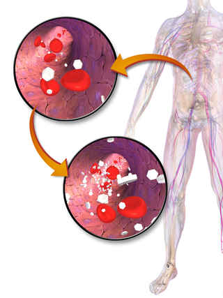
Hyperglycemia is a condition in which an excessive amount of glucose circulates in the blood plasma. This is generally a blood sugar level higher than 11.1 mmol/L (200 mg/dL), but symptoms may not start to become noticeable until even higher values such as 13.9–16.7 mmol/L (~250–300 mg/dL). A subject with a consistent fasting blood glucose range between ~5.6 and ~7 mmol/L is considered slightly hyperglycemic, and above 7 mmol/L is generally held to have diabetes. For diabetics, glucose levels that are considered to be too hyperglycemic can vary from person to person, mainly due to the person's renal threshold of glucose and overall glucose tolerance. On average, however, chronic levels above 10–12 mmol/L (180–216 mg/dL) can produce noticeable organ damage over time.
Gigantism, also known as giantism, is a condition characterized by excessive growth and height significantly above average. In humans, this condition is caused by over-production of growth hormone in childhood.
Cushing's disease is one cause of Cushing's syndrome characterised by increased secretion of adrenocorticotropic hormone (ACTH) from the anterior pituitary. This is most often as a result of a pituitary adenoma or due to excess production of hypothalamus CRH that stimulates the synthesis of cortisol by the adrenal glands. Pituitary adenomas are responsible for 80% of endogenous Cushing's syndrome, when excluding Cushing's syndrome from exogenously administered corticosteroids. The equine version of this disease is Pituitary pars intermedia dysfunction.
Insulin-like growth factor 1 (IGF-1), also called somatomedin C, is a hormone similar in molecular structure to insulin which plays an important role in childhood growth, and has anabolic effects in adults.
Acanthosis nigricans is a medical sign characterised by brown-to-black, poorly defined, velvety hyperpigmentation of the skin. It is usually found in body folds, such as the posterior and lateral folds of the neck, the armpits, groin, navel, forehead and other areas.
Hypopituitarism is the decreased (hypo) secretion of one or more of the eight hormones normally produced by the pituitary gland at the base of the brain. If there is decreased secretion of one specific pituitary hormone, the condition is known as selective hypopituitarism. If there is decreased secretion of most or all pituitary hormones, the term panhypopituitarism is used.
An insulinoma is a tumour of the pancreas that is derived from beta cells and secretes insulin. It is a rare form of a neuroendocrine tumour. Most insulinomas are benign in that they grow exclusively at their origin within the pancreas, but a minority metastasize. Insulinomas are one of the functional pancreatic neuroendocrine tumour (PNET) group. In the Medical Subject Headings classification, insulinoma is the only subtype of "islet cell adenoma".
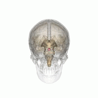
Pituitary adenomas are tumors that occur in the pituitary gland. Most pituitary tumors are benign, approximately 35% are invasive and just 0.1% to 0.2% are carcinomas. Pituitary adenomas represent from 10% to 25% of all intracranial neoplasms and the estimated prevalence rate in the general population is approximately 17%.
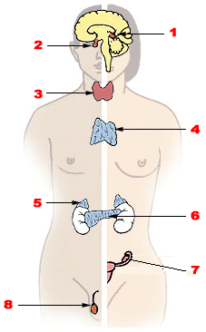
Endocrine glands are ductless glands of the endocrine system that secrete their products, hormones, directly into the blood. The major glands of the endocrine system include the pineal gland, pituitary gland, pancreas, ovaries, testicles, thyroid gland, parathyroid gland, hypothalamus and adrenal glands. The hypothalamus and pituitary glands are neuroendocrine organs.
Diabetes is a chronic disease in cats whereby either insufficient insulin response or insulin resistance leads to persistently high blood glucose concentrations. Diabetes affects up to 1 in 230 cats, and may be becoming increasingly common. Diabetes is less common in cats than in dogs. The condition is treatable, and if treated properly the cat can experience a normal life expectancy. In cats with type2 diabetes, prompt effective treatment may lead to diabetic remission, in which the cat no longer needs injected insulin. Untreated, the condition leads to increasingly weak legs in cats and eventually to malnutrition, ketoacidosis and/or dehydration, and death.
Multiple endocrine neoplasia type 1 (MEN-1) is one of a group of disorders, the multiple endocrine neoplasias, that affect the endocrine system through development of neoplastic lesions in pituitary, parathyroid gland and pancreas. Individuals suffering from this disorder are prone to developing multiple endocrine and nonendocrine tumors. It was first described by Paul Wermer in 1954.
Hyperpituitarism is a condition due to the primary hypersecretion of pituitary hormones; it typically results from a pituitary adenoma. In children with hyperpituitarism, disruption of growth regulation is rare, either because of hormone hypersecretion or because of manifestations caused by local compression of the adenoma.
Somatostatinomas are a tumor of the delta cells of the endocrine pancreas that produces somatostatin. Increased levels of somatostatin inhibit pancreatic hormones and gastrointestinal hormones. Thus, somatostatinomas are associated with mild diabetes mellitus, steatorrhoea and gallstones, and achlorhydria. Somatostatinomas are commonly found in the head of pancreas. Only ten percent of somatostatinomas are functional tumours [9], and 60–70% of tumours are malignant. Nearly two-thirds of patients with malignant somatostatinomas will present with metastatic disease.
Endocrine diseases are disorders of the endocrine system. The branch of medicine associated with endocrine disorders is known as endocrinology.
Acromegaly is a disorder that results in excess growth of certain parts of the human body. It is caused by excess growth hormone (GH) after the growth plates have closed. The initial symptom is typically enlargement of the hands and feet. There may also be an enlargement of the forehead, jaw, and nose. Other symptoms may include joint pain, thicker skin, deepening of the voice, headaches, and problems with vision. Complications of the disease may include type 2 diabetes, sleep apnea, and high blood pressure.
Diabetes mellitus is a disease in which the beta cells of the endocrine pancreas either stop producing insulin or can no longer produce it in enough quantity for the body's needs. The disease can affect humans as well as animals such as dogs.
Diabetes mellitus, often known simply as diabetes, is a group of common endocrine diseases characterized by sustained high blood sugar levels. Diabetes is due to either the pancreas not producing enough insulin, or the cells of the body becoming unresponsive to the hormone's effects. Classic symptoms include thirst, polyuria, weight loss, and blurred vision. If left untreated, the disease can lead to various health complications, including disorders of the cardiovascular system, eye, kidney, and nerves. Untreated or poorly treated diabetes accounts for approximately 1.5 million deaths every year.
Cushing's syndrome disease, also known as hyperadrenocorticism and spontaneous hypercortisolism, is a condition resulting from an endocrine disorder where too much adrenocorticotropic and cortisol hormones are produced, causing toxicity. It may arise in animals as well as in humans. Cushing's is an umbrella term for conditions caused by elevated cortisol and adrenocorticotropic hormone levels.
↑ Peterson, Mark E.; Taylor, R. Steven; Greco, Deborah S.; Nelson, Richard W.; Randolph, John F.; Foodman, Melissa S.; Moroff, Scott D.; Morrison, Susan A.; Lothrop, Clinton D. (1990). "Acromegaly in 14 Cats". Journal of Veterinary Internal Medicine. 4 (4): 192–201. doi:10.1111/j.1939-1676.1990.tb00897.x. ISSN0891-6640.
↑ Niessen, Stijn J.M.; Church, David B.; Forcada, Yaiza (2013). "Hypersomatotropism, Acromegaly, and Hyperadrenocorticism and Feline Diabetes Mellitus". Veterinary Clinics of North America: Small Animal Practice. 43 (2): 319–350. doi:10.1016/j.cvsm.2012.12.004.
↑ Keulen, L. J. M. van; Wesdorp, J. L.; Kooistra, H. S. (1996). "Diabetes Mellitus in a Dog with a Growth Hormone-producing Acidophilic Adenoma of the Adenohypophysis". Veterinary Pathology. 33 (4). SAGE Publications: 451–453. doi:10.1177/030098589603300417. ISSN0300-9858.
↑ Fracassi, F.; Gandini, G.; Diana, A.; Preziosi, R.; Ingh, T.S.G.A.M. van den; Famigli-Bergamini, P.; Kooistra, H.S. (2007). "Acromegaly due to a somatroph adenoma in a dog". Domestic Animal Endocrinology. 32 (1). Elsevier BV: 43–54. doi:10.1016/j.domaniend.2005.12.009. ISSN0739-7240.
↑ Dominici, Fernando P.; Argentino, Danila P.; Muñoz, Marina C.; Miquet, Johanna G.; Sotelo, Ana I.; Turyn, Daniel (2005). "Influence of the crosstalk between growth hormone and insulin signalling on the modulation of insulin sensitivity". Growth Hormone & IGF Research. 15 (5). Elsevier BV: 324–336. doi:10.1016/j.ghir.2005.07.001. ISSN1096-6374.
↑ Clemmons, David R. (2012). "Metabolic Actions of Insulin-Like Growth Factor-I in Normal Physiology and Diabetes". Endocrinology and Metabolism Clinics of North America. 41 (2). Elsevier BV: 425–443. doi:10.1016/j.ecl.2012.04.017. ISSN0889-8529.
↑ Zimmer, C.; Hörauf, A.; Reusch, C. (2000). "Ultrasonographic examination of the adrenal gland and evaluation of the hypophyseal‐adrenal axis in 20 cats". Journal of Small Animal Practice. 41 (4): 156–160. doi:10.1111/j.1748-5827.2000.tb03185.x. ISSN0022-4510.
↑ Kley, S.; Alt, M.; Zimmer, C.; Hoerauf, A.; Reusch, C. E. (2007-11-01). "Evaluation of the low-dose dexamethasone suppression test and ultrasonographic measurements of the adrenal glands in cats with diabetes mellitus". Schweizer Archiv für Tierheilkunde. 149 (11): 493–500. doi:10.1024/0036-7281.149.11.493. ISSN0036-7281.
↑ Meij, Björn P; Auriemma, Edoardo; Grinwis, Guy; Buijtels, Jenny J C W M; Kooistra, Hans S (2010). "Successful Treatment of Acromegaly in a Diabetic Cat with Transsphenoidal Hypophysectomy". Journal of Feline Medicine and Surgery. 12 (5): 406–410. doi:10.1016/j.jfms.2010.03.014. ISSN1098-612X.
↑ Blois, S. L; Holmberg, D. L (2008). "Cryohypophysectomy used in the treatment of a case of feline acromegaly". Journal of Small Animal Practice. 49 (11): 596–600. doi:10.1111/j.1748-5827.2008.00590.x. ISSN0022-4510.
1 2 3 Dunning, M.D.; Lowrie, C.S.; Bexfield, N.H.; Dobson, J.M.; Herrtage, M.E. (2009). "Exogenous Insulin Treatment after Hypofractionated Radiotherapy in Cats with Diabetes Mellitus and Acromegaly". Journal of Veterinary Internal Medicine. 23 (2): 243–249. doi:10.1111/j.1939-1676.2008.0242.x. ISSN0891-6640.
↑ Littler, R. M.; Polton, G. A.; Brearley, M. J. (2006). "Resolution of diabetes mellitus but not acromegaly in a cat with a pituitary macroadenoma treated with hypofractionated radiation". Journal of Small Animal Practice. 47 (7): 392–395. doi:10.1111/j.1748-5827.2006.00078.x. ISSN0022-4510.
This page is based on this Wikipedia article Text is available under the CC BY-SA 4.0 license; additional terms may apply. Images, videos and audio are available under their respective licenses.