
The inner ear is the innermost part of the vertebrate ear. In vertebrates, the inner ear is mainly responsible for sound detection and balance. In mammals, it consists of the bony labyrinth, a hollow cavity in the temporal bone of the skull with a system of passages comprising two main functional parts:

The middle ear is the portion of the ear medial to the eardrum, and distal to the oval window of the cochlea.
The ossicles are three bones in either middle ear that are among the smallest bones in the human body. They serve to transmit sounds from the air to the fluid-filled labyrinth (cochlea). The absence of the auditory ossicles would constitute a moderate-to-severe hearing loss. The term "ossicle" literally means "tiny bone". Though the term may refer to any small bone throughout the body, it typically refers to the malleus, incus, and stapes of the middle ear.
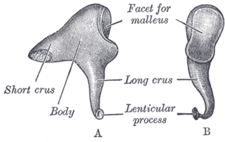
The incus or anvil in the ear is one of three small bones (ossicles) in the middle ear. The incus receives vibrations from the malleus, to which it is connected laterally, and transmits these to the stapes medially. The incus is named for its resemblance to an anvil.

The stapes or stirrup is a bone in the middle ear of humans and other animals which is involved in the conduction of sound vibrations to the inner ear. This bone is connected to the oval window by its annular ligament, which allows the footplate to transmit sound energy through the oval window into the inner ear. The stapes is the smallest and lightest bone in the human body, and is so-called because of its resemblance to a stirrup.

Otosclerosis is a condition of the middle ear where portions of the dense enchondral layer of the bony labyrinth remodel into one or more lesions of irregularly-laid spongy bone. As the lesions reach the stapes the bone is resorbed, then hardened (sclerotized), which limits its movement and results in hearing loss, tinnitus, vertigo or a combination of these. The term otosclerosis is something of a misnomer: much of the clinical course is characterized by lucent rather than sclerotic bony changes, so the disease is also known as otospongiosis.

An ear is the organ that enables hearing and body balance using the vestibular system. In mammals, the ear is usually described as having three parts: the outer ear, the middle ear and the inner ear. The outer ear consists of the pinna and the ear canal. Since the outer ear is the only visible portion of the ear in most animals, the word "ear" often refers to the external part alone. The middle ear includes the tympanic cavity and the three ossicles. The inner ear sits in the bony labyrinth, and contains structures which are key to several senses: the semicircular canals, which enable balance and eye tracking when moving; the utricle and saccule, which enable balance when stationary; and the cochlea, which enables hearing. The ear is a self cleaning organ through its relationship with earwax and the ear canals. The ears of vertebrates are placed somewhat symmetrically on either side of the head, an arrangement that aids sound localization.

The acoustic reflex is an involuntary muscle contraction that occurs in the middle ear in response to loud sound stimuli or when the person starts to vocalize.
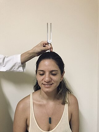
The Weber test is a screening test for hearing performed with a tuning fork. It can detect unilateral (one-sided) conductive hearing loss and unilateral sensorineural hearing loss. The test is named after Ernst Heinrich Weber (1795–1878). Conductive hearing ability is mediated by the middle ear composed of the ossicles: the malleus, the incus, and the stapes. Sensorineural hearing ability is mediated by the inner ear composed of the cochlea with its internal basilar membrane and attached cochlear nerve. The outer ear consisting of the pinna, ear canal, and ear drum or tympanic membrane transmits sounds to the middle ear but does not contribute to the conduction or sensorineural hearing ability save for hearing transmissions limited by cerumen impaction.

Tympanoplasty is the surgical operation performed to reconstruct hearing mechanism of middle ear.

Amblysomus is a genus of the golden mole family, Chrysochloridae, comprising five species of the small, insect-eating, burrowing mammals endemic to Southern Africa. All five species can be found in South Africa and some are also found in Eswatini and Lesotho.

The evolution of mammalian auditory ossicles was an evolutionary process that resulted in the formation of the bones of the mammalian middle ear. These bones, or ossicles, are a defining characteristic of all mammals. The event is well-documented and important as a demonstration of transitional forms and exaptation, the re-purposing of existing structures during evolution.

The following outline is provided as an overview of and topical guide to human anatomy:
The neural encoding of sound is the representation of auditory sensation and perception in the nervous system. The complexities of contemporary neuroscience are continually redefined. Thus what is known of the auditory system has been continually changing. The encoding of sounds includes the transduction of sound waves into electrical impulses along auditory nerve fibers, and further processing in the brain.

The incudomalleolar joint or articulatio incudomallearis is a small synovial joint between the malleus (hammer) and the incus (anvil). The joint's function is to transfer vibrations between the ossicles in the middle ear, which is perceived as sound. Contrary to other synovial joints the movement is very limited. All of the ossicles move more or less as a unit, at least at low frequencies.
In medicine, an ossicular replacement prosthesis is a device intended to be implanted for the functional reconstruction of segments of the ossicles and facilitates the conduction of sound waves from the tympanic membrane to the inner ear. There are two common types of ossicular replacement prostheses, the total ossicular replacement prosthesis (TORP) and partial ossicular replacement prosthesis (PORP). A TORP replaces the entire ossicular chain while a PORP replaces only the incus and malleus but not the stapes. Indications for use of an ossicular replacement prosthesis include:

The malleus, or hammer, is a hammer-shaped small bone or ossicle of the middle ear. It connects with the incus, and is attached to the inner surface of the eardrum. The word is Latin for 'hammer' or 'mallet'. It transmits the sound vibrations from the eardrum to the incus (anvil).
A middle ear implant is a hearing device that is surgically implanted into the middle ear. They help people with conductive, sensorineural or mixed hearing loss to hear.

Thickened earlobes-conductive deafness syndrome, also known as Escher-Hirt syndrome, or Schweitzer Kemink Graham syndrome, is a rare genetic disorder which is characterized by ear and jaw abnormalities associated with progressive hearing loss. Two families worldwide have been described with the disorder.
Chaoyangodens is an extinct genus of eutriconodont mammal from the Early Cretaceous of China. It includes a single species, Chaoyangodens lii, known from a single complete skeleton recovered from the Dawangzhangzi bed of the Yixian Formation, part of the fossiliferous Jehol biota. Chaoyangodens was a moderate-sized Mesozoic mammal. The generic name refers to Chaoyang Prefecture while the specific name honors the collector of the fossil, Hai-Jun Li. Chaoyangodens is intermediate in age between Liaoconodon and a diverse fauna of eutriconodonts from older beds of the Yixian Formation. Like Liaoconodon, it is not easily equated with other eutriconodonts, since it bears distinctive dental traits relative to recognized eutriconodont subgroups.

